UTERINE CERVICAL CYTOMORPHOLOGY IN SYMPTOMATIC POSTMENOPAUSAL WOMEN
Nisa Kaiho1, Laitonjam Sushila Devi2, Laiphrakpam Ranjit Singh3
1Tutor, Department of Pathology, Jawaharlal Nehru Institute of Medical Sciences, Imphal, Manipur.
2Associate Professor, Department of Pathology, Jawaharlal Nehru Institute of Medical Sciences, Imphal, Manipur. 3Professor & HOD, Department of Obstetrics & Gynaecology, Regional Institute of Medical Sciences, Imphal, Manipur.
ABSTRACT
BACKGROUND
In postmenopausal women, risk of uterine cervical dysplasia and malignancy is increased, especially in those with no history of previous Papanicolaou (Pap) smears. Therefore, routine screening can help in reducing morbidity and mortality.
AIM
To study the uterine cervical cytomorphology in symptomatic postmenopausal women, prevalence of dysplasia/malignancy, to observe the relation of cervical cytomorphology with urogenital symptoms, age of onset and duration of menopause.
MATERIALS AND METHODS
A total of 102 symptomatic postmenopausal patients underwent Pap smear examination and reporting was done based on The 2001 Bethesda System.
RESULTS
Out of 102 smears, 101(99.02%) cases were satisfactory for reporting. Age ranged from 44 to 79 years with mean and median age of 54.3±6.8 and 52.5 years respectively. Maximum cases were in the 50-59 age group, 57(56.5%) cases. Age of onset of menopause varied from 40 to 56 years with mean and median age of 48.6±3.4 and 49 years respectively. Duration of menopause ranged from 1 to 26 years with mean and median duration of 5.78±5.7 and 4.0 years respectively. Negative for intraepithelial lesion or malignancy (NILM) and epithelial cell abnormalities (ECA) were 86(85.1%) and 15(14.9%) cases respectively. ECA were most commonly seen in the 60-69 years’ age group, 7(46.7%) cases. Overall prevalence of cervical dysplasia was 9(8.9%), with highest prevalence in the 60-69 age group, 5(27.7%); and carcinoma was 3(2.9%) with highest in the 60-69 age group, 2(11.1%) cases. Vaginal discharge was the commonest urogenital symptom, 40 (39.8%) cases. Reactive cellular changes (RCC) was the commonest finding in vaginal discharge, 24(60%) cases. Maximum cases of ECA, 12(44.4%) cases, were associated with postmenopausal bleeding (PMB). Mean age in dysplasia and malignancy (58.6±7.3 years) was significantly higher (P<0.019), with higher proportion in women >54 years (P<0.030). Significant correlation of dysplasia and malignancy was seen with duration >5 years (P<0.011).
CONCLUSION
Pap smear examination in symptomatic postmenopausal women will definitely bring about better management of cases thereby improving the quality of life.
KEYWORDS
Pap smear, Postmenopausal women, Cervical dysplasia, HPV, Cervical cancer.
HOW TO CITE THIS ARTICLE: Kaiho N, Devi LS, Singh LR. Uterine cervical cytomorphology in symptomatic postmenopausal women. J. Evid. Based Med. Healthc. 2016; 3(27), 1237-1241. DOI: 10.18410/jebmh/2016/284
INTRODUCTION: Menopause occurs when menstruation has permanently stopped for a period of one year. Atrophic changes follow reduced oestrogen stimulation which predispose patients to vaginitis and cervicitis. Various urogenital symptoms may also occur in these women.
Most postmenopausal women do not avail medical consultation early due to social or cultural inhibitions and economic reasons. In such women, there is a risk of undetected malignancies of the urogenital tract, especially
cervical cancer. In Manipur, cervical cancer is the second leading site of cancer among females.1
The Papanicolaou (Pap) smear introduced by George N Papanicolaou has helped in cancer prevention and reduction of morbidity and mortality rates. The slow progression of cervical cancer, cytologically identifiable precursors, and effective treatments have made it one of the most preventable cancers.2
The present study is a cytological study especially in regard to atrophy related changes of the uterine cervix which will be of immense help in better management of symptomatic postmenopausal patients. It also serves as a screening agent for pre-malignant and malignant lesions of the cervix for those who have never undergone such tests. Financial or Other, Competing Interest: None.
Submission 03-03-2016, Peer Review 17-03-2016, Acceptance 25-03-2016, Published 02-04-2016. Corresponding Author:
Dr. Nisa Kaiho,
Department of Pathology,
Jawaharlal Nehru Institute of Medical Sciences, Imphal, Manipur. E-mail: [email protected]
AIMS AND OBJECTIVES:
1. To study the cytomorphology of uterine cervical Pap smears in postmenopausal women presenting with urogenital symptoms.
2. To find out the prevalence of dysplasia/malignancy in symptomatic postmenopausal women.
3. To study the relationship of cervical cytomorphology with urogenital symptoms, age of onset of menopause and duration of menopause.
MATERIALS AND METHODS: This cross-sectional study was carried out in the Department of Pathology, Regional Institute of Medical Sciences (RIMS), Imphal from August 2008 to July 2010 in collaboration with the Department of Obstetrics and Gynaecology, RIMS. A total of 102 symptomatic postmenopausal patients underwent uterine cervical Pap smear examination and the smears were studied using the Pap staining method. Women with natural menopause for a period of not less than one year duration
with urogenital symptoms like vaginal
discharge/dryness/bleeding, dysuria, etc. were included in the study. Known cases of malignancy/dysplasia of the urogenital tract, total hysterectomy, radiotherapy of pelvic region, chemotherapy and extreme debility were excluded. Detailed history was taken regarding symptoms, menopause, past illness, investigations and treatment. The patient was advised to avoid douching or use of any lubricant for 24 hours prior to sample collection. Per speculum examination of vagina and cervix was done, followed by Pap smear from the squamocolumnar junction using a sterile
Ayre’s spatula. The material was then evenly spread on a
clean glass slide which was fixed immediately in 95% ethanol for 30 minutes followed by Pap stain, mounting of the smears with DPX and examination under a compound microscope.
RESULTS: Of the 102 Pap smears taken, 101(99.02%) cases were satisfactory for reporting. One case (0.98%) was found unsatisfactory for evaluation and was excluded for statistical analysis.
Age-Wise Distribution: Age ranged from 44 to 79 years with mean age of 54.3±6.8 years and median age of 52.5 years. Maximum number was found in the 50-59 years’ age group, 57(56.5%) cases, followed by 40-49 years, 22(21.8%) cases, 60-69 years, 18(17.8%) cases and ≥70 years, 4(3.9%) cases respectively.
Age of Onset of Menopause: Age varied from 40 to 56 years with mean age of 48.6±3.4 years and median age at 49 years. Maximum patients, 44(43.6%) cases were seen in the 45-49 age group.
Duration of Menopause: Duration ranged from 1 to 26 years with mean and median duration of 5.78±5.7 and 4.0 years respectively. Maximum cases were seen in the first 5 years, 68(67.5%) cases.
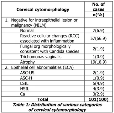
Cytological Results According To the Bethesda System 2001: 86 cases (85.1%) were reported as NILM. ECA were 15(14.9%) cases. The number of cases with various categories of cervical cytomorphology is given in Table 1.
Cervical cytomorphology
No. of cases n(%)
1. Negative for intraepithelial lesion or malignancy (NILM)
Normal 7(6.9)
Reactive cellular changes (RCC)
associated with inflammation 57(56.9) Fungal org morphologically
consistent with Candida species 2(1.9)
Trichomonas vaginalis 1(0.9)
Atrophy 19(18.9)
2. Epithelial cell abnormalities (ECA)
ASC-US 2(1.9)
ASC-H 1(0.9)
LSIL 5(4.9)
HSIL 4(3.9)
Ca 3(2.9)
Total 101(100)
Table 1: Distribution of various categories of cervical cytomorphology
ASC-US: Atypical Squamous Cells of Undetermined Significance.
ASC-H: Atypical Squamous Cells, Cannot Exclude HSIL. LSIL: Low-grade Squamous Intraepithelial Lesion. HSIL: High-grade Squamous Intraepithelial Lesion. Ca: Squamous Cell Carcinoma.
Maximum cases of NILM were in the 50-59 years’ age group, 51(59.3%) cases, whereas that of ECA occurred in the 60-69 years’ age group, 7 cases (46.7%). Mean age of cases with cervical dysplasia and malignancy was higher as compared to those cases without cervical dysplasia and malignancy and the difference was found to be statistically significant (P<0.019). Cases with cervical dysplasia & malignancy was more in cases >54 years, 9(19.6%) while the number of cases without dysplasia & malignancy was
more in those ≤54 years, 3(5.5%) and the difference was
statistically significant (P<0.030).
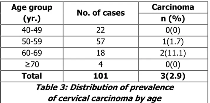
The prevalence of cervical dysplasia and carcinoma by age is given in Table 2 and 3.
Age gp (Yr.)
No. of cases
Cervical dysplasia
LSIL HSIL Total SIL*
n (%) n (%) N (%)
40-49 22 1(4.5) 1(4.5) 2(9)
50-59 57 1(1.7) 0 (0) 1(1.7)
60-69 18 2(11.1) 3(16.6) 5(27.7)
≥70 4 1(25) 0 (0) 1(25)
Total 101 5(4.9) 4(3.9) 9(8.9)
Table 2: Distribution of prevalence of cervical dysplasia by age
Age group
(yr.) No. of cases
Carcinoma n (%)
40-49 22 0(0)
50-59 57 1(1.7)
60-69 18 2(11.1)
≥70 4 0(0)
Total 101 3(2.9)
Table 3: Distribution of prevalence of cervical carcinoma by age
The present study reveals a higher prevalence of LSIL, 5(4.9%) as compared to HSIL, 4(3.9%) and carcinoma 3(2.9%). Of the various urogenital symptoms, vaginal discharge was the most common, 40 cases (39.8%) with genital prolapse as the least common, 4 cases (3.9%). Other symptoms are postmenopausal bleeding (PMB), pruritus vulvae, vaginal dryness, pain in lower abdomen, frequency, dysuria. Maximum cases were in the 50-59 years’ age group, 57 cases (56.9%).
Distribution of Cases by Urogenital Symptoms and Cervical Cytomorphology:
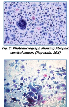
NILM: RCC was the most common urogenital symptom except in vaginal dryness and dysuria. In dryness, atrophic smear was the most common, 3(50%) cases. [Fig. 1] In dysuria, RCC and atrophic smears were equally seen, 2(40%) cases each. Cases with vaginal discharge, pruritus, dryness, prolapse, pain lower abdomen, frequency and dysuria were found to be more in cases with NILM as compared to those cases with ECA.
ECA: 2(5%) cases of ASC-US and 1(2.5%) case of HSIL were associated with vaginal discharge. Maximum cases, 12(44.4%), of ECA were associated with PMB: 1(3.7%) case of ASC-H, 5(18.5%) cases of LSIL and 3(11.1%) cases each of HSIL and SCC. [Fig. 2].
Distribution of cases with respect to duration of menopause and cervical cytomorphology:
NILM: Maximum cases, 61 (70.9%), were seen in the first 5 years of menopause.
ECA: 2(100%) cases of US and 1(100%) case of ASC-H were found in the first 5 years of menopause. In LSIL, 2(40%) cases occurred in the first 5 years and 1 case (20%) each at 6-10, 16-20 and 21-25 years respectively. In HSIL, 2(50%) cases occurred at 11-15 years and 1(25%) case each at 1-5 and 6-10 years’ duration. Carcinoma was seen in 1(33.3%) case each at 1-5, 6-10 and 16-20 years’ duration respectively. Mean duration of menopause was higher in those cases with ECA as compared to those cases with NILM and the difference was statistically significant (P<0.018). Significant correlation of dysplasia and malignancy was seen with duration of menopause >5 years, (P<0.011).
Distribution of Cases with Respect to Duration of Menopause and Urogenital Symptoms: Symptoms were most frequent in the first 5 years of postmenopause, 68(67.7%) cases, except for genital prolapse which occurred at 6-10 years postmenopause.
DISCUSSION: Pap smears are easy, cost effective screening procedures mostly done in women with clinical symptoms. Systematised mass screening programme to study the prevalence of dysplasia is unavailable in Manipur. In the present study, maximum cases were found in the 50-59 years’ age group, 57(56.5%). Thakur S and Roy S3 found maximum cases in the 50-60 age group (48%). Very elderly postmenopausal women are rarely tested due to low hospital attendance in concordance with the finding of Ferrante JM et al.4 The mean age of onset of menopause in the present study was 48.6±3.4 years and median age was 49 years, which are similar to the finding of Shah R et al.5 Similar to Thakur S and Roy S3, maximum number of cases attained menopause between 45-49 years, 44(43.6%) cases.
Cytology Results According to the Bethesda System 2001: NILM was seen in 86(85.1%) cases similar to that reported by Saad RS et al.6 RCC associated with inflammation were seen in 57(56.9%) cases, which is lower to that of Abati A et al,7 55(65.47%) cases. Normal smears were seen in 7(6.9%) cases in which 3(42.9%) were in the 40-49 age group which is similar to the finding of Cheung ANY et al.8 Atrophic smears were seen in 19(18.9%) cases, similar to Mulay K et al (16.15%),9 with the highest number of cases in the 50-59 age group, 12(63.1%). Postmenopausal related atrophy changes do not depend on the duration of menopause.10 Fungal organisms morphologically consistent with Candida species were seen in 2(1.9%) cases. Misra JS et al11 reported an incidence rate of 0.5%. Higher incidence of candidiasis may be due to causes like concomitant steroid or broad-spectrum antibiotic therapy. Partners may be a reservoir of infection as also stated by Thomas A et al.10Trichomonas vaginalis seen in 1(0.9%) case was lower than the finding of Misra JS and Pandey S12 (1.4% cases). The prevalence rate was 1.7% whereas Misra JS et al11 reported prevalence rate of 1.1% in women >40 years. In postmenopausal women, inflammatory changes do not always reflect cervical infection or result in detection of specific pathogens.10
also be reactivation of latent HPV infection, especially some types of yet unknown carcinogenic potential.17
Prevalence of Cervical Malignancy: Overall prevalence was 3(2.9%), similar to Okonda S et al18 (3.6%). The present study reveals a higher prevalence rate of LSIL, 5(4.9%), as compared to HSIL, 4(3.9%) and carcinoma 3(2.9%). This is similar to Okonda S et al18 and Misra JS and Pandey S.12
Urogenital Symptoms: Maximum cases were in the 50-59
years’ age group, 57(56.9%). Barlow DH et al19 reported
maximum cases in the 45-59 age group. Overall, most common symptom was vaginal discharge, 40 (39.8%) cases, similar to Thomas A et al3 and Misra JS and Pandey S.12 Second most common was PMB, 27(26.8%) cases, similar to Thakur S and Roy S3 (28%). Pruritus vulvae was seen in 7(6.9%) cases, similar to Thomas A et al (8% cases).10 He found that inflammation was present among all the various symptoms. In the present study, all 3 cases of carcinoma were associated with PMB similar to the finding of Niehe VG.20 The proportion of cases with PMB was found to be more in cases with ECA as compared to those with NILM and the difference was statistically significant (P<0.000), as also noted by Avasthi K et al.21 Therefore, thorough diagnostic evaluation is mandatory in PMB to facilitate early detection of dysplastic or malignant cases.
Duration of Menopause: Duration (1 to 26 years) was similar to that of Thomas A et al10 (1 to >25 years). Mean duration was higher in ECA (9.07±7.3 years) as compared to NILM and the difference was statistically significant (P<0.018). Significant correlation of dysplasia and malignancy was seen with duration >5 years (P<0.011). Most complaints, 68(67.7%) cases were encountered during the first 5 years of menopause.Maximum cases of PMB, 10 cases, were seen immediately after the first 12 months of amenorrhoea as also reported byAstrup K and Olivarius NF et al.22 Singh A and Arora S23 reported 42% cases within the first 5 years of menopause. Variations in findings in comparison with other studies may be due to differences in the sample size and duration of study period. There is an increased risk of rapidly progressing cancers surfacing clinically at unfavourable stages. Misapplication of age as criteria for reducing importance of Pap testing in this age group will deny them the opportunity for timely disease detection. In the absence of mass cervical screening programmes in our state, this study shows the importance of conducting routine Pap smear tests in postmenopausal women attending gynaecological outpatient departments, especially in those who have never or rarely been tested.
CONCLUSION: A significant number of dysplastic and malignant smears were seen in symptomatic postmenopausal women with no history of abnormal smears. In view of the absence of systematised uterine cervical screening programs in our state, these findings stress the importance of routine Pap smears in such women,
especially in the older age groups, for the timely detection of pre-malignant lesions which would help in reducing morbidity and mortality due to uterine cervical cancer. Knowledge about prevalence of dysplasia and malignancy in this age group would also be invaluable in developing strategies for Pap smear testing and improvement of cytodiagnosis.
Fig. 1: Photomicrograph showing Atrophic cervical smear. (Pap stain, 10X)
Fig. 2: Photomicrograph of cervical Pap smear showing SCC. (Pap stain, 40X)
BIBLIOGRAPHY:
1. Population based cancer registry, Imphal, National cancer registry programme, ICMR, Annual report, 2008.
2. Leyden WA, Manos MM, Geiger AM, et al. Cervical cancer in women with comprehensive health care access: attributable factors in the screening process. J Natl Cancer Inst 2005;97(9):675-683.
3. Thakur S, Roy S. A study of gynaecological conditions in postmenopausal women. Obs & Gynae Today 2005;10:472-474.
4. Ferrante JM, Gonzalez EC, Roetzheim RG, et al. Clinical and demographic predictors of late-stage cervical cancer. Arch Fam Med 2000;9(5):439-445.
5. Shah R, Kalgutkar S, Savardekar L, et al. Menopausal symptoms in urban Indian women. Obs & Gynae Today 2004;11(10):667-670.
7. Abati A, Jaffurs W, Wilder AM. Squamous atypia in the atrophic cervical vaginal smear: a new look at an old problem. Cancer 1998;84(4):218-225.
8. Cheung ANY, Szeto EF, Ng K, et al. Atypical squamous cells of undetermined significance on cervical smears. Cancer 2004;102(2):74-80.
9. Mulay K, Swain M, Patra S, et al. A comparative study of cervical smears in an urban hospital in India and a population-based screening program in Mauritius. Indian J Pathol Microbiol 2009;52(1):34-37.
10.Thomas A, Correa MMA, Kumar KR. Clinical profile and
cervical cytomorphology in symptomatic
postmenopausal women. Indian J Pathol Microbiol 2003;46(2):176-179.
11.Misra JS, Das K, Chandrawati. Cytological detection of sexually transmitted diseases (STDs) during routine outpatient screening. J Obstet Gynecol Ind 2001;51(4):133-136.
12.Misra JS, Pandey S. Cervical cytology in menopausal women. J Obstet Gynecol Ind 2003;53(5):468-472. 13.Wong QC, Collins R, Kalkstein K, et al. Cervical cancer
screening among elderly urban women in a primary care setting. Proc West Pharmacol Soc 2005;48:154-156.
14.Howell LP, Tabnak F, Tudury AJ, et al. Role of pap test terminology and age in the detection of carcinoma invasive and carcinoma in situ in medically underserved California women. Diagn Cytopathol 2004;30(4):227-234.
15.Datta SD, Koutsky LA, Ratelle S, et al. Human papillomavirus infection and cervical cytology in women screened for cervical cancer in the United States, 2003-2005. Ann Intern Med 2008;148(7):493-500.
16.Colgan TJ, Clarke A, Hakh N, Seidenfeld A. Screening for cervical disease in mature woman: strategies for improvement. Cancer 2002;96(4);195-203.
17.Zietkowiak W, Zimna K, Sroka L, et al. Frequency of HPV infection of the uterine cervix among perimenopausal women in Wielkopolska region. Ginekol Pol 2002;73(11):939-944.
18.Okonda S, Wright C, Michelow P. The status of cervical cytology in Swaziland, Southern Africa: a descriptive study. Cytojournal 2009;6(14):1-7.
19.Barlow DH, Brockie JA, Rees CMP. Study of general practice consultations and menopausal problems. BMJ 1991;302:274-276.
20.
Niehe VG. Clinical profile of patients with carcinoma cervix in All India Institute of Medical Sciences (AIIMS), in New Delhi, India. Obs & Gynae Today 2006;11:459-460.
21.Avasthi K, Jindal P, Garg S. Epithelial abnormalities of the cervix in menopausal and perimenopausal women. Obs & Gynae Today 2009;14:344-347.
22.Astrup K, Olivarius NF. Frequency of spontaneously occurring postmenopausal bleeding in the general
population. Acta Obstet Gynecol Scand
2004;83(2):203-207.