Detection and genotyping of HPV in women
with indeterminate cytology and low-grade
squamous intraepithelial lesions
Detecção e genotipagem do HPV em mulheres com citologia
indeterminada e lesão intraepitelial escamosa de baixo grau
Francisca A. Queiroz1; Danielle A. P. Rocha2; Roberto Alexandre A. B. Filho2; Cristina Maria B. Santos2 1. Fundação Alfredo da Mata (FUAM) – Manaus, Amazonas. 2. Universidade Federal do Amazonas (UFAM).
First submission on 29/10/14; last submission on 08/05/15; accepted for publication on 10/05/15; published on 20/06/15
ABSTRACT
Introduction: The human papillomavirus (HPV) is the main risk factor related to cervical cancer, the third most frequent type of cancer in Brazilian women. Early identiication of high-risk HPV types in the normal cervix, or cervix with premalignant lesions may help prevent the progression of these lesions to cancer. Objective: This study aimed to detect and genotype HPV in women with low-grade intraepithelial lesion (LSIL) and atypical squamous cells of undetermined signiicance (ASC-US). Methods: Patients were selected from iles of the cytology laboratory of Fundação Alfredo da Mata (FUAM), between January 2009 and July 2011, for cytological reassessment and HPV molecular detection with genotyping. Results: Out of the100 eligible patients, 70% (70/100) participated in the study; initially, 34 of them had ASC-US and 36 had LSIL. After cytological reassessment, eight (11.4%) patients showed normal cytology; 33 (47.2%), inlammatory lesions; 22 (31.4%), ASCUS; six (8.6%), LSIL; and one (1.4%), high-grade squamous intraepithelial lesions (HSIL). HPV was detected in 28.6% (20/70) of the samples. Out of the 20 HPV-positive patients, one had normal cytology, six showed inlammatory cytology, 10 showed ASCUS, two had LSIL and one, HSIL. After genotyping, the HPV types identiied were: 6, 16, 58, 61, 70, 83, 84 and 85. The most prevalent HPV type was the 58. Conclusion: The presence of high-risk HPV in women with old cervical lesions, whether they have evolved or not, indicates the need to improve patient monitoring and surveillance.
Key words: cervical cytology; HPV; PCR.
INTRODUCTION
Cervical cancer is a condition that causes great impact in the Brazilian public health context because it is the third most frequent malignancy in women in the country. In the state of Amazonas, however, the problem is even more serious, since this kind of cancer ranks irst among the most prevalent tumors affecting women(1). Concerning the etiology of cervical cancer,
human papillomavirus (HPV) is considered an essential, but not a suficient risk factor for malignant transformation, as cancer is a multifactorial disease(2).
There are no oficial data on the prevalence of HPV infection in Brazil. Most of the estimates come from regional studies, which although limited in scope, when considered together can provide
a picture of infection by this microorganism. A systematic review study showed an overall prevalence of 13.7%-54.4% HPV infection of the cervix in Brazilian women(3, 4).
Currently, there are over 100 described HPV types, 40 of which can cause genital infections(5). HPVs are classiied according to
their association with cancer. The low-risk (non-oncogenic) types, of which the most prevalent are HPVs 6 and 11, cause benign exophytic lesions (such as warts and condyloma) and low-grade intraepithelial lesions (LSIL) of the uterine cervix. The high-risk (oncogenic) types, including the most prevalent 16 and 18 HPV types, can cause high-grade intraepithelial cervical lesions(6, 7).
Many HPV infections are asymptomatic, and most of them progress to a state of spontaneous remission, becoming undetectable within two years. However, persistent infection with
high-risk HPV can result in morphological changes in the ecto- and endocervical epithelium, which may favor the development of precancerous lesions, and later, neoplastic lesions. These precursor changes can be detected by cytological screening or Pap smear(8).
The use of molecular methods for deoxyribonucleic acid (DNA) detection and genotyping of HPV in cervical samples along with cytology may be an important strategy in the identiication of patients predisposed to malignant processes, contributing to reduce morbidity and mortality from cervical cancer(9). Molecular
tests for HPV detection are important especially in cases of lesions that are indeterminate by cytology (squamous atypia of undetermined signiicance [ASC-US]), because cervical cytology has low reproducibility, with sensitivity ranging from 50% to 99%, and up to 29.7% of false-negative results(10, 11). The aim
of this study was to detect and genotype HPV among women with LSIL and ASC-US, and correlate the socioeconomic data and risk factors associated with cervical cancer with molecular detection of the pathogen.
METHODS
Sample collection
This cross-sectional study, held at Fundação Alfredo da Mata (FUAM) in Manaus (Amazonas, Brazil), from January 2009 to July 2011, analyzed women with positive Pap smear results for ASC-US and LSIL. Data collection was based on reports of the cervical cancer information system ([SISCOLO], Federal Health Department, Brazil) used by the cytology laboratory of FUAM. The women were contacted by telephone and invited to participate in the study. Out of the 100 invited women, 70 agreed to participate. Participants were interviewed, they provided informed consent, and were examined by the nursing staff. Cervical samples were obtained for the Pap test to evaluate lesion progression, and for the polymerase chain reaction (PCR) to detect HPV DNA. The cytology specimens were classiied as normal (negative for malignancy), reactive changes, ASC-US, LSIL, high-grade squamous intraepithelial lesions (HSIL) and invasive carcinoma.
DNA extraction
A 400-µl proteolytic tris(hydroxymethyl)aminomethane (Tris) + proteinase K (TPK) buffer (Tris buffer + ethylenediaminetetraacetic acid [EDTA] [TE] {Tris HCl 50 mM + EDTA 50 mM pH = 8.0}, Tween 20% and proteinase K
10 mg/ml) was added to 400 µl of each sample, and the samples were incubated for 60 minutes at 56ºC and for 10 minutes at 95ºC in a dry bath. Then, DNA was extracted by the phenol/ chloroform method(12), precipitated with absolute ethanol
and resuspended in 50 µl of ultrapure water (pH = 7.6). Quantiication of DNA was performed in a NanoDrop (Thermo Scientiic) equipment to check the extraction eficiency.
PCR for HPV DNA detection
Nested-PCR was performed for HPV DNA detection. The irst reaction was performed using MY09 and MY11(13) primers, which
amplify a 450-bp fragment. The reaction inal volume was 25 µl, containing 5 U of Platinum Taq DNA Polymerase (Invitrogen, Brazil), 5 pmol of each primer, 2.5 µl of reaction buffer 10×, 50 mM of MgCl2, 10 mM of deoxyribonucleotides phosphate
(dNTP), 2.5 µl of sample and water. The second reaction was performed using GP5+ and GP6+(14) primers, which amplify a
150-bp fragment. The reaction inal volume was 25 µl, containing 5 U of Platinum Taq DNA Polymerase (Invitrogen, Brazil), 5 pmol of each primer, 2.5 µl of reaction buffer 10×, 50 mM of MgCl2, 10 mM of dNTP, 0.5 µl of sample (product resulting from the irst ampliication with MY09 and MY11) and water. Both reactions obeyed the following thermocycler conditions: 95ºC for one minute, 40 cycles of 95ºC for one minute, 55ºC for one minute and 72ºC for one minute, ending with 72ºC for ive minutes. In the reactions of this study, water was included as a negative control; and a previously sequenced sample, as positive control. Reactions were performed in a Veriti thermocycler (Applied Biosystems). The ampliications products were subjected to electrophoresis on agarose gel 2.0% stained with ethidium bromide (1 µg/µl), then visualized with the aid of a transilluminator, and the image was captured by a digital camera Olympus SP-500UZ.
HPV genotyping by gene sequencing
one minute, 15 cycles de 96ºC for 10 seconds, 50ºC for 15 seconds, 60ºC for 120 seconds. Precipitation of the sequencing plate was accomplished, and then the sequences were sequenced on an ABI 3130XL automated sequencer (Applied Biosystems) according to manufacturer’s instructions. For genotyping, the iles generated by the automated sequencer were compared with the sequences deposited in GenBank, using the BLAST program.
Data analysis
The correlation between the results obtained in Pap smear, clinical and socioeconomic data with molecular biology was established. For the quantitative variables, the mean and standard deviation (SD) were calculated. In data analysis, Pearson’s chi-squared test was also used. The analysis of quantitative variables was carried out by comparing the means and through Fisher’s exact test. When data normality was veriied, Student’s t test was used. The signiicance level ixed in the application of the tests was 5%.
Ethics
This research was conducted in compliance with all ethical issues concerning research with human beings, and the research project was approved by the ethical research committee of FUAM.
RESULTS
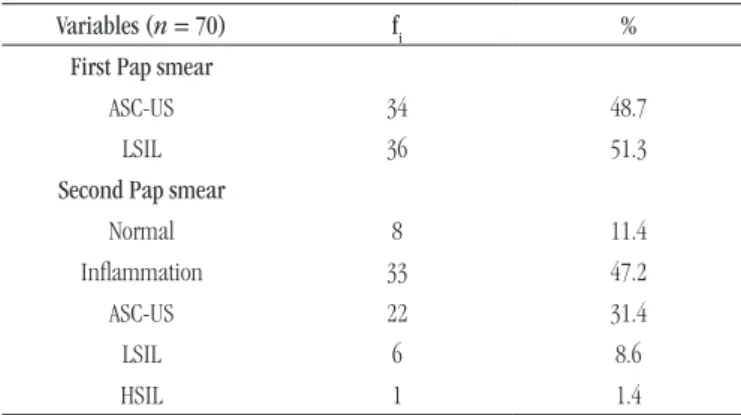
Out of the 70 women who agreed to participate in this research, 34 had ASC-US and 36 had LSIL in the irst Pap smear test. The mean age of these women was 29.9 years (SD = 11.1). In the second Pap smear, some women showed a different cytological diagnosis: eight had normal (11.4%) results, 33 had inlammation (47.2%), 22 showed ASC-US (31.4%), six presented LSIL (8.6%) and one, HSIL (1.4%) (Table 1). Table 2 details the modiications in Pap smear diagnoses in each case.
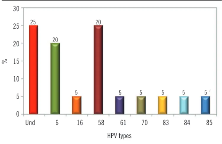
HPV infection was found in 28.6% (20/70) of the samples, and in these samples eight different types of HPV were identiied, with a predominance of HPV 58 and HPV 6 (Figure). It was not possible to
identify HPV type in ive HPV-positive samples (25%). Table 3 shows the distribution of HPV genotypes found in the second Pap smear.
In an interview conducted with patients, data on socioeconomic, clinical and sexual behavioral conditions of women, such as education status, family income, marital status, number of sexual partners, condom use, history of sexually transmitted diseases (STD),
TABLE 1 – Distribution by frequency according to the results of the irst and
second cytology, Manaus (AM)
Variables (n = 70) fi %
First Pap smear
ASC-US 34 48.7
LSIL 36 51.3
Second Pap smear
Normal 8 11.4
Inlammation 33 47.2
ASC-US 22 31.4
LSIL 6 8.6
HSIL 1 1.4
fi: simple absolute frequency; ASC-US: squamous atypia of undetermined significance; LSIL: low-grade squamous intraepithelial lesions; HSIL: high-grade squamous intraepithelial lesions.
TABLE 2 – Comparative distribution of Pap results in the irst and second
samples, Manaus (AM)
Second Pap smear (n = 70)
First Pap smear
ASC-US LSIL
Total
fi % fi %
Normal 3 37.5 5 62.5 8
Inlammation 18 54.5 15 45.5 33
ASC-US 10 45.5 12 54.5 22
LSIL 2 33.3 4 66.7 6
HSIL 1 100 - - 1
fi: simple absolute frequency; ASC-US: squamous atypia of undetermined significance; LSIL: low-grade squamous intraepithelial lesions; HSIL: high-grade squamous intraepithelial lesions.
TABLE 3 – Distribution of HPV genotypes according to the
second Pap Smear, Manaus (AM) HPV genotype
(n = 20)
Second Pap smear
Normal Inlammation ASC-US LSIL HSIL Total
6 - 1 3 - - 4
16 - - - - 1 1
58 1 2 1 1 - 5
61 - - 1 - - 1
70 - - - 1 - 1
83 - - 1 - - 1
84 - 1 - - - 1
85 - 1 - - - 1
Undetermined - 1 4 - - 5
fi: simple absolute frequency; HPV: human papillomavirus; ASC-US: squamous atypia
of undetermined significance; LSIL: low-grade squamous intraepithelial lesions; HSIL: high-grade squamous intraepithelial lesions.
DISCUSSION
The development of cervical cancer is directly related to persistent infection of high-risk HPV, and is preceded by pre-malignant lesions, called squamous intraepithelial lesion (SIL), which are classiied into categories (low SIL or high SIL), depending on the morphological changes of cells seen by the cytological exam. The low-grade lesions represent mainly the changes associated with active replication of HPV, and in these stages some lesions may undergo spontaneous remission. The high-grade lesions indicate cell transformation, mainly characterized by nuclear changes (lost nucleus/cytoplasm ratio, nuclear hyperchromatism, nuclear pleomorphism and chromatin architecture), and in these cases it is possible to treat and monitor patients, preventing lesions from becoming invasive(15-17). Squamous changes of undetermined
signiicance refer more to striking cytological abnormalities than to reactive changes, being suggestive of intraepithelial lesion, but are neither quantitatively nor qualitatively suficient for a deinitive result. For this diagnosis, the cells must present squamous differentiation, increased nuclear/cytoplasmic ratio, irregular chromatin distribution and irregular nucleus shape(18).
In this study, in most cases there was regression of abnormal cytology, with only three cases of progression from ASC-US to LSIL (two cases) and HSIL (one case). It is important to consider, however, that in this study the time between the irst and the second sample collection was short (maximum of three years).
Since 1950, the screening of cervical lesions has been based only on cytology, which is a simple and inexpensive test. In this examination, it is possible to identify the likely presence of HPV by its cytopathic effect, known as koilocytosis. However, the widespread use of this technique as a method of preventing cervical cancer presents some questions, mainly related to the high rate of false-TABLE 4 – Comparison between clinical variables and distribution
according to socioeconomic data regarding the outcome of PCR women seen at the clinic for STD at FUAM, Manaus (AM)
Variables (n = 70)
PCR
Positive Negative
Total p
fi % fi %
Age
Mean ± SD 29 ± 9.6 30.3 ± 11.8 0.674***
Marital status
Single 13 28.9 32 71.1 45 ****
Married 6 27.3 16 72.7 22
Divorced 1 50 1 50 2
Widow - - 1 100 1
Age at irst intercourse
< 16 years old 12 26.7 33 73.3 45 ≥ 16 years old 8 32 17 68 25 0.636*
Age at irst pregnancy
< 16 years old 6 40 9 60 15
≥ 16 years old 4 14.3 24 85.7 28 0.066*
Condom use with regular partner (n = 58)
Always 8 50 8 50 16
Not always 1 4.5 21 95.5 22 ****
Rarely 3 27.3 8 72.7 11
Never 6 30 3 6 9
Condom use with casual partner (n = 12)
Always 5 41.6 5 41.6 10
Never - - 2 100 2 ****
Lifelong number of partners
1 to 4 14 28.6 36 71.4 50
6 to 10 3 27.3 8 72.4 11 ****
More than 10 3 15 6 12 9
Contraceptive use
Yes 9 47.4 10 52.6 19
No 11 21.6 40 78.4 51 0.034*
Clinical complaints
Bleeding - - 1 100 1
Genital warts 11 42.3 15 57.7 26 ****
Discharge - - 4 100 4
Without complaints 9 23.1 30 76.9 39
Previous STD
Yes 16 25.8 46 74.2 62
No 4 50 4 50 8 0.156**
*Qui-squared test; **Fisher’s exact test; ***Student’s t test; ****it is not possible to apply the test due to restriction of qui-square; fi: simple absolute frequency; SD: standard-deviation; PCR: polymerase chain reaction; STD: sexually transmitted diseases; FUAM: Fundação Alfredo da Mata.
FIGURE − Distribution of HPV types found in HPV-positive samples HPV: human papillomavirus; Und: undetermined.
HPV types 30
25
20
15
10
5
0
%
Und 6 16 58 61 70 83 84 85
25
20
5 5 5 5 5 5
negative results(19). There is a tendency, however, to consider that
the best routine for identiication and monitoring of HPV lesions to prevent cervical cancer is the combination of Pap smear with PCR, in which there is ampliication of conserved regions of the viral genome and then identiication of the viral type by gene sequencing of the ampliied fragment, or by speciic probes. PCR would therefore be important to complement the Pap smear exam, mainly to increase the eficiency of screening programs for cervical cancer prevention. In epidemiological studies, PCR is the most widely used technique(17, 20-22).
The emergence of PCR as a method for HPV detection was extremely important because identiication and implementation of eficient virological diagnostic tests were hampered by the fact that HPV cannot be grown in the laboratory from clinical specimens (as done with some other viruses), for its species-speciic character (which limits the spread in animal models) and because immunological tests were not suitable for the detection of this virus(20, 21). In this study, we used the technique of nested
PCR to increase sensitivity of the reaction, since using only MY09/ MY11 primers, 8.6% of samples (6/70) resulted in false-negative diagnoses. The nested PCR technique, however, has the disadvantage of increasing the possibility of contamination. In order to prevent contamination, all necessary precautions were taken in all stages of the process, including the use of two negative controls in each PCR reaction. We found a 28.6% prevalence of HPV infection. Although it is a considerable value, it is not as high as other reported studies, and even studies of women with normal cytology found higher HPV rates(22-25). In our study, HPV 58 was found to be the most prevalent
among infected women (25%). This viral type is classiied as high-risk HPV and is closely related to HPV 16(26, 27). Global studies show
HPV 58 as the seventh most common type in women with precursor lesions and cervical cancer, and the sixth most common type in women without alterations. However, variations in prevalence have shown signiicant regional differences. The highest prevalence rates of HPV 58 are in Latin American and Asian countries. High prevalence rates of this type have been found in China, Japan, Mexico, Costa Rica, Colombia and Brazil(27-30). In the Amazon
region, studies of Rocha et al. (2013) and Santos et al. (2013) found
a signiicant prevalence of HPV 58 in women by studying routine pelvic examination. Considering the fact that the Northern region of Brazil is a popular region for Japanese immigrants and that the state borders are open to immigrants from neighboring countries (Colombia and Peru, mainly), our data corroborate the information on circulating viral types in these countries.
It is important to consider that HPV 16 was found only in one sample, and HPV 18 was not detected in any of the women. These two viruses are the most prevalent in most epidemiological studies related to cervical cancer and its precursor lesions. Unlike this study, most Brazilian studies have found HPV 16 as the most prevalent, both in women without cytological abnormalities and in women
with premalignant lesions and invasive cancer(22, 31-33). However,
in agreement with our work, other Brazilian studies have found low prevalence of HPV 18(22, 34, 35). We should point out that for viral
genotyping, the method of automated DNA sequencing was used. The advantage of direct sequencing is the identiication of any virus type present (unlike the methods using a pool of probes for hybridization); the main drawbacks are the dificulty of sequencing small DNA fragments, the impossibility of detecting coinfections, and the capacity of detecting only the type of higher viral load in the sample. Thus, it was not possible to identify HPV in ive of 20 samples (25%), which only got positive results after the second reaction (nested PCR using GP5+/GP+ primers), whose inal product is 150 bp.
During the period of the study (January 2009 to July 2011), the sample consisted of 100 women who had Pap results of ASC-US and LSIL. All women were sought, but only 70 (70%) agreed to be reassessed. In this sample, the majority of HPV-infected women were observed to be young, unmarried, and live on low incomes; about half of them had initiated sexual activity before 16 years of age. However, no association of socioeconomic factors and sexual behavior with HPV infection was found.
The state of Amazonas was the irst Brazilian state to provide the anti-HPV vaccination for all girls aged between 11 and 14 years, in 2013. On that occasion, the bivalent vaccine was chosen. In 2014, the anti-HPV vaccination oficially entered the Brazilian immunization schedule. Since then the vaccine has been administered to girls around the country, and the quadrivalent vaccine has been chosen. Although these vaccines have been implemented, the discussion regarding the need for vaccine development that takes into account the genotypic diversity of circulating HPVs regionally remains.
CONCLUSION
In this study, although we found a considerable category change of cytological abnormalities in a short time, with a prevalence of the cytological regression framework, we also found a signiicant prevalence of HPV infection in women. The high prevalence of HPV 58 infection among infected women emphasizes the need for further studies on the genotypic variety of HPV in this Brazilian region. It also stresses the need to develop other vaccines considering the regional genotypic diversity.
ACKNOWLEDGMENTS
RESUMO
Introdução: O papilomavírus humano (HPV) é o principal fator de risco relacionado com câncer cervical. A identificação precoce
de tipos de HPV de alto risco em cérvice normal ou com lesões precursoras pode auxiliar a prevenir que essas lesões progridam para neoplasias malignas. Objetivo: Este estudo teve como objetivo detectar e genotipar o HPV em mulheres com resultado citológico de lesão intraepitelial escamosa de baixo grau (LSIL) e células escamosas atípicas de significado indeterminado (ASC-US). Métodos: Mulheres diagnosticadas com LSIL e ASC-US foram selecionadas a partir do arquivo de exames do Laboratório de Citologia da Fundação Alfredo da Matta (FUAM), em Manaus (AM), no período de janeiro de 2009 a julho de 2011, para reavaliação citológica, detecção molecular e genotipagem do HPV. A detecção molecular foi realizada pela técnica de nested reação em cadeia da polimerase (PCR); a genotipagem, por sequenciamento automático do ácido desoxirribonucleico (DNA).
Resultados: Das 100 pacientes selecionadas, 70% (70/100) participaram do estudo, sendo que inicialmente 34 delas tinham
resultado citológico de ASC-US e 36 de LSIL. Após reavaliação citológica, oito (11,4%) apresentaram citologia normal; 33 (47,2%), citologia inflamatória; 22 (31,4%), ASC-US; seis (8,6%), LSIL e uma (1,4%), lesão intraepitelial de alto grau (HSIL). O HPV foi detectado em 28,6% (20/70) das amostras examinadas. Das 20 pacientes HPV positivas, uma apresentou citologia normal; seis apresentaram citologia inflamatória; 10 exibiram ASC-US; duas, LSIL e uma, HSIL. Foram identificados os tipos de HPV 6, 16, 58, 61, 70, 83, 84 e 85, sendo o HPV 58 o mais prevalente. Conclusão: A presença de HPV de alto risco em mulheres com lesões cervicais antigas, tendo elas evoluído ou não, mostra a necessidade de maior acompanhamento e vigilância dessas pacientes.
Unitermos: citologia cervical; HPV; PCR.
REFERENCES
1. Instituto Nacional do Câncer José Alencar Gomes da Silva. Estimativa 2012: incidência de câncer no Brasil. Instituto Nacional do Câncer. Rio de Janeiro, Brasil; 2011.
2. Muñoz N, Castellsagué X, de González AB, Gissmann L. HPV in the etiology of human cancer. Vaccine. 2006; 24(3): 1-10.
3. Ayres ARG, Silva GA. Cervical HPV infection in Brazil: systematic review. Rev Saúde Pública. 2010; 44(5): 963-74.
4. Oliveira GR, Vieira VC, Barral MFM, et al. Risk factors and prevalence of HPV infection in patients from Basic Health Units of an University Hospital in southern Brazil. Rev Bras Ginecol Obstet. 2013; 35(5): 226-32. 5. Hariri S, Dunne E, Saraiya M, Unger E, Markowitz L. Human papillomavirus. In: CDC, editors. Manual for the surveillance of vaccine-preventable diseases. 5 ed. Atlanta: CDC; 2011.
6. Louvanto K, Rintala MA, Syrjänen KJ, Grénman SE, Syrjänen SA. Incident cervical infections with high- and low-risk human papillomavirus (HPV) infections among mothers in the prospective Finnish Family HPV Study. BMC Infect Dis. 2011; 11: 179.
7. Zampronha RAC, Freitas-Júnior R, Murta EFC, et al. Human papillomavirus types 16 and 18 and the prognosis of patients with stage I cervical cancer. Clinics. 2013; 68(6): 809-14.
8. Araújo SCF, Caetano R, Braga JU, Silva FVC. Eficacy of commercially available vaccines against HPV infection in women: a systematic review and meta-analysis. Cad Saúde Pública. 2013; 29(Suppl 1): S32-S44. 9. Silveira LMS, Silva HA, Pinheiro VMF, Veloso AOL, Everton HFSN. Cytological abnormalities in cervix of women attended at Central Laboratory of Public Health of Maranhão. NewsLab. 2007; 81: 130-40.
10. Franco EL, Villa LL, Sobrinho JP, et al. Epidemiology of acquisition and clearance of cervical human papillomavirus infection in women from a high-risk area for cervical cancer. J Infect Dis. 1999; 180(5): 1415-23. 11. Nuovo J, Melnikow J, Howell LP. New tests for cervical cancer screening. Am Fam Physician. 2001; 64(5): 780-6.
12. Sambrook J, Fritsch EF, Maniatis T. Molecular cloning: a laboratory manual. 2 ed. New York: Cold Spring Harbor Lab; 1989.
13. Manos MN. Use of the polymerase chain reaction for the detection of genital human papillomaviruses. In: Furth M, editor. Molecular diagnostics of human cancer. New York: Cold Spring Harbor Laboratory; 1989. p. 209-14 [Cancer Cell Series, 7].
14. de Roda Husman AM, Walboomers JM, van de Brule AJ, Meijer CJ, Snijders PJ. The use of general primers GP5 and GP6 elongated at their 3’ ends with adjacent highly conserved sequences improves human papillomavirus detection by PCR. J Gen Virol. 1995; 76(Pt 4): 1057-62. 15. Finam RR, Tamim H, Almawi WY. Identiication of Chlamydia trachomatis DNA in human papillomavirus (HPV) positive women with normal and abnormal cytology. Arch Gynecol Obstet. 2002; 266(3): 168-71. 16. Steben M, Duarte-Franco E. Human papillomavirus infection: epidemiology and pathophysiology. Gynecol Oncol. 2007; 107(2 Suppl 1): S2-S5.
19. Amaral RG, Manrique EJC, Guimarães JV, et al. Inluence of adequacy of the sample on detection of the cervical cancer. Rev Bras Ginecol Obstet. 2008; 30(11): 556-60.
20. Magalhães IM, Moysés N, Afonso LA, Oliveira LHS, Cavalcanti SMB. Comparison of two pairs of primers used in polymerase chain reaction for the detection of human papillomaviruses in cervical smears. DST– J Bras Doenças Sex Transm. 2008; 20(2): 93-8.
21. Passos MRL, Almeida G, Giraldo PC, et al. Genital human papillomavirosis. Part I. DST – J Bras Doenças Sex Transm. 2008; 20(2): 108-24.
22. Rocha DAP, Barbosa-Filho RAA, Queiroz FA, Santos CMB. High prevalence and genotypic diversity of human papillomavirus in Amazonian women, Brazil. Infect Dis Obstet Gynecol. 2013; 2013: 1-7. 23. Tábora N, Ferrera A, Bakkers JMJE, et al. Human papillomavirus infection in Honduran women with normal cytology. Cancer Causes Control. 2009; 20: 1663-70.
24. Piras F, Piga M, Montis AD, et al. Prevalence of human papillomavirus infection in women in Benin, West Africa. J Virol J. 2011; 8: 514. 25. Lindemann MLM, Calvo JMS, Antonio JC, et al. Prevalence and distribution of high-risk genotypes of HPV in women with severe cervical lesions in Madrid, Spain: importance of detecting genotype 16 and other high-risk genotypes. Adv Prev Med. 2011; 2011: 1-4.
26. Villiers EM, Fauquet C, Broker TR, Bernard HU, zur Hausen H. Classiication of papillomaviruses. Virology. 2004; 324(1): 17-27. 27. Calleja-Macias IE, Villa LL, Prado JC, et al. Worldwide genomic diversity of the high-risk human papillomavirus types 31, 35, 52 and 58, four close relatives of human papillomavirus type 16. J Virol. 2005; 79(21): 13630-40.
28. Camara GNL, Cerqueira DM, Oliveira AP, Silva EO, Carvalho LGS, Marins CRF. Prevalence of human papillomavirus types in women with pre-neoplastic and neoplastic cervical lesions in the Federal District of Brazil. Mem Inst Oswaldo Cruz. 2003; 98(7): 879-83.
29. Canche JC, Lópes IR, Suárez NG, et al. High prevalence and low E6 genetic variability of human papillomavirus 58 in women with cervical cancer and precursor lesions in Southeast Mexico. Mem Inst Oswaldo Cruz. 2010; 105(2): 144-8.
30. Takehara K, Toda T, Nishimura T, et al. Human papillomavirus types 52 and 58 are prevalent in uterine cervical squamous lesions from Japanese women. Pathol Res Inter. 2011; 2011: 1-7.
31. Lippman AS, Sucupira MCA, Jones HE,et al. Prevalence, distribution and correlates of endocervical human papillomavirus types in Brazilian women. Int J STD AIDS. 2010; 21(2): 105-9.
32. Santos JC, Cezar MRS, Lisboa MR, Moura MMF. Occurrence of human papillomavirus in uterine cervix of women in the western Brazilian Amazon. Acta Amazônica. 2013; 43(2): 185-90.
33. Silva KC, Rosa MLG, Moyses N, Afonso LA, Oliveira LHS, Cavalcanti SMB. Risk factors associated with human papillomavirus infection in two populations from Rio de Janeiro, Brazil. Mem Inst Oswaldo Cruz. 2009; 104(6): 884-91.
34. Castro MM, Farias IP, Borboema-Santos CM, Correia G, Astolf-Filho S. Prevalence of human papillomavirus (HPV) type 16 variants and rare HPV types in a central Amazon region. Gen Mol Research. 2011; 10(1): 186-96.
35. Mattos AT, Freitas LB, Lima BMC, Miranda ES, Spano LC. Diversity and uncommon HPV types in HIV seropositive and seronegative women attending an STI clinic. Braz J Microbiol. 2011; 42: 786-93.
MAILING ADDRESS
Danielle Albuquerque Pires Rocha