Bifidobacterium longum
CCM 7952 Promotes
Epithelial Barrier Function and Prevents Acute
DSS-Induced Colitis in Strictly Strain-Specific
Manner
Dagmar Srutkova1, Martin Schwarzer1, Tomas Hudcovic1, Zuzana Zakostelska2, Vladimir Drab3, Alena Spanova4, Bohuslav Rittich4, Hana Kozakova1*,
Irma Schabussova5
1Laboratory of Gnotobiology, Institute of Microbiology of the Czech Academy of Sciences, v.v.i., Novy Hradek, Czech Republic,2Laboratory of Cellular and Molecular Immunology, Institute of Microbiology of the Czech Academy of Sciences, v.v.i., Prague, Czech Republic,3Dairy Research Institute Ltd., Prague, Czech Republic,4Brno University of Technology, Faculty of Chemistry, Brno, Czech Republic,5Institute of Specific Prophylaxis and Tropical Medicine, Center for Pathophysiology, Infectiology and Immunology, Medical University of Vienna, Vienna, Austria
Abstract
Background
Reduced microbial diversity has been associated with inflammatory bowel disease (IBD) and probiotic bacteria have been proposed for its prevention and/or treatment. Neverthe-less, comparative studies of strains of the same subspecies for specific health benefits are scarce. Here we compared twoBifidobacterium longumssp.longumstrains for their capac-ity to prevent experimental colitis.
Methods
Immunomodulatory properties of nine probiotic bifidobacteria were assessed by stimulation of murine splenocytes. The immune responses toB.longumssp.longumCCM 7952 (Bl 7952) and CCDM 372 (Bl 372) were further characterized by stimulation of bone marrow-derived dendritic cell, HEK293/TLR2 or HEK293/NOD2 cells. A mouse model of dextran sulphate sodium (DSS)-induced colitis was used to compare their beneficial effectsin vivo.
Results
The nine bifidobacteria exhibited strain-specific abilities to induce cytokine production. Bl 372 induced higher levels of both pro- and anti-inflammatory cytokines in spleen and dendritic cell cultures compared to Bl 7952. Both strains engaged TLR2 and contain ligands for NOD2. In a mouse model of DSS-induced colitis, Bl 7952, but not Bl 372, reduced clinical symptoms and preserved expression of tight junction proteins. Importantly, Bl 7952 improved intestinal barrier function as demonstrated by reduced FITC-dextran levels in serum.
OPEN ACCESS
Citation:Srutkova D, Schwarzer M, Hudcovic T, Zakostelska Z, Drab V, Spanova A, et al. (2015) Bifidobacterium longumCCM 7952 Promotes Epithelial Barrier Function and Prevents Acute DSS-Induced Colitis in Strictly Strain-Specific Manner. PLoS ONE 10(7): e0134050. doi:10.1371/journal. pone.0134050
Editor:Mathias Chamaillard, INSERM, FRANCE
Received:April 1, 2015
Accepted:July 3, 2015
Published:July 28, 2015
Copyright:© 2015 Srutkova et al. This is an open access article distributed under the terms of the
Creative Commons Attribution License, which permits unrestricted use, distribution, and reproduction in any medium, provided the original author and source are credited.
Data Availability Statement:All relevant data are within the paper and its Supporting Information files.
Conclusions
We have shown that Bl 7952, but not Bl 372, protected mice from the development of exper-imental colitis. Our data suggest that although some immunomodulatory properties might be widespread among the genusBifidobacterium, others may be rare and characteristic only for a specific strain. Therefore, careful selection might be crucial in providing beneficial outcome in clinical trials with probiotics in IBD.
Introduction
Inflammatory bowel disease (IBD), such as ulcerative colitis (UC) and Crohn’s disease (CD), comprises a variety of chronic immune-mediated inflammatory disorders of the gastrointesti-nal tract. Although their aetiology and pathogenesis are not completely understood, it is becoming clear that the combination of genetic, immunological, environmental, and microbial factors play an important role in the development and progression of these conditions [1,2].
Once considered rather rare, IBD has been raising dramatically over the past few decades in the high income countries [3,4]. It has been suggested that modern style of life, such as modern sanitation systems, modifications in diet, decline in endemic parasitism, smaller family size or overuse of antibiotics changed the structure and function of the intestinal microbiota [5]. Indeed, dysbiosis, such as reduction in mucosa-associatedBifidobacteriumspp. orLactobacillus
spp., along with an increased relative abundance in pathogenicEscherichia coliwas observed in IBD patients [6–8]. Current pharmaceutical treatment of IBD, which includes anti-inflamma-tory and immunosuppressive drugs, biological agents and antibiotics, induces or maintain remission, but is not curative [9]. Moreover, long-term use of these drugs can lead to substan-tial side effects such as allergic reactions or liver problems [10]. Since IBD is clearly multifacto-rial and results from complex host-microbiota interactions, preventive strategies targeting the aberrant composition of the intestinal microbiota may have the potential to open new possibili-ties to tackle these diseases.
Probiotic bacteria, most notably theBifidobacteriumandLactobacillusgenera have been used for the prevention and/or treatment of gastrointestinal inflammatory diseases [11,12]. Application of multispecies product VSL#3, which is a mixture ofLactobacillus, Bifidobacter-ium, andStreptococcusstrains, has been used successfully to treat UC [11,13,14]. Controver-sially, clinical trials using different strains have not confirmed the beneficial effects [15–18]. It is getting clear that not only the type of disease and the immunological status of the host, but also the selection of probiotic strain and the mode of application are important factors to be taken into consideration. Essentially, the immunomodulatory potential and beneficial effect of probiotic bacteria seems to be strictly strain-specific and cannot be automatically applied to another strain, or even if to another species.
Several animal models have been developed to understand aetiology and pathogenesis of IBD and to evaluate novel prophylactic/therapeutic strategies [19–21]. Colitis induced by dex-tran sulphate sodium (DSS) is one of the most extensively used experimental models due to its simplicity, reliability and applicability. Acute or chronic disease can be induced by administra-tion of adaptable concentraadministra-tion of DSS in drinking water [19,22,23]. Despite certain differ-ences, DSS model resembles crucial clinical and histopathological features of human IBD; such as changes in morphology of inflamed colon, body weight loss, bloody diarrhoea and aberrant regulatory mechanisms in colon followed by cytokine dysregulation [19,21,22,24].
Other important factors which are shared by human IBD and DSS-induced colitis are reduced expression and/or reorganization of tight junction proteins (e.g. zonulin-1 or occlu-din) in the epithelium, and increased intestinal permeability for luminal bacteria [25]. The breakdown of the gut barrier function have been shown to precede the clinical relapses in UC patients and also the development of intestinal inflammation in DSS-induced colitis [26].
The genusBifidobacteriumis considered as a key member of the human gut microbiota which has been shown to exert a range of beneficial effects on the immune system [27–29]. Notably, the representatives of the speciesB.longumare one of the dominant bacterial mem-bers of the gut microflora of healthy breast-fed infants [30,31]. Interestingly, a double blinded randomised controlled clinical trial showed thatB.longumreduced clinical appearance of chronic mucosal inflammation in patients with active UC [32]. Moreover, Fujiwaraet al. described inhibitory effect ofB.longumstrain on DSS-induced experimental colitis [33]. Along these lines, we have shown recently that mucosal application ofB.longumprevented the devel-opment of experimental allergy in mice [34,35]. Although these data suggest thatB.longum
might be a promising candidate for prevention/treatment of immune-mediated inflammatory diseases, question remains whether differentB.longumstrains, belonging to one subspecies, are equal in their health-beneficial effect.
In the present study, nine different probiotic strains of the genusBifidobacterium, which were originally collected from healthy children and adults, were tested for their ability to induce cytokine production by murine splenocytes. Based on the cytokine profiles, two candi-dates of one subspecies: i)Bifidobacterium longumssp.longumCCDM 372 (Bl 372), the strain with high stimulatory capacity and ii)B.longumssp.longumCCM 7952 (Bl 7952), the strain with low stimulatory capacity, were selected for further comparativein vitroandin vivostudies. In experimental model of acute ulcerative colitis, administration of Bl 7952, but not Bl 372, pre-vented the disruption of gut barrier function by enhanced expression of tight junction proteins in epithelial layer which was associated with reduced development of DSS-induced symptoms.
Materials and Methods
Bacterial strains and culture conditions
NineBifidobacteriumstrains:B.longumssp.longumCCDM 372 (Bl 372) and CCM 7952 (Bl 7952);B.longumssp.infantisCCDM 369 (Bi 369);B.animalisCCDM 218 (Ban 218) and CCDM 366 (Ban 366);B.adolescentisCCDM 368 (Bad 368), CCDM 370 (Bad 370), CCDM 371 (Bad 371) and CCDM 373 (Bad 373) were isolated from faecal samples of healthy adults and breast-fed infants and kindly provided by Prof. V. Rada (Department of Microbiology, Nutrition and Dietetics, Czech University of Agriculture, Prague, Czech Republic) and Prof. J. Nevoral (Department of Internal Medicine, 1stFaculty of Medicine, Charles University in Prague, Czech Republic). These strains were deposit in Culture Collection of Dairy Microor-ganisms (Milcom, Prague, Czech Republic) and Czech Collection of MicroorMicroor-ganisms (Brno, Czech Republic). The isolates were cultivated in MRS medium (Oxoid, Hampshire, UK) sup-plemented with 0.05% L-cysteine-hydrochloride (MRSC) at 37°C in anaerobic conditions for 48 to 72 hours.
Selection and identification of probiotic strains
Inactivation of bacterial strains
Nine probioticBifidobacteriumstrains were cultivated in MRSC medium at 37°C in anaerobic condition to the end of exponential growth phase. Bacterial cells were inactivated with 1% phosphate-buffered formalin for 3 h at room temperature, washed 3 times with sterile phos-phate buffered saline (PBS) and stored at 4°C as previously described [38].
Stimulation of mouse splenocytes with inactivated
Bifidobacterium
strains
Immunomodulatory potential ofBifidobacteriumstrains was testedin vitroon splenocytes derived from naïve BALB/c mice (8 weeks of age; n = 5) in two independent experiments. Spleens were removed aseptically and single cell suspensions were prepared by disruption of the tissues through a cell strainer into culture medium (RPMI 1640 medium supplemented with 10% heat-inactivated FCS, 10mM HEPES, 100 U/ml penicillin and 100μg/ml
streptomy-cin). Spleen cells (6 x 10⁵/well) were stimulated with formalin-inactivatedBifidobacterium
strains (6 x 10⁶/well), Pam3CSK4 (1μg/ml, InvivoGen, USA) or media alone in 96-well plates
at 37°C and 5% CO2for 48 h. Concentration of IFN-γ, TNF-α, IL-6 and IL-10 was determined in cell supernatants by the MILLIPLEX MAP Mouse Cytokine/Chemokine Panel (Millipore Corporation, Billerica, MA, USA) according to manufacturer’s instructions and analysed with Bio-Plex System (Bio-Rad Laboratories, USA).
Preparation and activation of mouse bone marrow-derived dendritic cells
Mouse bone marrow-derived dendritic cells (BM-DC) derived from naïve BALB/c mice (8 weeks of age; n = 3) were prepared as previously described [34,39]. Briefly, bone marrow pre-cursors isolated from femurs and tibias were seeded at 2 x 10⁵cells/ml in RPMI 1640 culture medium containing 10% FCS, 150μg/ml gentamycin, and 20 ng/ml mouse GM-CSF
(Sigma-Aldrich, Germany) and incubated for 8 days. BM-DC (106cells/well) were stimulated with for-malin-inactivatedB.longumstrains Bl 7952 and Bl 372 (107CFU/well), Pam3CSK4 (1μg/ml),
ultrapure LPS (1μg/ml, InvivoGen, USA) or left untreated for 18 h. The levels of 10,
IL-12p70, IL-6 and TNF-αwere analysed in supernatants of stimulated cells by ELISA using Ready-Set-Go! kits (eBioscience, USA) according to manufacturer’s instructions. For cell sur-face marker analysis, BM-DC were labelled for 30 min at 4°C with anti-mouse FITC-conju-gated CD11c, APC-conjuFITC-conju-gated MHC II and PE-conjuFITC-conju-gated CD40, CD80 or CD86 monoclonal antibodies (eBioscience, USA). The data were acquired on a BD FACSAria III flow cytometer (BD Biosciences, USA) and analysed with FlowJo software 7.6.2 (TreeStar, USA).
Stimulation of Human embryonic kidney 293 cells stably transfected with
TLRs and NOD2
Human embryonic kidney (HEK) 293 cells stably transfected with plasmid carrying human (h) TLR2/CD14 gene were kindly provided by Prof. M. Yazdanbakhsh (Leiden, Netherlands), hTLR4/MD2/CD14 were a gift of Prof. B. Bohle, PhD (Vienna, Austria) and hNOD2 express-ing cells were purchased from InvivoGen (USA). Cells were stimulated for 20 h with
Pam3CSK4 (1μg/ml), LPS (1μg/ml), muramyl dipeptide (100 ng/ml, InvivoGen) and
Animals
All experimental mice (female 8-week-old BALB/c) were kept in IVC cages (Tecniplast, Italy), exposed to 12: 12-h light-dark cycles, and fed with standard pellet diet (ST1, Bergman, Kocanda, Czech Republic) and tap waterad libitum. All experiments were approved by the Animal Experimentation Ethics Committee of the Institute of Microbiology of the Academy of Sciences of the Czech Republic and conducted in accordance with the“European Convention for the Protection of Vertebrate Animals used for Experimental and other Scientific Purposes (CETS No.: 123)”.
Experimental design and induction of colitis
Female 8-week-old BALB/c mice were divided into 4 experimental groups. Two groups were treated with 200μl of live bacterial suspension containing 2 x 10⁸CFUs of Bl 7952 (Bl 7952/
DSS; n = 10) or Bl 372 (Bl 372/DSS; n = 8) in PBS by intragastric gavage. Controls (PBS/DSS; n = 10) received PBS only. The administration of bacterial suspensions or PBS was repeated daily for 10 days followed by treatment with 2.5% w/v DSS in drinking water in order to induce acute colitis. Age-matched untreated mice (Naïve; n = 5) were used as healthy controls. Dex-tran sulphate sodium (DSS, molecular weight 40 kDa; ICN Biomedicals, Cleveland, OH, USA) was administered in the drinking water (2.5% w/v) for 7 days. Clinical symptoms of inflamma-tion were evaluated daily, degree of colitis was determined as disease activity index (DAI) according to Cooperet al. [40] with minor modifications (S1 Table). Animals were sacrificed by CO2inhalation and cervical dislocation. The colon was aseptically removed, the length was measured and segments of colon descendens (approximately 0.5 cm in length located 1 cm proximal to the anus) were fixed in 4% buffered paraformaldehyde (Sigma Aldrich, Germany) for histological and immunohistochemical analysis.
Determination of cytokine response of mesenteric lymph node cells
Mesenteric lymph nodes (MLN) were excised aseptically from all experimental mice; cell sus-pensions in concentration 6 x 10⁶cells/ml were prepared and cultivated as described previously [34,39]. Production of IFN-γ, TNF-αand IL-10 was determined in cell supernatants by the MILLIPLEX MAP Mouse Cytokine/Chemokine Panel (Millipore Corporation, Billerica, MA, USA) according to manufacturer’s instruction and analysed with Bio-Plex System (Bio-Rad Laboratories, USA).
Histopathological evaluation of inflammation in colonic mucosa
Colon descendens from all experimental mice (one segment from each mouse) were fixed in 4% paraformaldehyde and processed to paraffin blocks as previously described [41]. For determina-tion of inflammadetermina-tion in colonic mucosa and mucin producdetermina-tion, the tissue specimens were sliced to 5-μm thickness, deparaffined and stained with haematoxylin and eosin (H&E) or Alcian Blue
with post-staining by Nuclear Fast Red (All from Vector, Burlingame, CA, USA). The degree of pathophysiology of the tissue was characterized by presence of ulcerations, damage to the surface epithelium, crypt distortion, signs of oedema, infiltration of inflammatory cells into lamina pro-pria or submucosa and reduction of goblet cells and mucin production according to Cooper
et al. [40]. The histopathological evaluation was performed blindly by two investigators.
Immunohistochemical determination of zonulin-1 and occludin in colon
For immunohistochemical staining, the 5-μm deparaffined colon sections (3 sections from
min at 37°C. Endogenous peroxidase was blocked with 0.3% hydrogen peroxide in 100% meth-anol for 15 min. Nonspecific adsorption was eliminated by incubation of the sections in 10% normal goat serum in PBS for 30 min. Samples were incubated overnight with polyclonal rabbit anti-zonulin-1 (2.5μg/ml) or anti-occludin (2.5μg/ml) (ZYMED Laboratories Inc., San
Fran-cisco, CA, USA) at 4°C. After washing in PBS, section were incubated with goat anti-rabbit IgG conjugated with horseradish peroxidase (1: 200 in PBS) (Jackson, ImmunoLabs.,West Grove, PA, USA) for 1 hour and stain by AEC chromogen solution (Dako, Carpinteria, CA, USA) for 5 min. The counterstain was carried out with haematoxilin and samples were mounted in the Histotec Aqueous Mountant (Serotec, UK) and viewed under an Olympus BX 40 microscope equipped with an Olympus DP 70 digital camera. Photographs were taken on proposal of Camedia Master 2.5 and DP-Soft (Olympus, Germany).
Western blot analysis of zonulin-1 and occludin
Segment of colon descendens (approximately 0.5 cm in length located 1.5 cm proximal to the anus) from all experimental mice was homogenized on ice in protein extract buffer (Pierce, Rockford, IL, USA) with a protease inhibitor cocktail (Pierce) for 10 min and sonicated. Sam-ples were centrifuged at 10,000 x rpm for 10 min at 4°C and stored at−80°C until use. Protein concentration was measured using the BCA Protein Assay Kit (Pierce). Western blotting was performed as described by Cinovaet al. [42] using antibodies against occludin (1:1000) (Invi-trogen, Carlsbad, CA, USA), zonulin-1 (1:1000) (ZYMED Laboratories Inc., San Francisco, CA, USA), andβ-actin (1:5000) (Abcam, Cambridge, CA, USA). After incubation with the respective primary antibodies, secondary staining was performed using horseradish peroxi-dase-conjugated species-specific antibodies (1:1000) (ZYMED Laboratories). The reaction was developed using the SuperSignal Weat Femto Maximum Sensitivity Substrate (Thermo Scien-tific, USA) and the signal intensities were measured on the G:BOX (Syngene, UK) and pro-cessed with Adobe Photoshop CS5.
Evaluation of the intestinal permeability
in vivo
The intestinal permeability was measured by determination of the amount of FITC-dextran in blood after oral administration as described previously [43]. Briefly, female BALB/c mice were intragastrically gavaged by Bl 7952 (Bl 7952/DSS; n = 5) or PBS (PBS/DSS; n = 5) for ten conse-cutive days prior to intestinal inflammation was induced by drinking of 2.5% DSS in water for 7 days. Naïve mice served as healthy controls (Naïve; n = 5). On the last day of DSS administra-tion, each mouse received 360 mg/kg of the body weight of FITC-dextran (molecular weight 4.0 kDa; Sigma-Aldrich) by intragastric gavage. Blood samples were obtained after 5 hours, centrifuged at 3,000 x rpm for 30 min, and serum was collected. The concentration of FITC-dextran was determined by spectrophotofluorometry (Safire2, Tecan Group Ltd., Mannedorf, Switzerland) with an excitation wavelength of 483 nm and an emission wavelength of 525 nm using serially diluted FITC-dextran as standard.
Statistical analysis
Results
Identification of probiotic
Bifidobacterium
isolates to the species,
subspecies and strain level
Nine bacterial isolates of the genusBifidobacteriumfrom healthy children and adults have been selected for their probiotic properties based on resistance to low pH and resistance to bile salt (data not shown). These isolates were identified to the species, subspecies and strain level. PCR-based methods (S1 Materials and Methods) let to identification of three species and two subspecies of the genusBifidobacterium:B.longumssp.longumCCDM 372 (Bl 372) and CCM 7952 (Bl 7952);B.longumssp.infantisCCDM 369 (Bi 369),B.animalisCCDM 218 (Ban 218) and CCDM 366 (Ban 366) andB.adolescentisCCDM 368 (Bad 368), CCDM 370 (Bad 370); CCDM 371 (Bad 371), and CCDM 373 (Bad 373). Discrimination of theB.longumstrains into subspecieslongumandinfantiswas performed on the basis of Amplified ribosomal DNA restriction analysis by the enzymeSau3AI (S1 Fig) as described previously [36].
Immunostimulatory properties of probiotic strains from the genus
Bifidobacterium
are strictly strain-specific
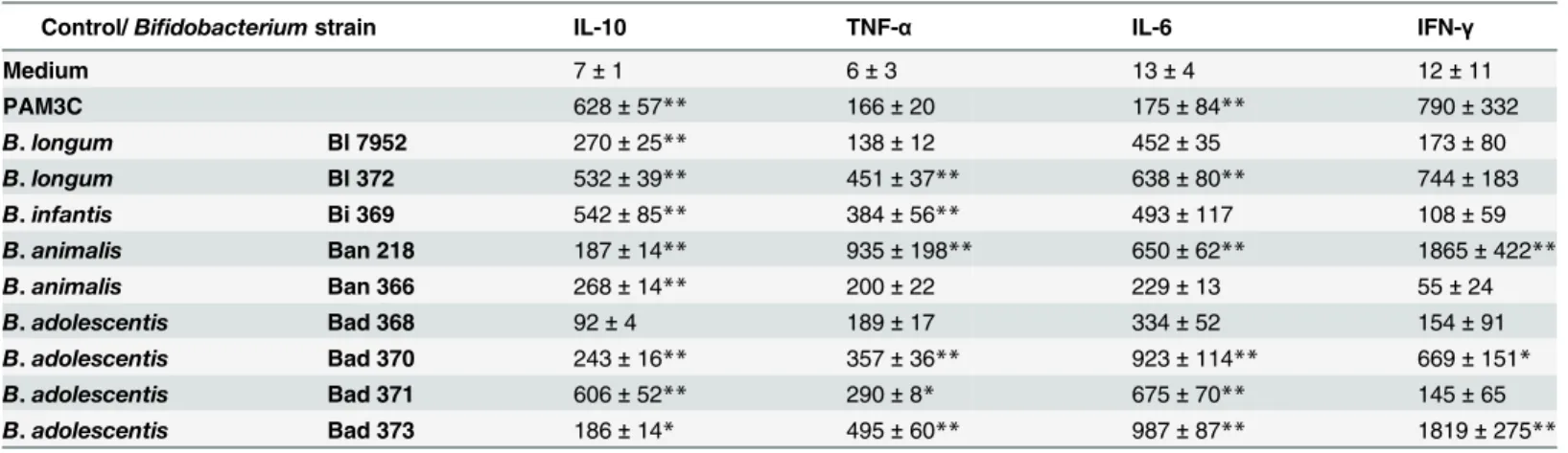
Thein vitrostimulation of spleen cells collected from naïve BALB/c mice with nine Bifidobac-teriumstrains revealed distinct and strain-specific pattern of cytokine production (Table 1). Production of IL-10 by probiotic strains has been associated with their protective effect on inflammatory diseases [44]. Various levels of IL-10 were detected in spleen cultures with values ranging between 100–700 pg/ml, depending on the used strain, where Bi 369, Bad 371, and Bl 372 were the most robust inducers of this anti-inflammatory cytokine (Table 1). On the other hand, Ban 218 and Bad 373 induced low levels of IL-10, but high levels of TNF-α, IL-6 and IFN-γ. Strains Bl 372 and Bad 370 induced substantial production of pro-inflammatory cyto-kines TNF-α, IL-6, and IFN-γ. Strains Bl 7952 and Ban 366 led to only moderate production of TNF-αand IFN-γ, but significantly elevated levels of IL-10. According to thisin vitroanalysis, we selected two strains of one subspeciesB.longumssp.longum: Bl 372 and Bl 7952, which provided contrasting cytokines pattern and used them for further characterization.
Table 1. Cytokine production by splenocytes stimulated with inactivated bacteria of differentBifidobacteriumstrains.
Control/Bifidobacteriumstrain IL-10 TNF-α IL-6 IFN-γ
Medium 7±1 6±3 13±4 12±11
PAM3C 628±57** 166±20 175±84** 790±332
B.longum Bl 7952 270±25** 138±12 452±35 173±80
B.longum Bl 372 532±39** 451±37** 638±80** 744±183
B.infantis Bi 369 542±85** 384±56** 493±117 108±59
B.animalis Ban 218 187±14** 935±198** 650±62** 1865±422**
B.animalis Ban 366 268±14** 200±22 229±13 55±24
B.adolescentis Bad 368 92±4 189±17 334±52 154±91
B.adolescentis Bad 370 243±16** 357±36** 923±114** 669±151*
B.adolescentis Bad 371 606±52** 290±8* 675±70** 145±65
B.adolescentis Bad 373 186±14* 495±60** 987±87** 1819±275**
Splenocytes isolated from naïve mice (n = 5) were stimulated with formalin-inactivated bifidobacteria (6 x 10⁷CFU/ml) for 48 h. Pam3CSK4 (PAM3C, 1μg/
ml) was use as a positive control. Non-stimulated splenocytes (Medium) were evaluated as control of basal cytokine levels. Concentration of cytokines in supernatants was determined by multiplex assay. Data are expressed as mean±SEM. Results are representatives of two repeat experiments. Significant
difference to medium was calculated using One-way ANOVA and Dunnett’s multiple comparison post-hoc test*p<0.05;**p<0.01.
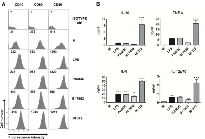
Strains Bl 7952 and Bl 372 have differential ability to activate dendritic
cells
in vitro
Dendritic cells (DC) have been shown to play a central role in regulating intestinal immune homeostasis by induction of tolerance to harmless antigens and commensals or initiating pro-tective immunity against pathogens, contributing to control of intestinal diseases such as inflammatory bowel diseases [45]. In our study, BM-DCs derived from naïve BALB/c mice were used asin vitromodel to investigate the immunostimulatory potential of bothB.longum
spp.longumstrains. Expression of co-stimulatory markers CD40, CD80, and CD86 were measured to investigate the activation status of BM-DC after stimulation with each bacterial strain. The induction of these surface markers differed between the tested strains. Higher lev-els of CD40, CD80 and CD86 were observed in DC incubated with Bl 372 than with Bl 7952 (Fig 1A). Levels of IL-10, TNF-α, IL-6, and IL-12p70 were measured in the supernatants of BM-DC stimulated with bothB.longumspp.longumstrains (Fig 1B). The data show that stimulation of BM-DC with Bl 372 resulted in significantly higher levels of secreted cytokines than stimulation with Bl 7952.
Fig 1. Stimulation of bone marrow-derived dendritic cells with Bl 7952 and Bl 372.Bone marrow-derived dendritic cells (BM-DC) from naïve mice were cultured with formalin-inactivated Bl 7952 or Bl 372 (107CFU/ml) for 18 h. Ultra-pure lipopolysaccharide fromE.coli(LPS, 1μg/ml) and Pam3CSK4 (PAM3C, 1μg/ml) were used as positive controls. Untreated cells (M) served as negative control. (A) Expression of CD40, CD80 and CD86 was assessed by means of flow cytometry. BM-DCs were gated as MHCII+CD11c+. Numbers represent florescence units from one representative experiment out of three. (B)
Cytokines in cell culture supernatants were determined by ELISA. Results are representative of three repeat experiments. Data are expressed as mean±SEM. Significant differences between cytokine levels of experimental group to negative control (M) was calculated using One-way ANOVA and
Dunnett’s multiple comparison post-hoc test (**p<0.01,***p<0.001).
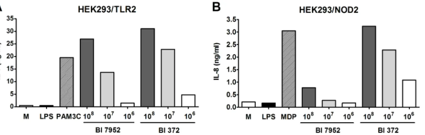
Both Bl 7952 and Bl 372 signal through TLR2 and NOD2 receptor
To assess the role of TLR2 and NOD2 in recognition of Bl 7952 and Bl 372, HEK293 cells stably transfected with TLR2/CD14 or NOD2 were stimulated with increasing concentrations of both strains. Pam3CSK4 and MDP were used as positive controls for TLR2 and NOD2, respectively. After 20 h of incubation, supernatants were harvested and analysed for IL-8 production. At all three tested concentrations, both Bl 7952 and Bl 372 activated TLR2 in an analogous and dose-dependent manner (Fig 2A). In contrast, stimulation of HEK293/NOD2 with Bl 372 induced markedly higher levels of IL-8 in comparison to stimulation with Bl 7952 (Fig 2B). These results suggest that both Bl 7952 and Bl 372 have similar pattern of usage of TLR2 but distinct patterns of interaction with NOD2. There was no stimulation of HEK293/TLR4 cells with any ofB.longumstrains (data not shown).
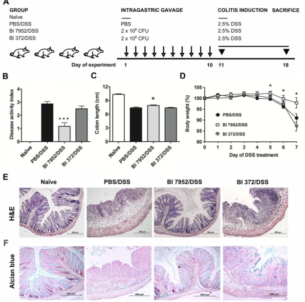
Prophylactic application of Bl 7952, but not Bl 372, ameliorates
DSS-induced colitis
Here we have shown that two strains of one subspeciesB.longumssp.longum, Bl 7952 and Bl 372, possess different immunomodulatory propertiesin vitro(Table 1, Figs1and2). To com-pare their propertiesin vivo, the mouse model of acute ulcerative colitis induced by administra-tion of 2.5% DSS in drinking water was used. Animals received Bl 7952 or Bl 372 on 10
consecutive days prior to colitis induction (Fig 3A). Disease progression was characterized by weight loss, appearance of diarrhoea or loose faeces and visible faecal blood and summarized in disease activity index (DAI) assessed according to the scale (0–4) of Cooperet al. [40] (S1 Table). DSS-treatment increased DAI, reduced the colon length, and reduced body weight in control mice (PBS/DSS) in comparison to naïve animals (Fig 3B–3D). Mice pre-treated with Bl 7952 showed improvement of DAI, and reduction of DSS-induced colon shortening and weight loss compare to PBS/DSS mice (Fig 3B–3D). In contrast, pre-treatment with Bl 372 had no impact on any of these parameters and Bl 372-treated mice did not significantly differ from PBS/DSS group (Fig 3B–3D).
Histopathological evaluation of the colonic mucosa after DSS-treatment was performed to establish a score (0–4) as described before [40]. The scoring was based on infiltration of inflam-matory cells into lamina propria and submucosa, submucosa thickening, and loss of the entire
Fig 2. Activation of TLR2 and NOD2 by Bl 7952 and Bl 372.Human embryonic kidney cells (HEK293) stably transfected with an expression vector for human TLR2 (293-hTLR2/CD14) or with NOD2 (293-hNOD2) were stimulated with formalin-inactivated Bl 7952 or Bl 372 for 20 h. Stimulation was performed at concentrations of 106, 107or 108CFU/ml. Cells stimulated with ultra-pure lipopolysaccharide fromE.coli(LPS, 1μg/ml) or untreated cells (M) were used
as negative controls. Cells stimulated with Pam3CSK4 (PAM3C, 1μg/ml) or muramyl dipeptide (MDP, 1μg/ml) were used as positive controls for TLR2 or NOD2, respectively. Data are expressed as one representative experiment out of three.
crypt with retained surface epithelium [40,41]. On sacrifice, histological finding encompassed infiltration of inflammatory cells into lamina propria, thickening of submucosa, loss of epithe-lial layer and disappearance of mucosal crypt in colonic wall of DSS-treated controls (PBS/ DSS; grade 3.5 ± 0.5) and in Bl 372 pre-treated/DSS-treated mice (Bl 372/DSS; grade 3.5 ± 0.3) (Fig 4E). In contrast, Bl 7952-pre-treated mice displayed inhibitory effect on DSS-induced his-tological changes (Bl 7952/DSS; grade 0.75 ± 0.25) compared to controls. Bl 7952 reduced infil-tration of inflammatory cells and pathological changes in mucosa or epithelial layer (Fig 3E).
Fig 3. Impact of Bl 7952 and Bl 372 on DSS-induced colitis.(A) Mice were treated with Bl 7952 (n = 10), with Bl 372 (n = 8) or with PBS (n = 10) on ten consecutive days. Naïve animals (n = 5) were left untreated. Colonic inflammation was induced by the addition of 2.5% (w/v) DSS in the drinking water for 7
days. (B) Disease activity index and (C) colon length were evaluated at the end of the experiment. (D) Body weight of mice was evaluated throughout the experiment and the values are expressed as percentage of change of the initial value measured before DSS administration. Changes in colonic mucosa after DSS-treatment are shown on representative histological sections of healthy untreated mice (Naïve), mice treated with PBS (PBS/DSS), Bl 7952 (Bl 7952/
DSS) or Bl 372 (Bl 372/DSS). The sections were stained by H&E (E) to address the degree of inflammation and by alcian blue (F) to show the changes in production of mucus in colonic tissue. Graphs show mean±SEM and represent one out of two experiments. Unpaired Student’s t-test was used for comparison of experimental groupsvs. control PBS/DSS group (*p<0.05,***p<0.001).
In control mice with colitis (PBS/DSS) and in mice treated with Bl 372 (Bl 372/DSS), colonic mucin production by goblet cells (Alcian Blue staining) was decreased in comparison to naïve mice (Fig 3F). Markedly, application of Bl 7952 preserved the thinning of the mucus layer and goblet cell depletion (Bl 7952/DSS).
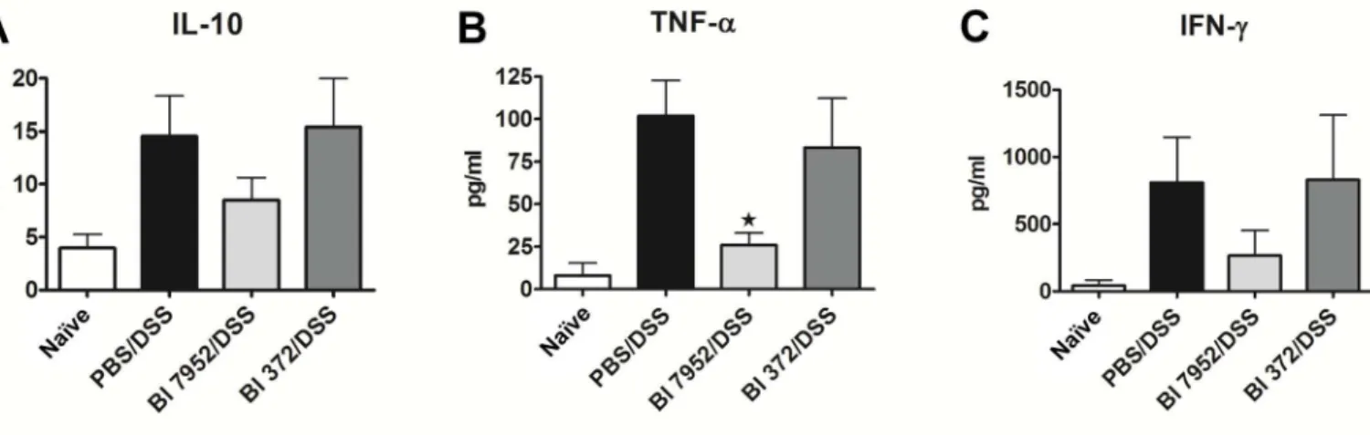
Administration of Bl 7952 has an impact on the production of cytokines in
the mesenteric lymph node cells
Changes in cytokine microenvironment in the gut associated lymphoid tissue, such as mesen-teric lymph nodes (MLN), might impact the development of intestinal inflammation in colitis. Therefore, we investigated whether the protective effect ofB.longumBl 7952 on colitis is asso-ciated with changes in production of pro- and anti-inflammatory cytokines. MLN cells col-lected from naïve, PBS/DSS, Bl 7952/DSS or Bl 372/DSS animals were cultivated for 48 h. Level of cytokines in supernatant was measured by ELISA. We found that pre-treatment with Bl 7952 but not with Bl 372 decreased significantly the production of TNF-α(Fig 4B). Although the levels of IL-10 and IFN-γwere reduced in MLN cell cultures by Bl 7952 in comparison to DSS-controls, the difference did not reach significant level (Fig 4A and 4C).
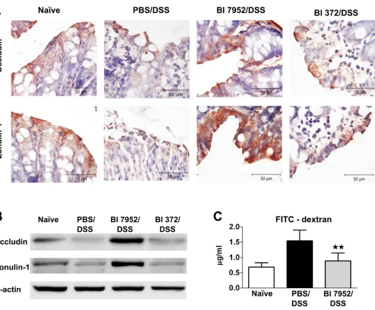
Bl 7952 preserves the expression of zonulin-1 and occludin and
decreases colon permeability in DSS-treated mice
Altered intestinal barrier function (breakdown or impairment of the epithelial barrier), which is associated with increased intestinal permeability through decreased expression of tight junc-tion proteins, has been implicated as a critical factor in the development of intestinal inflam-mation in mouse models of colitis or in human IBD. Occludin and zonulin-1 are proteins involved in the maintenance of the integrity of intact tight junction complexes and barrier function [46]. Here we investigated whether pre-treatment with Bl 7952 interferes with the dis-ruption of these tight junction proteins induced by DSS-treatment. As shown by immunohis-tochemistry staining (Fig 5A) and Western blotting (Fig 5B), expression of occludin and zonulin-1 was reduced in PBS/DSS mice in comparison to naïve animals. There were no
Fig 4. Bl 7952 strain downregulated the secretion of pro-inflammatory cytokines in mesenteric lymph node cells of mice with DSS-induced colitis. Spontaneous production of anti-inflammatory IL-10 (A) or pro-inflammatory cytokines TNF-α(B) and IFN-γ(C) were analysed by multiplex assay in
supernatants of mesenteric lymph node cells incubated with media only for 48 h. Data are expressed as mean±SEM of untreated (n = 5), PBS/DSS (n = 10), Bl 7952/DSS (n = 10) or Bl 372/DSS (n = 8) mice. Unpaired Student’s t-test was used for comparison of experimental groupsvs. PBS/DSS groups
(*p<0.05).
differences between levels of these tight junction proteins in PBS/DSS mice and mice pre-treated with Bl 372 (Fig 5A and 5B). In contrast, application of Bl 7952 preserved the loss of expression and alteration of distribution of both proteins (Fig 5A and 5B).
In order to investigate whether preventive application of Bl 7952 could improve the altered gut barrier function in DSS-colitis, single dose of FITC-dextran was administered by gavage and the intensity of fluorescence was measured in serum 5 h later. The data show that pre-treatment with Bl 7952 markedly decreased FITC-dextran serum concentration in comparison to PBS/DSS mice, reaching similar levels found in naïve mice (Fig 5C). Thus, Bl 7952 preserved
Fig 5. Bl 7952 induces upregulation of zonulin-1 and occludin in colon.Mice were treated with Bl 7952 (n = 10), with Bl 372 (n = 8) or with PBS (n = 10) on ten consecutive days or were left untreated (Naïve; n = 5). Colonic inflammation was induced by addition of 2.5% (w/v) DSS in the drinking water for 7
days. (A) Immunohistochemical detection of occludin and zonulin-1 proteins on representative paraffin-embedded sections of colon. (B) Representative western blotting of occludin and zonulin-1 proteins in the colonic mucosa. Expression ofβ-actin was used as internal control. (C) Evaluation of intestinal permeability by FITC-dextran. Serum levels of 4.0-kDa FITC-dextran were measured in naïve controls (n = 5), PBS/DSS-treated (n = 5) and Bl
7952/DSS-treated mice (n = 5) 5 hours after its intragastric administration. Data are express as mean±SEM. Unpaired Student’s t-test was used for comparison of Bl
7952/DSS groupvs. control PBS/DSS group (**p<0.01).
the expression of tight junction proteins, which was associated with improved intestinal barrier function in DSS-treated mice.
Discussion
I00t is clearly established that the altered composition of the intestinal microbiota plays a role in initiating and maintaining of IBD [47]. Several studies have reported reduction in potentially beneficialBifidobacteriumspecies in IBD patients [6,48]. Moreover, recent study has shown thatB.longumwas one of the dominant species decreased in paediatric patients with new-onset Crohn’s disease [49]. Therefore, modulation of the gut habitat with probiotics, particularly spe-cies from the generaBifidobacteriumandLactobacillus, represents a novel and exciting strategy in prevention/treatment of microbial dysbiosis associated with mucosal inflammation [50].
In this study we investigated immunomodulatory properties of nineBifidobacteriumstrains which were obtained from faeces of healthy breast-fed infants and healthy adults, and which possess probiotic properties, such as resistance to gastric acidity (low pH) and bile toxicity, conditions simulating those of the gut environment (data not shown). These probiotic strains were classified by PCR based methods on the species, subspecies, and strain level asB.longum
ssp.longumandB.longumssp.infantis,B.adolescentisandB.animalis. In order to call a bacte-rial strain“probiotic”, it should be well-characterized, classified and specified for its effect on human health. Probiotic bacteria exert their beneficial effects in different ways, among which the immunomodulation of local and systemic immune responses is an important mechanism [51]. In this respect, we tested all strains for the ability to induce cytokine production in spleen cell cultures derived from naïve mice. Results indicate that all strains possess intrinsic immu-nostimulatory potential, but their ability to induce cytokine expression varies significantly from one bacterial strain to other. We have shown that someBifidobacteriumstrains, such as Bad 368, Ban 366 or Bl 7952, are poor inducers of both pro- and anti-inflammatory cytokines, while other strains belonging to the same species or subspecies, such as Bad 370, Ban 218 or Bl 372, can stimulate high levels of all evaluated cytokines. These strain-specific effects are in accordance with previous observations on human immunocompetent cells [52–55]. Nonethe-less, comparative studies on the immunomodulatory properties ofBifidobacteriumstrains of the same species or subspecies are limited [54–56].
Regulatory cytokine IL-10, which can be produced by multiple cell types, has been shown to play an important role in the maintenance of intestinal homeostasis [44]. Mice with defects in IL-10 production spontaneously develop severe intestinal inflammation in conventional condi-tions [57]. Relevant to this point, it has been reported that intragastric administration of IL-10-producing recombinantLactococcus lactisreduced colitis in DSS-treated or IL-10-/- mice [58]. Therefore, the capacity of probiotic strain to induce production of IL-10 may be one fac-tor contributing to their beneficial effects [44]. We observed that the majority of tested Bifido-bacteriumstrains, except for Bad 368, induced significant levels of IL-10 from naïve spleen cell cultures. Along these lines, bifidobacteria have been shown to induced IL-10 also in human monocyte-derived DC [53], PBMC [59,60] or in human colonic lamina propria DC [52].
Although there have been several studies linking immunomodulatory properties of probi-otic strainin vitroand its ability to prevent experimental colitis in mice, no clear association has been established so far. In an experimental model of TNBS-induced colitis, Foligneat al. demonstrated that probiotic strains with high IL-10/IL-12 ratioin vitroprovided the best pro-tectionin vivo[63]. Similarly, Kwonet al. demonstrated that administration of probiotic mix-ture with potent anti-inflammatory properties (high levels of the IL-10/IL-12 production ratio) suppressed the progression of experimental colitis in mice [64]. Along these lines, we have shown recently thatB.longumNCC 3001, a probiotic strain with high IL-10/IFN-γratio, offered long term protection in a mouse model of birch pollen allergy [35]. Moreover, we have demonstrated that neonatal colonization of germ-free mice withB.longumssp.longumCCM 7952 prevented experimental sensitization in a mouse model of allergy [34].
In this study we employed bothin vitroculture system andin vivomouse model of DSS-induced colitis to compare the immunomodulatory potential of two probiotic strains of the genusBifidobacteriumwhich belong to the same subspeciesB.longumssp.longum. We found that the activity of these two strains, Bl 372 and Bl 7952, differs significantly. While Bl 7952, the strain with low stimulatory potentialin vitro(as demonstrated on stimulated splenocytes and BM-DC), was able to prevent clinical symptoms in a mouse model of DSS-induced colitis, preserved the tight junction proteins expression and protected epithelial barrier function, the strain Bl 372, which induced high levels of cytokinesin vitro, had no beneficial effect. Our results are consistent with those reported by Miletiet al. who tested the effect of application of three different probiotic strains before exposure to DSS, and observed that only one strain, which was characterized by low levels of induced cytokines, reduced severity of DSS-induced colitis [65].
It has recently been shown, that certain probiotic strains exert their immunomodulatory effects through the interaction with TLRs. Administration ofL.plantarumto healthy subjects increased levels of tight junction-associated zonulin-1 and occludin in the duodenal epithelium and this beneficial effect was shown to be dependent on TLR2-signalling [66]. In our study, both Bl 7952 and Bl 372 have been shown to signal through the TLR2. The fact that only Bl 7952 but not Bl 372 was protective, suggests that TLR2 is not the key player in preserving the gut epithelial barrier in our model.
There is a strong body of evidence suggesting the link between NOD2 and the development of IBD [67,68]. Within the colonic mucosa, NOD2 can be expresses by various cell popula-tions, such as epithelial cells [69]. In a mouse model,Nod2deficiency led to an altered compo-sition of the gut microbiota, predisposing mice to colitis [70]. NOD2 senses many types of peptidoglycan-derived muropeptides, which can vary significantly in their capacity to stimulate NOD2 [71]. Fernandezet al. showed that anti-inflammatory capacity of probiotic-derived pep-tidoglycan was linked to the presence of a NOD2 ligand [72]. In our present study, we demon-strate that although both Bl 7952 and Bl 372 possess ligands which are recognised by NOD2, they potential to stimulate NOD2 differ. Still, the role of NOD2 in recognition of these strains
in vivoremains to be evaluated.
and occludin in the intestinal epithelium which was associated with reduced leakiness of colonic epithelium. Thus, our data show that the choice ofBifidobacteriumstrain for specific benefit, such as maintenance of healthy and functional gut barrier should be considered on a strain-by-strain basis, and interspecies extrapolations are not valid.
In conclusion, our data show strictly strain-specific immune effects ofB.longumsubspecies. Thus, it is getting clear, that the beneficial effects of one probiotic strain cannot be extended to other bacteria of the same genus, species or even subspecies. Our work shows that prophylactic administration of probiotic strainB.longumssp.longumCCM 7952 is capable to preserve the disruption of tight junctions proteins associated with ulcerative colitis pathophysiology. There-fore, this bacterial strain plays the role as regulator of the integrity of the intestinal barrier, which might have important implications for understanding of probiotic mechanisms and for the control of intestinal homeostasis.
Supporting Information
S1 Fig. Amplified ribosomal DNA restriction analysis profile.Amplified ribosomal DNA restriction analysis profile of nine studiedBifidobacteriumstrains () and six type/collection control strains of corresponding species and subspecies. Dendrogram is generated from restric-tion of 914 bpamplicon by different enzymes (BamHI,NciI,Sau3AI) and based on UPGMA analysis of Pearson correlation coefficients.
(PDF)
S1 Materials and Methods. Identification ofBifidobacteriumstrain by PCR-based methods (PDF)
S1 Table. Scoring of disease activity index.Scoring of disease activity index is combined score of weight loss, stool consistency and bleeding divided by 3.Normal stool= well-formed pellets;
Loose stool= pasty and semi-formed stool that does not adhere to the anus;Diarrhoea= liquid stool that adheres to the anus. Modified according to Cooperet al. [1].
(PDF)
Acknowledgments
Supported by grants 303/09/0449 and P303/12/0535 of the Czech Science Foundation, grants CZ.3.22/2.1.00/09.01574 and CZ.3.22/2.1.00/13.03892, OeAD-GmbH grant CZ 15/2015 and grant SFB F46 from Austrian Science Fund. The financial support of internal grant FCH-S-14-2325 is gratefully acknowledged. Excellent technical assistance of A. Smolova, J. Jarkovska, I. Grimova, B. Drabonova and D. Drasnarova is gratefully acknowledged.
Author Contributions
Conceived and designed the experiments: DS MS TH HK IS. Performed the experiments: DS MS TH ZZ. Analyzed the data: DS MS TH ZZ HK IS. Contributed reagents/materials/analysis tools: VD AS BR. Wrote the paper: DS MS HK IS.
References
1. Baumgart DC, Sandborn WJ. Crohn's disease. The Lancet. 2012; 380(9853):1590–605. doi:10.1016/ s0140-6736(12)60026-9
2. Boirivant M, Cossu A. Inflammatory bowel disease. Oral Dis. 2012; 18(1):1–15. Epub 2011/05/14. doi: 10.1111/j.1601-0825.2011.01811.xPMID:21564424.
Gastroenterology. 2012; 142(1):46–54 e42; quiz e30. Epub 2011/10/18. doi:10.1053/j.gastro.2011.10. 001PMID:22001864.
4. Weinstock JV, Elliott DE. Translatability of helminth therapy in inflammatory bowel diseases. Int J Para-sitol. 2013; 43(3–4):245–51. Epub 2012/11/28. doi:10.1016/j.ijpara.2012.10.016PMID:23178819; PubMed Central PMCID: PMC3683647.
5. Manichanh C, Borruel N, Casellas F, Guarner F. The gut microbiota in IBD. Nat Rev Gastroenterol Hepatol. 2012; 9(10):599–608. Epub 2012/08/22. doi:10.1038/nrgastro.2012.152PMID:22907164. 6. Favier C, Neut C, Mizon C, Cortot A, Colombel JF, Mizon J. Fecal beta-D-galactosidase production and
Bifidobacteria are decreased in Crohn's disease. Dig Dis Sci. 1997; 42(4):817–22. PMID:9125655. 7. Sartor RB. Microbial influences in inflammatory bowel diseases. Gastroenterology. 2008; 134(2):577–
94. Epub 2008/02/05. doi:10.1053/j.gastro.2007.11.059PMID:18242222.
8. Guarner F, Malagelada JR. Gut flora in health and disease. Lancet. 2003; 361(9356):512–9. doi:10. 1016/S0140-6736(03)12489-0PMID:12583961.
9. Triantafillidis JK, Merikas E, Georgopoulos F. Current and emerging drugs for the treatment of inflam-matory bowel disease. Drug Des Devel Ther. 2011; 5:185–210. Epub 2011/05/10. doi:10.2147/DDDT. S11290PMID:21552489; PubMed Central PMCID: PMC3084301.
10. Zenlea T, Peppercorn MA. Immunosuppressive therapies for inflammatory bowel disease. World J Gastroenterol. 2014; 20(12):3146–52. Epub 2014/04/04. doi:10.3748/wjg.v20.i12.3146PMID: 24696600; PubMed Central PMCID: PMC3964386.
11. Bibiloni R, Fedorak RN, Tannock GW, Madsen KL, Gionchetti P, Campieri M, et al. VSL#3 probiotic-mixture induces remission in patients with active ulcerative colitis. Am J Gastroenterol. 2005; 100 (7):1539–46. Epub 2005/06/30. doi:10.1111/j.1572-0241.2005.41794.xPMID:15984978.
12. Verbeke KA, Boesmans L, Boets E. Modulating the microbiota in inflammatory bowel diseases: prebio-tics, probiotics or faecal transplantation? Proc Nutr Soc. 2014; 73(4):490–7. Epub 2014/06/28. doi:10. 1017/S0029665114000639PMID:24969143.
13. Venturi A, Gionchetti P, Rizzello F, Johansson R, Zucconi E, Brigidi P, et al. Impact on the composition of the faecal flora by a new probiotic preparation: preliminary data on maintenance treatment of patients with ulcerative colitis. Aliment Pharmacol Ther. 1999; 13(8):1103–8. PMID:10468688.
14. Miele E, Pascarella F, Giannetti E, Quaglietta L, Baldassano RN, Staiano A. Effect of a probiotic prepa-ration (VSL#3) on induction and maintenance of remission in children with ulcerative colitis. Am J Gas-troenterol. 2009; 104(2):437–43. Epub 2009/01/29. doi:10.1038/ajg.2008.118PMID:19174792. 15. Prantera C, Scribano ML, Falasco G, Andreoli A, Luzi C. Ineffectiveness of probiotics in preventing
recurrence after curative resection for Crohn's disease: a randomised controlled trial with Lactobacillus GG. Gut. 2002; 51(3):405–9. PMID:12171964; PubMed Central PMCID: PMC1773351.
16. Rahimi R, Nikfar S, Rahimi F, Elahi B, Derakhshani S, Vafaie M, et al. A meta-analysis on the efficacy of probiotics for maintenance of remission and prevention of clinical and endoscopic relapse in Crohn's disease. Dig Dis Sci. 2008; 53(9):2524–31. doi:10.1007/s10620-007-0171-0PMID:18270836. 17. Rolfe VE, Fortun PJ, Hawkey CJ, Bath-Hextall F. Probiotics for maintenance of remission in Crohn's
disease. Cochrane Database Syst Rev. 2006;(4: ):CD004826. doi:10.1002/14651858.CD004826. pub2PMID:17054217.
18. Wildt S, Nordgaard I, Hansen U, Brockmann E, Rumessen JJ. A randomised double-blind placebo-con-trolled trial with Lactobacillus acidophilus La-5 and Bifidobacterium animalis subsp. lactis BB-12 for maintenance of remission in ulcerative colitis. J Crohns Colitis. 2011; 5(2):115–21. Epub 2011/04/02. doi:10.1016/j.crohns.2010.11.004PMID:21453880.
19. Perse M, Cerar A. Dextran sodium sulphate colitis mouse model: traps and tricks. J Biomed Biotechnol. 2012; 2012:718617. Epub 2012/06/06. doi:10.1155/2012/718617PMID:22665990; PubMed Central PMCID: PMC3361365.
20. Neurath MF. Animal models of inflammatory bowel diseases: illuminating the pathogenesis of colitis, ileitis and cancer. Dig Dis. 2012; 30 Suppl 1:91–4. Epub 2012/10/25. doi:10.1159/000341131PMID: 23075875.
21. Alex P, Zachos NC, Nguyen T, Gonzales L, Chen TE, Conklin LS, et al. Distinct cytokine patterns identi-fied from multiplex profiles of murine DSS and TNBS-induced colitis. Inflamm Bowel Dis. 2009; 15 (3):341–52. Epub 2008/10/24. doi:10.1002/ibd.20753PMID:18942757; PubMed Central PMCID: PMC2643312.
23. Strober W, Fuss IJ, Blumberg RS. The immunology of mucosal models of inflammation. Annual review of immunology. 2002; 20:495–549. doi:10.1146/annurev.immunol.20.100301.064816PMID: 11861611.
24. Wirtz S, Neufert C, Weigmann B, Neurath MF. Chemically induced mouse models of intestinal inflam-mation. Nat Protoc. 2007; 2(3):541–6. doi:10.1038/nprot.2007.41PMID:17406617.
25. Poritz LS, Garver KI, Green C, Fitzpatrick L, Ruggiero F, Koltun WA. Loss of the tight junction protein ZO-1 in dextran sulfate sodium induced colitis. The Journal of surgical research. 2007; 140(1):12–9. Epub 2007/04/10. doi:10.1016/j.jss.2006.07.050PMID:17418867.
26. Wyatt J, Vogelsang H, Hubl W, Waldhoer T, Lochs H. Intestinal permeability and the prediction of relapse in Crohn's disease. Lancet. 1993; 341(8858):1437–9. PMID:8099141.
27. Turroni F, Foroni E, Pizzetti P, Giubellini V, Ribbera A, Merusi P, et al. Exploring the diversity of the bifi-dobacterial population in the human intestinal tract. Appl Environ Microbiol. 2009; 75(6):1534–45. doi: 10.1128/AEM.02216-08PMID:19168652; PubMed Central PMCID: PMC2655441.
28. Turroni F, Marchesi JR, Foroni E, Gueimonde M, Shanahan F, Margolles A, et al. Microbiomic analysis of the bifidobacterial population in the human distal gut. ISME J. 2009; 3(6):745–51. doi:10.1038/ismej. 2009.19PMID:19295640.
29. Ventura M, Turroni F, Lugli GA, van Sinderen D. Bifidobacteria and humans: our special friends, from ecological to genomics perspectives. J Sci Food Agric. 2014; 94(2):163–8. doi:10.1002/jsfa.6356 PMID:23963950.
30. Matsuki T, Watanabe K, Tanaka R, Fukuda M, Oyaizu H. Distribution of bifidobacterial species in human intestinal microflora examined with 16S rRNA-gene-targeted species-specific primers. Appl Environ Microbiol. 1999; 65(10):4506–12. PMID:10508082; PubMed Central PMCID: PMC91600. 31. Makino H, Kushiro A, Ishikawa E, Muylaert D, Kubota H, Sakai T, et al. Transmission of intestinal
Bifido-bacterium longum subsp. longum strains from mother to infant, determined by multilocus sequencing typing and amplified fragment length polymorphism. Applied and environmental microbiology. 2011; 77 (19):6788–93. doi:10.1128/AEM.05346-11PMID:21821739; PubMed Central PMCID: PMC3187114. 32. Furrie E, Macfarlane S, Kennedy A, Cummings JH, Walsh SV, O'Neil D A, et al. Synbiotic therapy
(Bifi-dobacterium longum/Synergy 1) initiates resolution of inflammation in patients with active ulcerative colitis: a randomised controlled pilot trial. Gut. 2005; 54(2):242–9. doi:10.1136/gut.2004.044834PMID: 15647189; PubMed Central PMCID: PMC1774839.
33. Fujiwara M, Kaneko T, Iwana H, Taketomo N, Tsunoo H, Kanno J, et al. Inhibitory effects of Bifidobac-terium longum on experimental ulcerative colitis induced in mice by synthetic dextran sulfate sodium. Digestion. 2003; 67(1–2):90–5. 69704. PMID:12743446.
34. Schwarzer M, Srutkova D, Schabussova I, Hudcovic T, Akgun J, Wiedermann U, et al. Neonatal coloni-zation of germ-free mice with Bifidobacterium longum prevents allergic sensiticoloni-zation to major birch pol-len allergen Bet v 1. Vaccine. 2013; 31(46):5405–12. doi:10.1016/j.vaccine.2013.09.014PMID: 24055352.
35. Schabussova I, Hufnagl K, Wild C, Nutten S, Zuercher AW, Mercenier A, et al. Distinctive anti-allergy properties of two probiotic bacterial strains in a mouse model of allergic poly-sensitization. Vaccine. 2011; 29(10):1981–90. doi:10.1016/j.vaccine.2010.12.101PMID:21216308.
36. Srutkova D, Spanova A, Spano M, Drab V, Schwarzer M, Kozakova H, et al. Efficiency of PCR-based methods in discriminating Bifidobacterium longum ssp. longum and Bifidobacterium longum ssp. infan-tis strains of human origin. J Microbiol Methods. 2011; 87(1):10–6. doi:10.1016/j.mimet.2011.06.014 PMID:21756944.
37. Krizova J, Spanova A, Rittich B. Evaluation of amplified ribosomal DNA restriction analysis (ARDRA) and species-specific PCR for identification of Bifidobacterium species. Syst Appl Microbiol. 2006; 29 (1):36–44. doi:10.1016/j.syapm.2005.07.003PMID:16423654.
38. Repa A, Grangette C, Daniel C, Hochreiter R, Hoffmann-Sommergruber K, Thalhamer J, et al. Mucosal co-application of lactic acid bacteria and allergen induces counter-regulatory immune responses in a murine model of birch pollen allergy. Vaccine. 2003; 22(1):87–95. PMID:14604575.
39. Schabussova I, Hufnagl K, Tang ML, Hoflehner E, Wagner A, Loupal G, et al. Perinatal maternal admin-istration of Lactobacillus paracasei NCC 2461 prevents allergic inflammation in a mouse model of birch pollen allergy. PloS one. 2012; 7(7):e40271. doi:10.1371/journal.pone.0040271PMID:22792257; PubMed Central PMCID: PMC3391241.
40. Cooper HS, Murthy SN, Shah RS, Sedergran DJ. Clinicopathologic study of dextran sulfate sodium experimental murine colitis. Lab Invest. 1993; 69(2):238–49. PMID:8350599.
42. Cinova J, De Palma G, Stepankova R, Kofronova O, Kverka M, Sanz Y, et al. Role of intestinal bacteria in gliadin-induced changes in intestinal mucosa: study in germ-free rats. PloS one. 2011; 6(1):e16169. doi:10.1371/journal.pone.0016169PMID:21249146; PubMed Central PMCID: PMC3020961. 43. Zakostelska Z, Kverka M, Klimesova K, Rossmann P, Mrazek J, Kopecny J, et al. Lysate of probiotic
Lactobacillus casei DN-114 001 ameliorates colitis by strengthening the gut barrier function and chang-ing the gut microenvironment. PloS one. 2011; 6(11):e27961. doi:10.1371/journal.pone.0027961 PMID:22132181; PubMed Central PMCID: PMC3222668.
44. Kole A, Maloy KJ. Control of intestinal inflammation by interleukin-10. Current topics in microbiology and immunology. 2014; 380:19–38. Epub 2014/07/10. doi:10.1007/978-3-662-43492-5_2PMID: 25004812.
45. Persson EK, Scott CL, Mowat AM, Agace WW. Dendritic cell subsets in the intestinal lamina propria: ontogeny and function. Eur J Immunol. 2013; 43(12):3098–107. doi:10.1002/eji.201343740PMID: 23966272; PubMed Central PMCID: PMC3933733.
46. Groschwitz KR, Hogan SP. Intestinal barrier function: molecular regulation and disease pathogenesis. J Allergy Clin Immunol. 2009; 124(1):3–20; quiz 1–2. doi:10.1016/j.jaci.2009.05.038PMID:19560575; PubMed Central PMCID: PMC4266989.
47. Kostic AD, Xavier RJ, Gevers D. The microbiome in inflammatory bowel disease: current status and the future ahead. Gastroenterology. 2014; 146(6):1489–99. doi:10.1053/j.gastro.2014.02.009PMID: 24560869; PubMed Central PMCID: PMC4034132.
48. Schwiertz A, Jacobi M, Frick JS, Richter M, Rusch K, Kohler H. Microbiota in pediatric inflammatory bowel disease. J Pediatr. 2010; 157(2):240–4 e1. Epub 2010/04/20. doi:10.1016/j.jpeds.2010.02.046 PMID:20400104.
49. Gevers D, Kugathasan S, Denson LA, Vazquez-Baeza Y, Van Treuren W, Ren B, et al. The treatment-naive microbiome in new-onset Crohn's disease. Cell Host Microbe. 2014; 15(3):382–92. Epub 2014/ 03/19. doi:10.1016/j.chom.2014.02.005PMID:24629344; PubMed Central PMCID: PMC4059512. 50. Geier MS, Butler RN, Howarth GS. Inflammatory bowel disease: current insights into pathogenesis and
new therapeutic options; probiotics, prebiotics and synbiotics. Int J Food Microbiol. 2007; 115(1):1–11. Epub 2006/12/02. doi:10.1016/j.ijfoodmicro.2006.10.006PMID:17137666.
51. Hill C, Guarner F, Reid G, Gibson GR, Merenstein DJ, Pot B, et al. Expert consensus document. The International Scientific Association for Probiotics and Prebiotics consensus statement on the scope and appropriate use of the term probiotic. Nat Rev Gastroenterol Hepatol. 2014; 11(8):506–14. doi:10. 1038/nrgastro.2014.66PMID:24912386.
52. Hart AL, Lammers K, Brigidi P, Vitali B, Rizzello F, Gionchetti P, et al. Modulation of human dendritic cell phenotype and function by probiotic bacteria. Gut. 2004; 53(11):1602–9. Epub 2004/10/14. doi:10. 1136/gut.2003.037325PMID:15479680; PubMed Central PMCID: PMC1774301.
53. Latvala S. Potentially probiotic bacteria induce efficient maturation but differential cytokine production in human monocyte-derived dendritic cells. World Journal of Gastroenterology. 2008; 14(36):5570. doi: 10.3748/wjg.14.5570PMID:18810777
54. Lopez P, Gueimonde M, Margolles A, Suarez A. Distinct Bifidobacterium strains drive different immune responses in vitro. Int J Food Microbiol. 2010; 138(1–2):157–65. Epub 2010/01/15. doi:10.1016/j. ijfoodmicro.2009.12.023PMID:20071048.
55. Medina M, Izquierdo E, Ennahar S, Sanz Y. Differential immunomodulatory properties of Bifidobacter-ium logum strains: relevance to probiotic selection and clinical applications. Clinical and experimental immunology. 2007; 150(3):531–8. Epub 2007/10/25. doi:10.1111/j.1365-2249.2007.03522.xPMID: 17956582; PubMed Central PMCID: PMC2219384.
56. Preising J, Philippe D, Gleinser M, Wei H, Blum S, Eikmanns BJ, et al. Selection of bifidobacteria based on adhesion and anti-inflammatory capacity in vitro for amelioration of murine colitis. Appl Environ Microbiol. 2010; 76(9):3048–51. doi:10.1128/AEM.03127-09PMID:20228095; PubMed Central PMCID: PMC2863435.
57. Kuhn R, Lohler J, Rennick D, Rajewsky K, Muller W. Interleukin-10-deficient mice develop chronic enterocolitis. Cell. 1993; 75(2):263–74. PMID:8402911.
58. Steidler L. Treatment of Murine Colitis by Lactococcus lactis Secreting Interleukin-10. Science. 2000; 289(5483):1352–5. doi:10.1126/science.289.5483.1352PMID:10958782
59. Dong H, Rowland I, Yaqoob P. Comparative effects of six probiotic strains on immune function in vitro. Br J Nutr. 2012; 108(3):459–70. Epub 2011/11/08. doi:10.1017/S0007114511005824PMID: 22054064.
61. Mann ER, Landy JD, Bernardo D, Peake ST, Hart AL, Al-Hassi HO, et al. Intestinal dendritic cells: their role in intestinal inflammation, manipulation by the gut microbiota and differences between mice and men. Immunol Lett. 2013; 150(1–2):30–40. Epub 2013/01/29. doi:10.1016/j.imlet.2013.01.007PMID: 23352670.
62. Steinbach EC, Plevy SE. The role of macrophages and dendritic cells in the initiation of inflammation in IBD. Inflamm Bowel Dis. 2014; 20(1):166–75. Epub 2013/08/27. doi:10.1097/MIB.0b013e3182a69dca PMID:23974993; PubMed Central PMCID: PMC4098861.
63. Foligne B, Nutten S, Grangette C, Dennin V, Goudercourt D, Poiret S, et al. Correlation between in vitro and in vivo immunomodulatory properties of lactic acid bacteria. World J Gastroenterol. 2007; 13 (2):236–43. PMID:17226902; PubMed Central PMCID: PMC4065951.
64. Kwon HK, Lee CG, So JS, Chae CS, Hwang JS, Sahoo A, et al. Generation of regulatory dendritic cells and CD4+Foxp3+ T cells by probiotics administration suppresses immune disorders. Proc Natl Acad Sci U S A. 2010; 107(5):2159–64. doi:10.1073/pnas.0904055107PMID:20080669; PubMed Central PMCID: PMC2836639.
65. Mileti E, Matteoli G, Iliev ID, Rescigno M. Comparison of the immunomodulatory properties of three pro-biotic strains of Lactobacilli using complex culture systems: prediction for in vivo efficacy. PloS one. 2009; 4(9):e7056. Epub 2009/09/17. doi:10.1371/journal.pone.0007056PMID:19756155; PubMed Central PMCID: PMC2738944.
66. Karczewski J, Troost FJ, Konings I, Dekker J, Kleerebezem M, Brummer RJ, et al. Regulation of human epithelial tight junction proteins by Lactobacillus plantarum in vivo and protective effects on the epithe-lial barrier. American journal of physiology Gastrointestinal and liver physiology. 2010; 298(6):G851–9. doi:10.1152/ajpgi.00327.2009PMID:20224007.
67. Ogura Y, Bonen DK, Inohara N, Nicolae DL, Chen FF, Ramos R, et al. A frameshift mutation in NOD2 associated with susceptibility to Crohn's disease. Nature. 2001; 411(6837):603–6. doi:10.1038/ 35079114PMID:11385577.
68. Hugot JP, Chamaillard M, Zouali H, Lesage S, Cezard JP, Belaiche J, et al. Association of NOD2 leu-cine-rich repeat variants with susceptibility to Crohn's disease. Nature. 2001; 411(6837):599–603. doi: 10.1038/35079107PMID:11385576.
69. Biswas A, Kobayashi KS. Regulation of intestinal microbiota by the NLR protein family. International immunology. 2013; 25(4):207–14. doi:10.1093/intimm/dxs116PMID:23325116; PubMed Central PMCID: PMC3597849.
70. Couturier-Maillard A, Secher T, Rehman A, Normand S, De Arcangelis A, Haesler R, et al. NOD2-medi-ated dysbiosis predisposes mice to transmissible colitis and colorectal cancer. The Journal of clinical investigation. 2013; 123(2):700–11. doi:10.1172/JCI62236PMID:23281400; PubMed Central PMCID: PMC3561825.
71. Inohara N, Ogura Y, Fontalba A, Gutierrez O, Pons F, Crespo J, et al. Host recognition of bacterial mur-amyl dipeptide mediated through NOD2. Implications for Crohn's disease. The Journal of biological chemistry. 2003; 278(8):5509–12. doi:10.1074/jbc.C200673200PMID:12514169.
72. Macho Fernandez E, Valenti V, Rockel C, Hermann C, Pot B, Boneca IG, et al. Anti-inflammatory capacity of selected lactobacilli in experimental colitis is driven by NOD2-mediated recognition of a spe-cific peptidoglycan-derived muropeptide. Gut. 2011; 60(8):1050–9. doi:10.1136/gut.2010.232918 PMID:21471573.
73. Pastorelli L, De Salvo C, Mercado JR, Vecchi M, Pizarro TT. Central role of the gut epithelial barrier in the pathogenesis of chronic intestinal inflammation: lessons learned from animal models and human genetics. Front Immunol. 2013; 4:280. Epub 2013/09/26. doi:10.3389/fimmu.2013.00280PMID: 24062746; PubMed Central PMCID: PMC3775315.
74. Schulzke JD, Ploeger S, Amasheh M, Fromm A, Zeissig S, Troeger H, et al. Epithelial tight junctions in intestinal inflammation. Ann N Y Acad Sci. 2009; 1165:294–300. Epub 2009/06/23. doi:10.1111/j. 1749-6632.2009.04062.xPMID:19538319.
75. Buning C, Geissler N, Prager M, Sturm A, Baumgart DC, Buttner J, et al. Increased small intestinal per-meability in ulcerative colitis: rather genetic than environmental and a risk factor for extensive disease? Inflamm Bowel Dis. 2012; 18(10):1932–9. Epub 2012/02/22. doi:10.1002/ibd.22909PMID:22344959. 76. Neurath MF. New targets for mucosal healing and therapy in inflammatory bowel diseases. Mucosal
Immunol. 2014; 7(1):6–19. doi:10.1038/mi.2013.73PMID:24084775.
77. Gyires K, Toth EV, Zadori SZ. Gut inflammation: current update on pathophysiology, molecular mecha-nism and pharmacological treatment modalities. Curr Pharm Des. 2014; 20(7):1063–81. PMID: 23782146.
79. Zyrek AA, Cichon C, Helms S, Enders C, Sonnenborn U, Schmidt MA. Molecular mechanisms underly-ing the probiotic effects of Escherichia coli Nissle 1917 involve ZO-2 and PKCzeta redistribution result-ing in tight junction and epithelial barrier repair. Cell Microbiol. 2007; 9(3):804–16. Epub 2006/11/08. doi:10.1111/j.1462-5822.2006.00836.xPMID:17087734.