Anaerobic bacteria in the intestinal microbiota of
Brazilian children
Silvia T. Talarico,IFlorenza E. Santos,IKatia Galea˜o Brandt,II Marina B. Martinez,ICarla R. TaddeiI,III,* IUniversidade de Sa˜o Paulo, Faculdade de Cieˆncias Farmaceˆuticas, Departamento de Ana´lises Clı´nicas e Toxicolo´gicas, Sa˜o Paulo/SP, Brazil.IIInstituto
Me´dico Professor Fernando Figueira, Recife/PE, Brazil.IIIUniversidade de Sa˜o Paulo, Escola de Artes, Cieˆncias e Humanidades, Sa˜o Paulo/SP, Brazil.
OBJECTIVE:Changes in the neonatal gut environment allow for the colonization of the mucin layer and lumen
by anaerobic bacteria. The aim of the present study was to evaluate Bifidobacterium, Lactobacillus and
Lactococcuscolonization through the first year of life in a group of 12 Brazilian infants and to correlate these
data with the levels ofEscherichia coli. The presence of anaerobic members of the adult intestinal microbiota,
includingEubacterium limosumandFaecalibacterium prausnitzii, was also evaluated.
METHODS: Fecal samples were collected during the first year of life, and 16S rRNA from anaerobic and facultative bacteria was detected by real-time PCR.
RESULTS:Bifidobacteriumwas present at the highest levels at all of the studied time points, followed byE. coli
andLactobacillus.E. limosumwas rarely detected, andF. prausnitziiwas detected only in the samples from the latest time points.
CONCLUSION:These results are consistent with reports throughout the world on the community structure of the intestinal microbiota in infants fed a milk diet. Our findings also provide evidence for the influence of the
environment on intestinal colonization due to the high abundance of E. coli. The presence of important
anaerobic genera was observed in Brazilian infants living at a low socioeconomic level, a result that has already been well established for infants living in developed countries.
KEYWORDS: Anaerobic Bacteria; Intestinal Microbiota; Brazilian Infants; Real-Time PCR.
Talarico ST, Santos FE, Brandt KG, Martinez MB, Taddei CR. Anaerobic bacteria in the intestinal microbiota of Brazilian children. Clinics. 2017; 72(3):154-160
Received for publication onFebruary 24, 2016;First review completed onJune 29, 2016;Accepted for publication onDecember 20, 2016 *Corresponding author. E-mail: [email protected]
’ INTRODUCTION
Intestinal microbiota play an important role in immunity development (1), nutrition (2) and health (3). The intestinal environment is known to change during the first weeks of a child́s life (4). Soon after birth, the child’s gut has classically been described as initially dominated by a range of facultative bacteria, such as representatives of Enterobacteriaceae, Strepto-coccus, and Staphylococcus (5). Once the available oxygen is consumed, strict anaerobes, including species ofBifidobacterium, Bacteroides, andClostridium, proliferate (6,7). At the end of the first year of life, the intestinal microbiota is mostly composed of anaerobic bacteria.
At the time of weaning, the populations ofBifidobacterium and Lactobacillus remain highly abundant in the intestinal microbiota, even after the introduction of solid foods to milk-fed infants (9,10). Indeed, some probiotic species from these
genera are able to control the composition of the microbiota due to their production of lactate and acetate in the gut environment, and these products play beneficial roles in host health maintenance (11,12).
Faecalibacterium prausnitziibelongs to theC. leptumgroup of bacteria (Firmicutes) and is a highly active member of the adult intestinal microbial community that exhibits anti-inflammatory effects (13). Studies of intestinal microbiota based on DNA methodologies have reported a high abundance of F. prausnitziiin healthy adults (13), but this species is rarely pre-sent in the microbiota of newborns (14).
Our group described the microbial profiles of Brazilian newborns and infants after constructing a16S rRNAlibrary (8,15). Phylogenetic analysis based on16S rRNAlibrary con-struction has been widely used to characterize human fecal microbiota over the last two decades (16-18). However,16S rRNA library construction may result in a less sensitive assessment of bacterial diversity (19, 20), possibly due to bias involved in PCR-dependent methodologies (8).
Indeed, in our previous reports (8,15), we were unable to detectBifidobacteriumand some members of adult-like intestinal microbiota, such asFaecalibacterium prausnitzii(13) and Eubac-terium limosum(21), even in older children. Lactobacillus was detected with low frequency and abundancy, and Bifidobac-terium was only detected using qPCR methodology (8,15). DOI:10.6061/clinics/2017(03)05
Copyright&2017CLINICS–This is an Open Access article distributed under the terms of the Creative Commons License (http://creativecommons.org/licenses/by/ 4.0/) which permits unrestricted use, distribution, and reproduction in any medium or format, provided the original work is properly cited.
qPCR has been widely applied for the quantification of bacterial DNA in different human samples, such as feces (22) and human milk (23), due its specificity and accuracy.
Brazilian newborns exhibited high relative abundances of EscherichiaandClostridiumspp. in neonatal samples, whereas StaphylococcusandBacteroidesspp. were detected at low fre-quencies and abundances (15). In infants, the microbial com-munity was composed of aerobic species of Bifidobacterium andClostridium, with a high abundance of facultative anaerobe Escherichia(15). The pattern of colonization has been noted to differ from that observed for neonates living in developed countries (5,24).
Due to the importance ofBifidobacterium andLactobacillus in the intestinal microbiota and because of the bias resulting from the methodology used (8), the aim of the present study was to use qPCR to investigate the process ofBifidobacterium andLactobacilluscolonization through the first year of life in a group of Brazilian infants living in low socio-economic conditions. We then correlated these data with the levels of Escherichia coli. The presence of anaerobic members of the adult-like intestinal microbial community (Eubacterium limo-sumandF. prausnitzii) was also evaluated to determine when these species are introduced into the intestinal microbiota of Brazilian children.
’ METHODS
Subjects and Study Design
Twelve infants, selected as previously described (15), were enrolled in this study. Briefly, all infants born by vaginal delivery at the University Hospital were followed monthly throughout the first 12 months of life. During the follow up period, information on breastfeeding, types of food con-sumed, eventual illnesses, antibiotic treatments, and social or economic disorders were gathered for each child during medical visits with a pediatrician. Fecal samples were col-lected at the hospital on the 2ndday after delivery (time point
1) and by the mother at home on the 7th(time point 2) and 30th days (time point 3) and at 3, 6 and 12 months of age (time points 4, 5 and 6, respectively). Among the 12 infants enrolled in this study, 8 of the mothers collected fecal samples at the 13th, 14thand 15thmonths of age, and those samples were stored at -80o
C. The mothers were instructed to collect the fecal sample with a standardized spoon on the day of the medical appointment, immediately after elimina-tion in a diaper, place it in a sterile plastic container, and store it in a freezer (-20o
C) until the appointment a number of hours later. The samples were transported to the laboratory in an ice-filled polystyrene container.
Study population
All of the infants were exclusively breastfed (100%) for the first 30 days after delivery. During the remaining study period, a portion of the infants were exclusively breast-fed until the end of the 5thmonth, whereas another portion were partially breastfed and supplemented with formula milk without prebiotic components (25% at the end of the 3rdmonth). All of the infants were eating solid food by the end of the 12thmonth of age (8). All of the infants lived in a low socioeconomic community and attended a day-care facility. Eight infants received oral antibiotics during the study period. These antibiotic treatments were prescribed for 7 or 10 days to treat respiratory infections, such as bronchiolitis, pneumonia, sinusitis and otitis (8).
DNA extraction
DNA was extracted from the stool samples using the QIAamp DNA Stool Mini-Kit (Qiagen, Canada) according to the manufacturer’s instructions. The extracted DNA was stored at -20o
C until qPCR analysis.
Real-Time PCR Assays
Real-time PCR (qPCR) was performed on DNA isolated from fecal samples to detect and quantify the presence and abundance of the following anaerobic and facultative anaero-bic bacteria: Bifidobacterium, Lactococcus, Lactobacillus, E. coli, E. limosum and F. prausnitzii. The reactions were performed using a 7500 Fast Real-Time PCR System (Applied Biosystems, Foster City, CA). The Bifidobacterium animalis subsp. lactis HN019,Lactococcus lactis subsp.lactis and Lactobacillus acido-philus ATCC 4356 strains were cultured in Lactobacilli MRS Broth (Difco) at 37o
C under anaerobic conditions,Escherichia coliATCC 25922 were cultured in TSB (Difco) at 37o
C under aerobic conditions. F. prausnitziiATCC 27766 were cultured following ATCCsinstructions. The genomic DNA from each bacterium was used to generate the standard curves and was extracted with the Wizard Genomic DNA Purification Kit (Promega) following the manufacturer’s instructions. The bacterial genomic DNA was used as a positive control for each corresponding reaction. Genomic DNA fromEubacterium limosumATCC 8486 was obtained directly from the ATCCs. To quantify the copy numbers of the 16S rRNAgene in the fecal samples, standard curves for the relationship between 16S rRNAgene copy number and threshold cycle (Ct) values were created by analyzing 10-fold serial dilutions of species-specific bacterial strains. Only the results with a reaction efficiency ratio above 95% were considered. DNA of Bifido-bacterium animalis subsp. lactis HN019 and Escherichia coli ATCC 25922 were used to construct the standard curves for the total bacteria experiments as described by Furret et al. (25). The results were expressed as log10 of the 16S rRNA copy
number/g of feces. Technical triplicates were prepared for the amplification reactions for all of the bacterial strains. The total reaction volume of 20mL contained 2mL of DNA extracted
from feces, as described earlier, and the remaining volume was composed of master mix (1x), primers and probes (Table 1). The amplification reactions forBifidobacteriumspp. and total
bacteria were performed in a TaqMans
system using the following program: 50o
C for 2 min, 95o
C for 10 min, and 40 cycles of 95o
C for 15 s and 60o
C for 1 min. The amplifica-tion reacamplifica-tions for Lactococcus, Lactobacillus, E. limosum and F. prausnitziiwere performed using the following program on a SYBRsGreen I system: 50o
C for 2 min, 95o
C for 10 min, and 40 cycles of 95o
C for 15 s and 60o
C for 1 min. A melting step was added to evaluate and optimize the amplification speci-ficity (95o
C for 15 s, 60o
C for 1 min, 95o
C for 15 s and 60o
C for 15 s).
Ethical considerations
This research was approved by the Ethics Committee of the HU-USP (under registration number 574/05). All of the mothers enrolled in the research signed an informed consent form.
’ RESULTS
Inter-individual variation
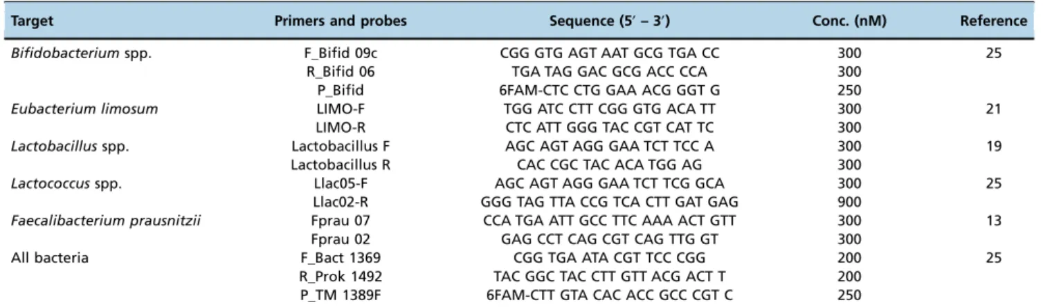
enrolled in this study. Not all requested samples were delivered for children #6 and #16; thus, these children were excluded from the individual variation analysis. The individual results showed a distinct pattern of colonization in the initial days after birth (Figure 1), with a predominance ofBifidobacterium.
The bacterial load differed among the children, with varia-tions of 3 log units (Figure 1), and 5 log units for children #7 and #17 in the first days of life. During the following months, the bacterial load of each studied genera increased, with some inter-individual variation.
Bifidobacterium was undetectable on the second day for children #1 and #15 and on the seventh day for child #8. A predominance of E. coli was observed on the second or seventh day for child #3, child #8, child #12 and child #14. The pediatrician reports for those six children indicated important external factors, such as the usage of antibiotics by mother during pregnancy (#1 and #12) or by the child at the 7thday (#3) or poor sanitary conditions (#8, #14 and #15).
Despite some intra-individual variations in the16S rRNA copy number, after the 3rdmonth, the microbial pattern was similar for all children, with a predominance of Bifidobacter-ium followed by E. coli and Lactobacillus and the lowest counts of 16S rRNA copy number for Lactococcusuntil the end of the first year of age.Bifidobacteriumwas undetectable at the sixth month of age for child #17, and the pediatrician records indicated a respiratory infection and antibiotic pre-scription at that time.
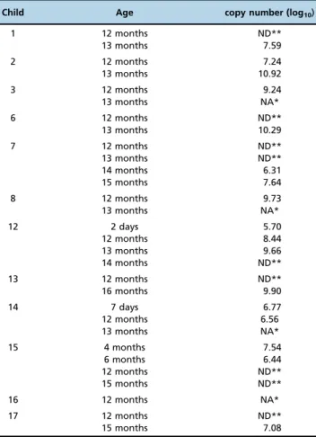
F. prausnitziiwas detected at some time points (Table 3). The abundance of this species had increased remarkably by the end of the first year of life, when it was detected in 45% of the infants. For the infants from whom fecal samples were collected after 12 months of age, the abundance of the 16S rRNAgene ofF. prausnitziiwas evaluated to determine its presence and abundance at these later time points. Of the 12 infants enrolled in this study, five of them were posi-tive forF. prausnitziiduring the first year, with values rang-ing from 5.7 log10to 15.39 log10copies/g of feces (Table 3).
Of the eight infants from who samples were collected after the 13th month of age, 7 had higher detectable levels of F. prausnitzii(Table 3).E. limosumexhibited the lowest abun-dances and frequencies among the evaluated anaerobic bacteria. The few samples in which this species was detected were collected from child #8 at time point 1 (4.83 log10
copies/g of feces), from child #15 at time point 2 (4.0 log10
copies/ g of feces) and from child #6 at time point 4 (7.5 log10
copies/g of feces). After 12 months of age, none of the children had detectableE. limosum.
Time point variation
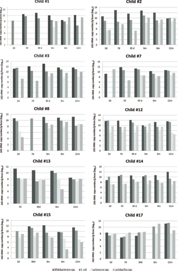
The quantification of the total bacteria at each time point revealed the highest values on the second day of life, with levels that were one or two log units higher than the average values (Figure 2, Table 2). At time point 1 (2 days of age) and throughout the first year, the mean values of total bacteria at each time point did not change significantly, and the differ-ences between time points were on the order of 1 log unit (Figure 2, Table 2).
Bifidobacteriumwas not detected at some time points for a few children, as mentioned previously (Figure 1). Of the anaerobic genera evaluated, Bifidobacterium was present in the greatest numbers at all of the time points tested, with maximum values after time point 3 (3 months of age). The mean values of the abundance ofBifidobacteriumwere similar at time points 3, 4 and 5 (Table 2). The abundance of Bifidobacterium did not appear to be associated with varia-tions in diet (proportion of breast milk or formula milk).
E. coliwas found in all of the children and at all time points (Table 2), and the16S rRNAcopy numbers were the second most abundant among the study subjects. The maximum 16S rRNAvalues were detected at time point 1 (Figure 2), and then the mean values ranged from 9.75 log10copies/g
of feces on the 7thday to 10.04 log
10copies/g of feces at the
12thmonth, with a minimum value at time point 4 (Table 2). At time points 3 and 4, the mean values forLactobacilluswere higher than those forEscherichia; however, this was seen in all of the children.
Lactobacillusspp. andLactococcusspp. were not detected in some children, (Figure 1, Table 2), with mean abundances that were between 1 and 3 log units lower than the abun-dances of Bifidobacterium (Figure 2). The maximum abun-dance ofLactobacilluswas observed at time point 1 (Table 2), and the minimum abundance was observed at time point 2 (Figure 2). The mean abundance values ofLactococcusat each time point did not change significantly over the course of the study, and the differences among them were on the order of 1 log unit (Figure 2, Table 2).
’ DISCUSSION
The results of quantification of the total bacteria16S rRNA copy number in the feces of the children enrolled in this study were similar to those reported in the literature (22,26), and some inter-individual variation was observed throughout the observation period. The data revealed that the bacteria population in the gut was highest at the time the delivery.
Table 1-Primers and probes used in this study.
Target Primers and probes Sequence (5’– 3’) Conc. (nM) Reference
Bifidobacteriumspp. F_Bifid 09c R_Bifid 06 P_Bifid
CGG GTG AGT AAT GCG TGA CC TGA TAG GAC GCG ACC CCA 6FAM-CTC CTG GAA ACG GGT G
300 300 250
25
Eubacterium limosum LIMO-F LIMO-R
TGG ATC CTT CGG GTG ACA TT CTC ATT GGG TAC CGT CAT TC
300 300
21
Lactobacillusspp. Lactobacillus F Lactobacillus R
AGC AGT AGG GAA TCT TCC A CAC CGC TAC ACA TGG AG
300 300
19
Lactococcusspp. Llac05-F Llac02-R
AGC AGT AGG GAA TCT TCG GCA GGG TAG TTA CCG TCA CTT GAT GAG
300 900
25
Faecalibacterium prausnitzii Fprau 07 Fprau 02
CCA TGA ATT GCC TTC AAA ACT GTT GAG CCT CAG CGT CAG TTG GT
300 300
13
All bacteria F_Bact 1369
R_Prok 1492 P_TM 1389F
CGG TGA ATA CGT TCC CGG TAC GGC TAC CTT GTT ACG ACT T 6FAM-CTT GTA CAC ACC GCC CGT C
200 200 250
Figure 1 - Inter-individual quantification of anaerobic and facultative bacteria in the intestinal microbiota, expressed as log10 of
However, theBifidobacteriumcounts increased and the E. coli counts decreased at the subsequent time point. These data are consistent with reports throughout the world that the intesti-nal ecosystem shifts toward an anaerobic environment after birth, and the levels of anaerobic bacteria increase (6).
Bifidobacterium is an important genus of gut microbiota, and some species play beneficial roles in maintaining host health (12). Breastmilk is both an important source of Bifido-bacteriumfor infants (29) and also a source of carbohydrates, which promote Bifidobacterium colonization, even in mixed feeding diets (10). The infants enrolled in this study were breastfed during their early life, and even during the intro-duction of formula milk, the mother’s milk was also present in their diet, characterizing the diet as mixed feeding (8). Bifidobacterium was not detected at some time points for a few children. Interestingly, the pediatrician reports indicated the use of antibiotics by some of these children before sample collection or the prescription of antibiotic treatment to the mothers for urinary tract infection during the last trimester of
pregnancy. The ability of antibiotic treatment to disturb the microbiota composition, including a decrease in Bifidobacter-iumabundance, has been reported previously (10). Although our sample size is limited, our results suggested that such changes may also occur in children.
However, Bifidobacterium was the predominant species detected in this group of infants at twelve months of age, corroborating the known benefits of breastfeeding as a source of Bifidobacterium and its maintenance in the gut mucosa (9,10). This genus has been described throughout the world as the predominant bacterial group detected in the feces of infants (26-28). Our data showed a lower16S rRNA copy number of Bifidobacterium after six months of age, potentially because of the introduction of new genera of bacteria via solid foods (30).
Lactobacilluscolonization at birth has been attributed to the maternal vaginal flora (31) and, possibly, to the presence of Lactobacillusin the womb environment (32). These findings may explain the observation that the maximum abundance
Table 2-Minimum, maximum and mean16S rRNAcopy number of bacteria in feces of infants. Values expressed as log10.
Bifidobacterium Lactobacillus Lactococcus Escherichia coli All bacteria
min. values
max values
average (SD)
min. values
max values
average (SD)
min. values
max values
average (SD)
min. values
max values
average (SD)
min. values
max values
average (SD)
2 d ND 11.18 10.9 (±1.27) ND 11.04 10.10 (±2.1) ND 7.74 7.16 (±0.52) 8.53 11.14 10.48 (±1.37) 9.76 14.12 13.13 (±1.28)
7 d ND 12.98 12.06 (±1.78) ND 9.45 8.51 (±0.92) ND 8.08 7.38 (±0.97) 6.33 10.16 9.75 (±1.46) 10.37 12.56 12.19 (±0.88)
1 mo 10.09 12.08 11.68 (±1.54) ND 9.74 9.00 (±1.6) ND 7.95 7.75 (±0.39) 6.51 10.52 9.98 (±1.28) 10.76 13.25 12.57 (±0.88)
3 mo 10.00 12.81 11.98 (±0.87) ND 10.79 9.96 (±0.99) 7.22 8.67 8.03 (±0.56) 8.45 10.28 9.72 (±0.70) 10.53 12.38 11.85 (±0.72)
6 mo ND 12.06 11.25 (±0.82) ND 8.03 10.08 (±2.49) ND 12.06 7.69 (±0.20) 8.18 10.18 9.02 (±0.72) 9.87 12.54 11.74 (±0.85)
12 mo 8.43 12.03 11.42 (±1.05) 5.95 10.34 9.64 (±1.47) ND 8.83 8.33 (±0.77) 7.53 10.69 10.04 (±1.26) 11.45 12.55 12.10 (±0.51)
ND – not detected
Figure 2 -Quantification of anaerobic and facultative bacteria in the intestinal microbiota of infants, expressed as log10of16S rRNA
ofLactobacillusoccurred on the second day of life in Brazilian infants, followed by a decrease at the seventh day and an increase after one month of age. These data suggest that known environmental changes (6) cause a decrease in the initial levels of the maternal microbiota and that an infant’s microbiota has begun to become established by this time; the increase in the abundance ofLactobacillusafter the first month of life highlights the fact that breast milk is an important natural promoter of this bacterial genus.
The inter-individual analysis showed that, in the first days of life, microbiota colonization is affected by individual exposure to environmental factors; in subsequent months, the pattern of anaerobic and facultative genera colonization appears to be mediated by the milk diet, with a predomi-nance of Bifidobacterium and, with lower abundance, Lacto-bacillus, supporting global knowledge about the role of dietary milk in infant intestinal colonization (9,10).
Because our previous study detected Escherichia in high abundance in this group of children based on library construction (8), we quantified Escherichia at different time points. At some time points, particularly at the second and 12thmonths, the copy numbers ofEscherichia 16S rRNAwere
the second highest, consistent with our previous results (8,15). However, due to the presence of Bifidobacterium and Lactobacillus and their protective properties (11) in these childreńs feces, lower values ofEscherichiathan those obser-ved in the present study were expected. In a study of healthy
children in Africa (33), a high proportion ofEscherichiawas detected in the intestinal microbiota in children ranging be-tween zero and 11 months of age. The environmental forces controlling the establishment of the fecal microbiota may favor the maintenance ofE. coliin this community (8,15), as well as in other developing countries (33).
F. prausnitzii is a well-established member of the adult intestinal microbiota with anti-inflammatory properties (13). A few papers have described the presence of this species in children’s feces in other populations (28,33,34), but this adult-like bacterium has not been detected in Brazilian infants’feces. In the present study, this bacterial species was only observed in the fecal microbiota of two newborns on the second and seventh days of life, suggesting maternal trans-mission. These results suggest that the intestinal environ-ment is unfavorable for its maintenance. After the sixth month of life, a gradual increase in the colonization quantity and frequency was observed. Interestingly, the majority of the fecal samples collected from children after one year (at 13-15 months of age) had greater abundances ofF. prausnitzii. These findings are in accordance with those published by Hopkins et al. (28) and Pop et al. (33), in which the abun-dance of this species increased at the end of the first year of life. These data suggest that until the sixth month of life, the intestinal environment is unfavorable for the establishment ofF. prausnitziiand that with the introduction of solid food and the development of a more stable environment, this genus becomes an important intestinal colonizer. Lin et al. (34) reported the presence ofF. prausnitzii in older children from both the USA and Bangladesh, with a higher abun-dance in American children. However, the role of environ-mental factors in the abundance of F. prausnitzii was not discussed, and more studies are needed to determine whether there is any relationship with external factors.
E. limosum is an anaerobic Gram-positive rod present in the colon of adult humans. This species has a butyrate-producing capacity and consequently has beneficial effects in inflammatory bowel disease (35). In this study, E. limo-sum was detected in four infants at different time points, indicating its presence in the environment and, therefore, demonstrating that exposure to this genus occurs early in life, although members of this genus have a low capacity for colonization of the intestinal milieu until the 12thmonth of life.
The present study reports the characterization of the fecal microbiota in Brazilian infants, which is dominated by BifidobacteriumandLactobacillus. The absence ofEubacterium limosum and the late colonization of F. prausnitzii are also notable. These findings suggest a lack of adult-like micro-biota in infants, corroborating the results of a previous study by Ringel-Kuka et al. (36). These results contribute to obser-vations throughout the world of the establishment of the intestinal microbiota of infants fed milk diets. The high abun-dance ofE. colisuggests a pattern related to unhygienic con-ditions, as reported previously in developing countries (34). These results complement analyses of the composition of the gut microbiota in this group of Brazilian breastfed infants living in low socio-economic conditions (8,15) and highlight the influence of both diet and the environment.
’ ACKNOWLEDGMENTS
This work was supported by grants from the São Paulo Research Foundation (FAPESP 2011/51196-7) awarded to CRT.
Table 3-Quantification ofFaecalibacterium prausnitziiin feces of infants.
Child Age copy number (log10)
1 12 months ND**
13 months 7.59
2 12 months 7.24
13 months 10.92
3 12 months 9.24
13 months NA*
6 12 months ND**
13 months 10.29
7 12 months ND**
13 months ND**
14 months 6.31
15 months 7.64
8 12 months 9.73
13 months NA*
12 2 days 5.70
12 months 8.44
13 months 9.66
14 months ND**
13 12 months ND**
16 months 9.90
14 7 days 6.77
12 months 6.56
13 months NA*
15 4 months 7.54
6 months 6.44
12 months ND**
15 months ND**
16 12 months NA*
17 12 months ND**
15 months 7.08
’ AUTHOR CONTRIBUTIONS
Talarico ST performed the experiments and participated in data analysis. Santos FE performed some experiments. Brandt KG selected the children, followed the medical appointments and collected the samples. Martinez MB designed the study and participated in manuscript writing. Taddei CR designed the study, followed the experiments, conducted the data analysis and wrote the manuscript.
’ REFERENCES
1. Garrett WS, Gordon JI, Glimcher LH. Homeostasis and Inflamation in the intestine. Cell. 2010;140(6):859-70, http://dx.doi.org/10.1016/j.cell.2010. 01.023.
2. Vrieze A, Holleman F, Zoetendal EG, De Vos WM, Hoekstra JB, Nieuwdorp M. The environment within: how gut microbiota may influence metabolism and body composition. Diabetologia. 2010;53(4):606-13, http://dx.doi.org/ 10.1007/s00125-010-1662-7.
3. Clemente JC, Ursell LK, Parfrey LW, Knight R The impact of the gut microbiota on human health: an integrative view. Cell. 2012;148(6):1258-70. 4. Fanaro S, Chierici R, Guerrini P, Vigi V. Intestinal microflora in early infancy: composition and development. Acta Paediatr Suppl. 2003; 91(441):48-55.
5. Adlerberth I, Lindberg E, Aberg N, Hesselmar B, Saalman R, Strannegard IL, et al. Reduced enterobacterial and increased staphylococcal coloniza-tion of infantile bowel: an effect of hygienic lifestyle? Pediatr Res. 2006; 59(1):96-101, http://dx.doi.org/10.1203/01.pdr.0000191137.12774.b2. 6. Rotimi VO, Duerden BI. The development of the bacterial flora in normal
neonates. J Med Microbiol. 1981;14(1):51-62, http://dx.doi.org/10.1099/ 00222615-14-1-51.
7. Palmer C, Bik EM, DiGiulio DB, Relman DA, Brown PO. Development of the human infant intestinal microbiota. PLoS Biol. 2007;5(7):e177, http://dx.doi.org/10.1371/journal.pbio.0050177.
8. Taddei CR, Oliveira FF, Duarte RT, Talarico ST, Takagi EH, Ramos Car-valho II, et al. High abundance ofEscherichiaduring the establishment of fecal microbiota in Brazilian children. Microb Ecol. 2014;67(3):624-34, http://dx.doi.org/10.1007/s00248-014-0381-x.
9. Salminen S, Gueimonde M. Gut microbiota in infants between 6 and 24 months of age. Nestle Nutr Workshop Ser Pediatr Program. 2005;56: 43-51, http://dx.doi.org/10.1159/000086235.
10. Fallani M, Young D, Scott J, Norin E, Amarri S, Adam R, et al. Intestinal microbiota of 6-week-old infants across Europe: geographic influence beyond delivery mode, breast-feeding, and antibiotics. J Pediatr Gastro-enterol Nutr. 2010;51(1):77-84, http://dx.doi.org/10.1097/MPG.0b013 e3181d1b11e.
11. Guilloteau P, Martin L, Eeckhaut V, Ducatelle R, Zabielski R, Van Immerseel F. From the gut to the peripheral tissues: the multiple effects of butyrate. Nutr Res Rev. 2010;23(2):366-84, http://dx.doi.org/10.1017/ S0954422410000247.
12. Fukuda S, Toh H, Hase K, Oshima K, Nakanishi Y, Yoshimura K, et al. Bifidobacteria can protect from enteropathogenic infection trough produc-tion of acetate. Nature. 2011;469(7331):543-7, http://dx.doi.org/10.1038/ nature09646.
13. Sokol H, Seksik P, Furet JP, Firmesse O, Nion-Larmurier I, Beaugerie L,
et al. Low Counts of Faecalibacterium prausnitziiin Colitis Microbiota.
Inflamm Bowel Dis. 2009;15(8):1183-9, http://dx.doi.org/10.1002/ibd. 20903.
14. Roger LC, McCartney AL. Longitudinal investigation of the faecal microbiota of healthy full-term infants using fluorescence in situ hybri-dization and denaturing gradient gel electrophoresis. Microbiol. 2010; 156(Pt 11):3317-28.
15. Brandt K, Taddei CR, Takagi EH, Oliveira FF, Duarte RT, Irino I, et al. Establishment of the bacterial fecal community during the first month of life in Brazilian newborns. Clinics. 2012;67(2):113-23, http://dx.doi.org/ 10.6061/clinics/2012(02)05.
16. Hayashi H, Takahashi R, Nishi T, Sakamoto M, Benno Y. Molecular analysis of jejunal, ileal, caecal and recto-sigmoidal human colonic microbiota using 16S rRNA gene libraries and terminal restriction frag-ment length polymorphism. J Med Microbiol. 2005;54(Pt 11):1093-101, http://dx.doi.org/10.1099/jmm.0.45935-0.
17. Marchesi JR, Sato T, Weightman AJ, Martin TA, Fry JC, Hiom SJ, et al. Design and evaluation of useful bacterium-specific PCR primers that amplify genes coding for bacterial 16S rRNA. Appl Environ Microbiol. 1998;64(2):795-9.
18. Wang M, Ahrne S, Antonsson M, Molin G.T-RFLP combined with
prin-cipal component analysis and 16S rRNA gene sequencing: an effective strategy for comparison of fecal microbiota in infants of different ages. J Microbiol Methods. 2004;59(1):53-69, http://dx.doi.org/10.1016/j.mimet. 2004.06.002.
19. Rinttilä T, Kassinen A, Malinen E, Krogius L, Palva A. Development of an extensive set of 16S rDNA-targeted primers for quantification of patho-genic and indigenous bacteria in faecal samples by real-time PCR. J Appl Microbiol. 2004;97(6):1166-77, http://dx.doi.org/10.1111/j.1365-2672.2004. 02409.x.
20. Lemos LN, Fulthorpe RR, Roesch LF. Low sequencing efforts bias ana-lyses of shared taxa in microbial communities. Folia Microbiol. 2012; 57(5):409-13, http://dx.doi.org/10.1007/s12223-012-0155-0.
21. Maukonen J and Saarela M.Eubacterium. In: Liu D. Molecular Detection of
Human Bacterial Pathogens. CRC Press; 2011. p. 391-403.
22. Penders J, Thijs C, Vink C, Stelma FF, Snijders B, Kummeling I, et al. Factors influencing the composition of the intestinal microbiota in early infancy. Pediatrics. 2006;118(2):511-21, http://dx.doi.org/10.1542/peds.2005-2824. 23. De Leoz ML, Kalanetra KM, Bokulich NA, Strum JS, Underwood MA,
German JB, et al. Human milk glycomics and gut microbial genomics in infant feces show a correlation between human milk oligosaccharides and gut microbiota: a proof-of-concept study. J Proteome Res. 2015;14(1): 491-502, http://dx.doi.org/10.1021/pr500759e.
24. Azad MB, Konya T, Maughan H, Guttman DS, Field CJ, Chari RS, et al. Gut microbiota of healthy Canadian infants: profiles by mode of delivery and infant diet at 4 months. CMAJ. 2013;185(5):385-94, http://dx.doi.org/ 10.1503/cmaj.121189.
25. Furet JP, Firmesse O, Gourmelon M, Bridonneau C, Tap J, Mondot S, et al. Comparative assessment of human and farm animal faecal microbiota using real-time quantitative PCR. FEMS Microbiol Ecol. 2009;68(3):351-62, http://dx.doi.org/10.1111/j.1574-6941.2009.00671.x.
26. Suau A, Bonnet R, Sutren M, Godon JJ, Gibson GR, Collins MD, et al. Direct analysis of genes encoding 16S rRNA from complex communities reveals many novel molecular species within the human gut. Appl Environ Microbiol. 1999;65(11):4799-807.
27. Favier CF, Vaughan EE, de Vos WM, Akkermans ADL. Molecular mon-itoring of succession of bacterial communities in human neonates. Appl Environ Microbiol. 2002;68(1):219-26, http://dx.doi.org/10.1128/AEM. 68.1.219-226.2002.
28. Hopkins MJ, Macfarlane GT, Furrie E, Fite A, Macfarlane S. Character-isation of intestinal bacteria in infant stools using real-time PCR and northern hybridisation analyses. FEMS Microbiol Ecol. 2005;54(1):77-85, http://dx.doi.org/10.1016/j.femsec.2005.03.001.
29. Martín R, Jimenez E, Heilig H, Fernandez L, Marín ML, Zoetendal EG, et al. Isolation of bifidobacteria from breast milk and assessment of the bifidobacterial population by PCR-denaturing gradient gel electrophor-esis and quantitative real-time PCR. Appl Environ Microbiol. 2009; 75(4):965-9, http://dx.doi.org/10.1128/AEM.02063-08.
30. Scholtens PA, Oozeer R, Martin R, Amor KB, Knol J. The early settlers: intestinal microbiology in early life. Annu Rev Food Sci Technol. 2012;3: 425-47, http://dx.doi.org/10.1146/annurev-food-022811-101120. 31. Dominguez-Bello MG, Costello EK, Contreras M, Magris M, Hidalgo G,
Fierer N, et al. Delivery mode shapes the acquisition and structure of the initial microbiota across multiple body habitats in newborns. Proc Natl Acad Sci U S A. 2010;107(26):11971-5, http://dx.doi.org/10.1073/pnas. 1002601107.
32. Funkhouser LJ, Bordenstein SR. Mom knows best: the universality of maternal microbial transmission. PLoS Biol. 2013;11(8):e1001631, http:// dx.doi.org/10.1371/journal.pbio.1001631.
33. Pop M, Walker AW, Paulson J, Lindsay B, Antonio M, Hossain MA, et al. Diarrhea in young children from low-income countries leads to large-scale alterations in intestinal microbiota composition. Genome Biol. 2014;15(6):R76, http://dx.doi.org/10.1186/gb-2014-15-6-r76.
34. Lin A, Bik EM, Costello EK, Dethlefsen L, Haque R, Relman DA, et al. Distinct distal gut microbiome diversity and composition in healthy children from Bangladesh and the United States. PLoS One. 2013;8(1): e53838, http://dx.doi.org/10.1371/journal.pone.0053838.
35. Possemiers S, Rabot S, Espin JC, Bruneau A, Philippe C, González-Sarrías
A, et al. Eubacterium limosum activates isoxanthohumol from hops
(Humulus lupulus L.) into the potent phytoestrogen 8-prenylnaringenin in vitro and in rat intestine. J Nutr. 2008;138(7):1310-6.