Structural Analysis and Aggregation
Propensity of Pyroglutamate A
β
(3-40) in
Aqueous Trifluoroethanol
Christina Dammers
1, Lothar Gremer
1,2, Kerstin Reiß
1, Antonia N. Klein
1,
Philipp Neudecker
1,2, Rudolf Hartmann
1, Na Sun
1, Hans-Ulrich Demuth
3,
Melanie Schwarten
1, Dieter Willbold
1,2*
1Institute of Complex Systems (ICS-6) Structural Biochemistry, Forschungszentrum Jülich, 52425, Jülich, Germany,2Institut für Physikalische Biologie, Heinrich-Heine-Universität Düsseldorf, 40225, Düsseldorf, Germany,3Fraunhofer Institute for Cell Therapy and Immunology, Department of Drug Design and Target Validation, 06120, Halle (Saale), Germany
*d.willbold@fz-juelich.de
Abstract
A hallmark of Alzheimer
’
s disease (AD) is the accumulation of extracellular amyloid-
β
(A
β
)
plaques in the brains of patients. N-terminally truncated pyroglutamate-modified A
β
(pEA
β
)
has been described as a major compound of A
β
species in senile plaques. pEA
β
is more
resistant to degradation, shows higher toxicity and has increased aggregation propensity
and
β
-sheet stabilization compared to non-modified A
β
. Here we characterized
recombi-nant pEA
β
(3
–
40) in aqueous trifluoroethanol (TFE) solution regarding its aggregation
pro-pensity and structural changes in comparison to its non-pyroglutamate-modified variant A
β
(1
–
40). Secondary structure analysis by circular dichroism spectroscopy suggests that
pEA
β
(3
–
40) shows an increased tendency to form
β
-sheet-rich structures in 20% TFE
con-taining solutions where A
β
(1
–
40) forms
α
-helices. Aggregation kinetics of pEA
β
(3
–
40) in
the presence of 20% TFE monitored by thioflavin-T (ThT) assay showed a typical sigmoidal
aggregation in contrast to A
β
(1
–
40), which lacks ThT positive structures under the same
conditions. Transmission electron microscopy confirms that pEA
β
(3
–
40) aggregated to
large fibrils and high molecular weight aggregates in spite of the presence of the helix
stabi-lizing co-solvent TFE. High resolution NMR spectroscopy of recombinantly produced and
uniformly isotope labeled [
U
-
15N]-pEA
β
(3
–
40) in TFE containing solutions indicates that the
pyroglutamate formation affects significantly the N-terminal region, which in turn leads to
decreased monomer stability and increased aggregation propensity.
Introduction
The pathology of Alzheimer’s disease (AD) is characterized by the presence of intracellular
neurofibrillary tangles consisting of accumulated hyperphosphorylated tau protein and
extra-cellular plaques containing amyloid-
β
(A
β
) as major component [
1
,
2
]. A
β
is derived by
OPEN ACCESS
Citation:Dammers C, Gremer L, Reiß K, Klein AN, Neudecker P, Hartmann R, et al. (2015) Structural Analysis and Aggregation Propensity of
Pyroglutamate Aβ(3-40) in Aqueous Trifluoroethanol. PLoS ONE 10(11): e0143647. doi:10.1371/journal. pone.0143647
Editor:Surajit Bhattacharjya, Nanyang Technological University, SINGAPORE
Received:September 25, 2015
Accepted:November 6, 2015
Published:November 23, 2015
Copyright:© 2015 Dammers et al. This is an open access article distributed under the terms of the Creative Commons Attribution License, which permits unrestricted use, distribution, and reproduction in any medium, provided the original author and source are credited.
Data Availability Statement:All relevant data are within the paper and its Supporting Information files.
processing of the amyloid precursor protein by
β
- and
γ
-secretases [
3
,
4
]. Multiple possible
cleavage positions of
γ
-secretase combined with various posttranslational modifications of the
cleaved products result in different A
β
isoforms with heterogeneity in length as well as N- and
C-terminal sequences [
5
,
6
]. Pyroglutamate-modified A
β
(pEA
β
) peptides have been reported
to be the dominant component of all N-terminal truncated A
β
variants in AD plaques as up to
20% of the total A
β
are pEA
β
variants [
7
–
9
]. pEA
β
deposits in the brains of presymptomatic
AD patients [
10
] and was shown to play a dominant role in plaque formation and to provoke
neurodegeneration in mouse models [
11
,
12
]. Formation of pEA
β
is based on N-terminal
trun-cation leading to N-terminal E3 or E11 and subsequent cyclization of the accessible E amino
group with the side chain carboxy group leading to pyroglutamate.
In vivo
it was suggested the
cyclization is catalyzed by the enzyme glutaminyl cyclase [
13
,
14
]. pEA
β
starting at position 3
(pEA
β
(3-x)) represents the major fraction of N-pyroglutamate modified A
β
species in
intracel-lular, extracellular and vascular accumulations in AD brains [
15
–
18
]. The cumulative
deposi-tion of pEA
β
in AD brains coincides with increased protease-stability since the pyroglutamate
modified N-terminus is inaccessible to aminopeptidases [
19
,
20
]. The loss of two negative
char-ges (side chain carboxyl groups of D1 and E3) and one positive charge (E3 amino group) at
physiological pH result in higher hydrophobicity of pEA
β
(3-x) [
19
,
21
]. Aggregation of pEA
β
(3-x) is up to 250-fold accelerated [
22
] and its neurotoxicity is enhanced [
23
,
24
] when
com-pared with the corresponding wild type A
β
species independent of the C-terminal length.
Structural analysis of pEA
β
monomers is essential to uncover its aggregation mechanism
and to reveal, why it is more prone to self-assembly than A
β
wild type. In a previous NMR
study, we analyzed pEA
β
(3–40) in aqueous buffer conditions at neutral pH and compared it
with given A
β
(1–40) data [
25
]. Most chemical shifts were nearly identical except of the very
N-terminal region and both peptides were shown to be unstructured.
Since structural studies of A
β
peptides are often limited due to their high aggregation
ten-dency, solvent conditions need to be carefully chosen.
α
-helical A
β
monomers are
predomi-nantly formed in micelles [
26
], hexafluoroisopropanol (HFIP)/water [
27
] and
2,2,2-trifluoroethanol (TFE)/water [
28
] mixtures. In a previous NMR study we compared
pEA
β
(3–40) with A
β
(1–40), both chemically synthesized with natural carbon and nitrogen
iso-tope abundance, in aqueous solution containing 40% TFE [
29
]. The results indicated that both
A
β
isoforms formed
α
-helical structures in two regions (aa 14–22 and aa 30–36) connected by
a flexible and disordered linker. However, pEA
β
(3–40) exhibited a decreased helix propensity
in agreement with its higher hydrophobicity and faster aggregation kinetics.
Here, we analyzed the solvent conditions for the transition of pEA
β
(3–40) from
TFE-induced
α
-helices to
β
-sheets by gradually lowering the TFE concentrations. We expand the
data by investigations of recombinantly produced [
U
-
15N]-pEA
β
(3–40) regarding structure
and aggregation in 20% TFE, where A
β
(1–40) is still in a predominantly
α
-helical monomeric
state, while pEA
β
(3–40) forms a
β
-sheet dominated secondary structure, starts to aggregate
and forms large fibrils.
Materials and Methods
Peptides and sample preparation
Expression and purification of uniformly isotope labeled [
U
-
15N]-pEA
β
(3–40) and [
U
-
15N]-A
β
(1–40) as well as non-labeled pEA
β
(3–40) were performed as described recently [
25
,
30
].
Synthetic non-labeled A
β
(1–40) was purchased from Bachem (Heidelberg, Germany). Samples
were prepared in Protein LowBinding tubes (Eppendorf AG, 230 Hamburg, Germany). The A
β
peptides were dissolved in HFIP (1,1,1,3,3,3,-hexafluoro-2-propanol, Sigma-Aldrich,
Vernetzungs-Fonds der Helmholtzgemeinschaft”. The funders had no role in study design, data collection and analysis, decision to publish, or preparation of the manuscript.
Hannover, Germany) and monomerized for 3 days at room temperature. Samples were
lyophi-lized and stored at -20°C.
CD spectroscopy
Lyophilized peptides were dissolved in aqueous buffer containing different TFE concentrations
(20–25% TFE in 50 mM potassium phosphate, pH 2.8) to a final concentration of 25
μM. UV
CD measurements were performed in 1 mm path-length cuvettes at 20°C. CD spectra were
recorded on a Jasco J-1100 spectropolarimeter from 260 to 187 nm with 0.5 nm step size, 50
nm/min scan speed, 1 nm bandwidth and 10 scans per sample. Background correction was
per-formed by subtraction of corresponding buffer spectra.
ThT assay
Lyophilized peptides were dissolved to a final concentration of 25
μM in buffer (20–25% TFE
in 50 mM potassium phosphate, pH 2.8) containing 10
μM ThT. Aggregation assays were
per-formed in black non-binding 96-well plates (Sigma-Aldrich, Germany) at 20°C with a total
vol-ume of 100
μl per well. Each reaction was performed fivefold and background corrected by
subtraction of the buffer control. Fluorescence was monitored in 10 min steps using a
micro-plate reader (PolarStar Optima, BMG, Offenburg, Germany) with 440 excitation and 492 nm
emission filters, respectively, in bottom-read mode. Wells were shaken 30°s prior to
measurement.
TEM microscopy
Lyophilized pEA
β
(3–40) was dissolved in aqueous buffer (20% TFE in 50 mM potassium
phos-phate, pH 2.8) and incubated for five days at 20°C. Fibrils were absorbed on formval/carbon
coated copper grids (S162, Plano, Wetzlar, Germany) for 5 min and washed with water. Negative
staining was performed by incubation with 1% (w/v) uranylacetate for 1 min. Images were taken
with a Libra 120 transmission electron microscope (Zeiss, Oberkochen, Germany) at 120 kV.
NMR spectroscopy
Lyophilized [
U
-
15N]-pEA
β
(3–40) and [
U
-
15N]-A
β
(1–40) were directly dissolved in 100%
TFE-d2-OH and diluted with 50 mM potassium phosphate, pH 2.8 to a final concentration of
25
μM peptide in 20% TFE. NMR spectra were acquired using a TXI- or QCI-cryoprobe
equipped Bruker Avance III HD 600 MHz spectrometer.
1H,
15N-HSQC correlation spectra
were recorded according to standard Bruker pulse-sequences [
31
–
33
].
1H
Nand
15N Backbone
resonance assignments in 30% TFE were obtained by using BEST-TROSY (BT)
HNCA+-optimized pulse sequences [
34
,
35
]. Resonance assignments of spectra recorded at 30% TFE
concentration were transferred to the spectrum recorded at 20% TFE through recordings at
TFE concentrations of 27% and 23%, respectively. Spectra were processed with NMRPipe [
36
]
and evaluated with CCPNmr Analysis [
37
]. Analysis of C
α
secondary chemical shifts is based
on the publication by Zhang
et al
. [
38
] and sequence corrected according to Schwarzinger [
39
].
Results
Secondary structure analysis by CD spectroscopy
-helices inducing effect of TFE on pEA
β
(3–40) and the shift towards
β
-sheets by lowering the
TFE contents was analyzed by recording CD spectra in aqueous buffer in different TFE
concen-trations. CD spectra in solutions containing varying concentrations of TFE ranging from 25%
to 22% at pH 2.8 indicated a mainly
α
-helical conformation based on minima at 208 nm and
222 nm, a maximum at 193 nm and an x-axis intercept at 200 nm. Marginally loweringthe TFE
concentration from 22% to 20% or 21% was sufficient to alter the
α
-helical protein
conforma-tion of pEA
β
(3–40) to a
β
-sheet dominated secondary structure (
Fig 1a
) as indicated by the
evolvement of characteristic
β
-sheet features in the corresponding CD spectra with a single
minimum at 216 nm, a x-axis intercept at 202 nm and a shift of the maximum to a lower
wave-length, i.e. 190 nm. CD spectra of A
β
(1–40) recorded under the same condition (20% TFE)
dif-fered compared to the spectra of pEA
β
(3–40) by indicating
α
-helices (
Fig 1b
).
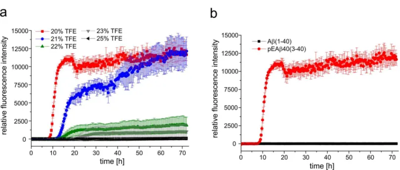
Aggregation kinetics and fibrillation
Aggregation analysis supported the CD data of pEA
β
(3–40) and A
β
(1–40) indicating different
secondary structural elements in aqueous solution containing 20% TFE.
Fig 2a
displays the
aggregation kinetics in TFE containing solutions ranging from 20% to 25% TFE at 20°C for
pEA
β
(3–40) as monitored by ThT assay. pEA
β
(3–40) aggregation reaches its maximum at TFE
concentrations of 20% and 21%. A distinct lag phase of 9h (20% TFE) or 14 h (21% TFE) is
observable following a clear growth phase where aggregation increased indicating that
β
-sheet
formation is not hindered by the presence of TFE. In the presence of 25% TFE no THT
fluores-cence increase was observed during 72 h, which indicate that pEA
β
(3–40) stays monomeric at
that condition. However, when the TFE concentration was lowered to 23% or 22% TFE a
sig-nificant delay as well as decreased maximum was detected within 72 h exploration time.
Aggre-gation of A
β
(1–40) could not be observed at all under the same conditions as no increase in
fluorescence intensity was detected within 72 h (
Fig 2b
). Thus, fibrillation and/or ThT positive
β
-sheet formation of A
β
(1–40) seemed to be inhibited or at least significantly delayed in the
presence of the helix-stabilizing fluoroalcohol TFE.
Fig 1. Far-UV CD spectra of pEAβ(3–40) and Aβ(1–40).Peptides were dissolved in buffer (20–25% TFE in 50 mM potassium phosphate, pH 2.8). CD spectra were recorded at 20°C from 260 to 187 nm, accumulated 10 times and background corrected. (a) CD spectra of 25μM pEAβ(3–40) in 25%, 23%, 22%, 21% and 20% TFE showed a shift fromα-helical structure towardsβ-sheets with decreasing TFE concentrations. (b) The CD spectrum of 25μM Aβ(1–
40) indicatedα-helices in 20% TFE while the spectrum of 25μM pEAβ(3–40) in 20% TFE showed mainlyβ-sheet rich structures.
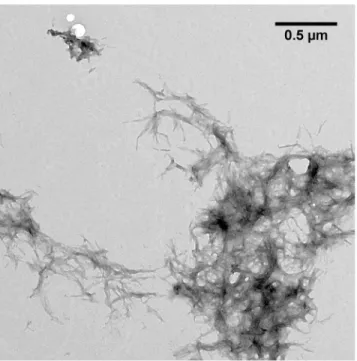
TEM microscopy was performed to analyze the structure of pEA
β
(3
–
40) aggregation
prod-ucts formed after incubation of the monomerized pepide in 20% TFE (
Fig 3
). pEA
β
(3–40) built
large fibrils accumulating into aggregates with several
μm in diameter. The fibrils were twisted,
exhibit a rather homogeneous morphology and accumulate into plaques comparable with
fibrils matured in aqueous solution [
47
].
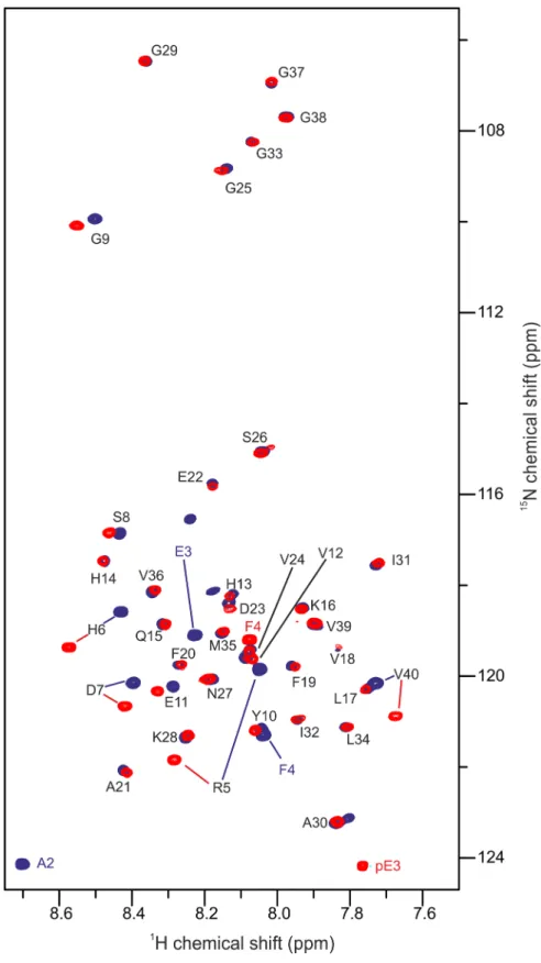
NMR spectroscopy
Since pEA
β
(3–40) started to aggregate at room temperature and built large fibrils in 20% TFE
(pH 2.8), 2D NMR spectroscopy was performed with only 25
μM recombinantly produced
[
U
-
15N]-pEA
β
(3–40). Nonetheless, considerably protein aggregation within 2 days even at the
low concentration applied impeded the recording of 3D experiments. To overcome this
prob-lem, backbone
1H
Nand
15N resonance assignments of pEA
β
(3–40) in 20% TFE were obtained
by a time optimized BT-HNCA+ pulse sequence performed with pEA
β
(3–40) in 30% TFE (
S1
Fig
). Transfer of the resonance assignments to the
1H,
15N-HSQC spectrum in 20% TFE was
per-formed gradually from 30% through 27% and 23% TFE (
S2 Fig
). The C
α
secondary chemical
shifts of pEA
β
(3–40) in 30% TFE were neighbor-corrected and plotted as a function of the aa
sequence (
S3 Fig
). There is evidence that aa Y10-A21 and K28-V36 are involved in the
α
-helix
formation as indicated by positive secondary chemical shifts. This is in accordance with the
sec-ondary structure of pEA
β
(3–40) in 40%TFE obtained from proton chemical shift data [
29
].
For reference, the spectrum of recombinant [
U
-
15N]-A
β
(1–40) was acquired under identical
conditions and both
1H,
15N-HSQC spectra were overlaid for comparison (
Fig 4
). The absence
of the first two amino acid residues D1 and A2 and the substitution of E3 to pE3 is the only
dif-ference between pEA
β
(3–40) and A
β
(1–40). The main changes in chemical shifts are therefore
expected to be primarily observed for the very N-terminal amino acid residues. Interestingly,
signals of the N-terminal region up to E11 differed significantly (
Fig 5
). Cross-peaks further
Fig 2. Aggregation kinetics of pEAβ(3–40) and Aβ(1–40) in TFE.(a) 25μM of monomerized pEAβ(3–40) were dissolved in buffer with various TFE contents (25%, 23%, 22%, 21% and 20% TFE in 50 mM potassium phosphate, pH 2.8) including 10μM ThT. pEAβ(3–40) aggregated in 20% and 21% TFE but was significantly decreased in aqueous solution with higher TFE concentration. (b) 25μM of monomerized pEAβ(3–40) and Aβ(1–40) were dissolved in buffer (20% TFE in 50 mM potassium phosphate, pH 2.8) including 10μM ThT. An increase in ThT fluorescence was observed for pEAβ(3–40) but not for Aβ
(1–40) within 72 h.
downstream towards the C-terminus, except of the C-terminal amino acid V40, were not or
only marginally changed.
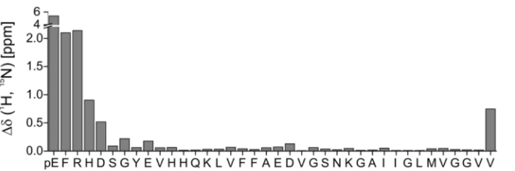
Discussion
Previous data indicated that pyroglutamate-modified A
β
shows increased tendency to form
β
-sheets compared to the unstructured A
β
in aqueous buffer at neutral pH [
22
,
48
,
49
]. Here, we
expand structural knowledge on pEA
β
(3–40) by showing that it forms
β
-sheets even in the
pres-ence of considerably amounts of the co-solvent TFE. This is consistent with data indicating that
the tendency to build
α
-helical structures is decreased for pEA
β
(3–40) as compared to
unstruc-tured A
β
(1–40) as analyzed by proton NMR spectroscopy at higher TFE concentrations, where
both peptides stay in monomeric conformation [
29
]. Our data indicate a change in
conforma-tion of pEA
β
(3–40) as a result of lowering the TFE concentration. We identified the exact TFE
concentration (20% at pH 2.8) where pEA
β
(3–40) already is in a
β
-sheet conformation, whereas
A
β
(1–40) exhibits still an overall
α
-helical conformation. This confirms, that TFE is not simply
favoring
α
-helical secondary structures, but secondary structures in general [
50
].
The differences in the secondary structure content observed by CD spectroscopy seem to be a
result of the modification from A
β
to pEA
β
, which modifies the N-terminal region significantly.
ThT and TEM data support this evidence by yielding characteristic aggregation kinetics and
large matured fibrils for pEA
β
(3–40) in 20% TFE but not for A
β
(1–40) under same conditions.
Using NMR spectroscopy, we analyzed pEA
β
(3–40) monomers in 20% TFE (pH 2.8).
Under these conditions, pEA
β
(3–40) slowly forms
β
-sheet containing aggregates that could not
be observed by solution NMR. Although it was shown in a model peptide starting with a
N-ter-minal E1 that conversion to pE1 results in altered chemical shifts only of the following two
amino acids [
51
], pE formation of A
β
peptides obviously affects not only the immediately
Fig 3. TEM image of pEAβ(3–40) in 50 mM potassium phosphate pH 2.8 containing 20% TFE.
Monomerized pEAβ(3–40) (25μM) was incubated for fibrillation at 20°C for five days and grids were prepared by negative staining. pEAβ(3–40) incubated in aqueous TFE solution formed large twisted fibrils up to several hundred nm in size which accumulate into large aggregates ranging from 1–5μm in diameter.
Fig 4.1H,15N-HSQC of pEAβ(3
–40) and Aβ(1–40).25μM of the monomerized peptides were dissolved in aqueous solution (50 mM potassium phosphate, pH 2.8) containing 20% TFE. Spectra were recorded on a 600 MHz Bruker spectrometer at 5°C. Overlay of the spectrum of pEAβ(3–40) (red) and Aβ(1–40) (blue) indicate that the pyroglutamate modification affects the N-terminal signals significantly towards E11 as well as V40.
adjacent N-terminus. It was previously shown by H
Nand H
αproton chemical shift differences
between pEA
β
(3
–
40) and A
β
(1
–
40) in higher TFE concentrations (40%), that mostly protons
of the six following amino acids from pE3 to S8 with decreasing influence towards the
C-termi-nus were affected [
29
]. In the presence of only 20% TFE, we observed many resonance
differ-ences of pEA
β
(3–40) compared to A
β
(1–40). Chemical shift differences of the N-terminal
amino acids towards E11 as well as the C-terminal V40 were mostly affected as shown in
Fig 5
.
Thus, it seems that the modification of A
β
(1–40) to pEA
β
(3–40) not only influence the
neigh-boring amino acids, but also change significantly the conformational state of at least 25% of all
amino acids. This result suggests that the N-terminal modification to pEA
β
has a significant
effect on secondary structure elements and thus is the driving force for pEA
β
(3–40) to be more
likely to build
β
-sheet structures under exactly the same conditions when compared to A
β
(1–
40). The propensity of pEA
β
(3
–
40) to aggregate in aqueous TFE solution seems to be
propa-gated by the modified N-terminus, but V40 obviously play also a central role, maybe due to
interactions of both termini. Since C-terminally truncated A
β
peptides are less prone to
aggre-gation, e.g. A
β
(1–38)
<
A
β
(1–40)
<
A
β
(1–42) [
52
,
53
], it is likely that their pE modified
iso-forms would show an increased aggregation propensity as compared to the non-pE-modified
isoforms, analogous to the results obtained within this study.
Supporting Information
S1 Fig.
15N-HSQC of pEA
β
(3
–
40) in 30% TFE.
(TIF)
S2 Fig.
15N-HSQC of pEA
β
(3
–
40) in different TFE concentrations.
(TIF)
S3 Fig. C
α
secondary chemical shifts of pEA
β
(3
–
40) in 30% TFE.
(TIF)
Acknowledgments
Matthias Stoldt, Justin Lecher and Maren Thomaier are highly acknowledged for fruitful
dis-cussions. We gratefully acknowledge support of CD from the International NRW Research
School BioStruct, granted by the Ministry of Innovation, Science and Research of the State
North Rhine-Westphalia, the Heinrich Heine University of Düsseldorf, and the Entrepreneur
Foundation at the Heinrich Heine University of Düsseldorf. The authors acknowledge access
to the Jülich-Düsseldorf Biomolecular NMR Center. DW was supported by grants from the
Fig 5. Chemical shift changesΔδ(1H,15N) = ((10
*Δδ1HN)2+ (Δδ15N)2)1/2between pEAβ(3
–40) and Aβ
(1–40) plotted as a function of the pEAβ(3–40) sequence.Pyroglutamate formation affects the N-terminal amino acids with decreasing effect towards the C-terminus and, interestingly, also the C-terminal V40.
“Portfolio Technology and Medicine”, the
“Portfolio Drug Research”
and the
Helmholtz-Vali-dierungsfonds of the
“Impuls und Vernetzungs-Fonds der Helmholtzgemeinschaft”.
Author Contributions
Conceived and designed the experiments: CD MS LG NS HUD DW. Performed the
experi-ments: CD ANK KR. Analyzed the data: CD MS RH PN. Wrote the paper: CD MS LG DW.
References
1. Walsh DM, Selkoe DJ. Deciphering the molecular basis of memory failure in Alzheimer's disease. Neu-ron. 2004; 44(1):181–93. doi:10.1016/j.neuron.2004.09.010PMID:15450169.
2. Ballard C, Khan Z, Clack H, Corbett A. Nonpharmacological treatment of Alzheimer disease. Can J Psychiatry. 2011; 56(10):589–95. PMID:22014691.
3. Haass C, Selkoe DJ. Cellular processing of beta-amyloid precursor protein and the genesis of amyloid beta-peptide. Cell. 1993; 75(6):1039–42. Epub 1993/12/17. PMID:8261505.
4. Weidemann A, Konig G, Bunke D, Fischer P, Salbaum JM, Masters CL, et al. Identification, biogenesis, and localization of precursors of Alzheimer's disease A4 amyloid protein. Cell. 1989; 57(1):115–26. Epub 1989/04/07. PMID:2649245.
5. De Strooper B, Vassar R, Golde T. The secretases: enzymes with therapeutic potential in Alzheimer disease. Nature reviews Neurology. 2010; 6(2):99–107. Epub 2010/02/09. doi:10.1038/nrneurol.2009. 218PMID:20139999; PubMed Central PMCID: PMC2879045.
6. De Strooper B. Proteases and proteolysis in Alzheimer disease: a multifactorial view on the disease process. Physiological reviews. 2010; 90(2):465–94. Epub 2010/04/16. doi:10.1152/physrev.00023. 2009PMID:20393191.
7. Gunn AP, Masters CL, Cherny RA. Pyroglutamate-Abeta: role in the natural history of Alzheimer's dis-ease. The international journal of biochemistry & cell biology. 2010; 42(12):1915–8. Epub 2010/09/14. doi:10.1016/j.biocel.2010.08.015PMID:20833262.
8. Mori H, Takio K, Ogawara M, Selkoe DJ. Mass spectrometry of purified amyloid beta protein in Alzhei-mer's disease. The Journal of biological chemistry. 1992; 267(24):17082–6. Epub 1992/08/25. PMID: 1512246.
9. Perez-Garmendia R, Gevorkian G. Pyroglutamate-Modified Amyloid Beta Peptides: Emerging Targets for Alzheimer s Disease Immunotherapy. Current neuropharmacology. 2013; 11(5):491–8. Epub 2014/ 01/10. doi:10.2174/1570159X11311050004PMID:24403873; PubMed Central PMCID: PMC3763757.
10. Sergeant N, Bombois S, Ghestem A, Drobecq H, Kostanjevecki V, Missiaen C, et al. Truncated beta-amyloid peptide species in pre-clinical Alzheimer's disease as new targets for the vaccination approach. Journal of neurochemistry. 2003; 85(6):1581–91. PMID:12787077.
11. Meissner JN, Bouter Y, Bayer TA. Neuron Loss and Behavioral Deficits in the TBA42 Mouse Model Expressing N-Truncated Pyroglutamate Amyloid-beta3-42. Journal of Alzheimer's disease: JAD. 2015; 45(2):471–82. Epub 2014/12/31. doi:10.3233/JAD-142868PMID:25547635.
12. Wirths O, Breyhan H, Cynis H, Schilling S, Demuth HU, Bayer TA. Intraneuronal pyroglutamate-Abeta 3–42 triggers neurodegeneration and lethal neurological deficits in a transgenic mouse model. Acta neuropathologica. 2009; 118(4):487–96. Epub 2009/06/24. doi:10.1007/s00401-009-0557-5PMID: 19547991; PubMed Central PMCID: PMC2737116.
13. Schilling S, Hoffmann T, Manhart S, Hoffmann M, Demuth HU. Glutaminyl cyclases unfold glutamyl cyclase activity under mild acid conditions. FEBS letters. 2004; 563(1–3):191–6. Epub 2004/04/06. doi: 10.1016/S0014-5793(04)00300-XPMID:15063747.
14. Cynis H, Rahfeld JU, Stephan A, Kehlen A, Koch B, Wermann M, et al. Isolation of an isoenzyme of human glutaminyl cyclase: retention in the Golgi complex suggests involvement in the protein matura-tion machinery. Journal of molecular biology. 2008; 379(5):966–80. doi:10.1016/j.jmb.2008.03.078 PMID:18486145.
15. Harigaya Y, Saido TC, Eckman CB, Prada CM, Shoji M, Younkin SG. Amyloid beta protein starting pyr-oglutamate at position 3 is a major component of the amyloid deposits in the Alzheimer's disease brain. Biochemical and biophysical research communications. 2000; 276(2):422–7. Epub 2000/10/12. doi: 10.1006/bbrc.2000.3490PMID:11027491.
17. Wirths O, Erck C, Martens H, Harmeier A, Geumann C, Jawhar S, et al. Identification of low molecular weight pyroglutamate Abeta oligomers in Alzheimer disease: a novel tool for therapy and diagnosis. The Journal of biological chemistry. 2010; 285(53):41517–24. Epub 2010/10/26. doi:10.1074/jbc. M110.178707PMID:20971852; PubMed Central PMCID: PMC3009878.
18. Hosoda R, Saido TC, Otvos L Jr., Arai T, Mann DM, Lee VM, et al. Quantification of modified amyloid beta peptides in Alzheimer disease and Down syndrome brains. J Neuropathol Exp Neurol. 1998; 57 (11):1089–95. PMID:9825946.
19. Jawhar S, Wirths O, Bayer TA. Pyroglutamate amyloid-beta (Abeta): a hatchet man in Alzheimer dis-ease. The Journal of biological chemistry. 2011; 286(45):38825–32. Epub 2011/10/04. doi:10.1074/ jbc.R111.288308PMID:21965666; PubMed Central PMCID: PMC3234707.
20. Cummins PM, O'Connor B. Pyroglutamyl peptidase: an overview of the three known enzymatic forms. Biochimica et biophysica acta. 1998; 1429(1):1–17. PMID:9920379.
21. Saido TC, Yamao-Harigaya W, Iwatsubo T, Kawashima S. Amino- and carboxyl-terminal heterogeneity of beta-amyloid peptides deposited in human brain. Neuroscience letters. 1996; 215(3):173–6. PMID: 8899741.
22. Schilling S, Lauber T, Schaupp M, Manhart S, Scheel E, Bohm G, et al. On the seeding and oligomeri-zation of pGlu-amyloid peptides (in vitro). Biochemistry. 2006; 45(41):12393–9. Epub 2006/10/13. doi: 10.1021/bi0612667PMID:17029395.
23. Bayer TA, Wirths O. Focusing the amyloid cascade hypothesis on N-truncated Abeta peptides as drug targets against Alzheimer's disease. Acta neuropathologica. 2014; 127(6):787–801. Epub 2014/05/08. doi:10.1007/s00401-014-1287-xPMID:24803226; PubMed Central PMCID: PMC4024135.
24. Russo C, Violani E, Salis S, Venezia V, Dolcini V, Damonte G, et al. Pyroglutamate-modified amyloid beta-peptides—AbetaN3(pE)—strongly affect cultured neuron and astrocyte survival. Journal of neuro-chemistry. 2002; 82(6):1480–9. PMID:12354296.
25. Dammers C, Gremer L, Neudecker P, Demuth HU, Schwarten M, Willbold D. Purification and Charac-terization of Recombinant N-Terminally Pyroglutamate-Modified Amyloid-beta Variants and Structural Analysis by Solution NMR Spectroscopy. PloS one. 2015; 10(10):e0139710. doi:10.1371/journal. pone.0139710PMID:26436664.
26. Shao H, Jao S, Ma K, Zagorski MG. Solution structures of micelle-bound amyloid beta-(1–40) and beta-(1–42) peptides of Alzheimer's disease. Journal of molecular biology. 1999; 285(2):755–73. Epub 1999/01/08. PMID:9878442.
27. Crescenzi O, Tomaselli S, Guerrini R, Salvadori S, D'Ursi AM, Temussi PA, et al. Solution structure of the Alzheimer amyloid beta-peptide (1–42) in an apolar microenvironment. Similarity with a virus fusion domain. European journal of biochemistry / FEBS. 2002; 269(22):5642–8. Epub 2002/11/09. PMID: 12423364.
28. Sticht H, Bayer P, Willbold D, Dames S, Hilbich C, Beyreuther K, et al. Structure of amyloid A4-(1– 40)-peptide of Alzheimer's disease. European journal of biochemistry / FEBS. 1995; 233(1):293–8. Epub 1995/10/01. PMID:7588758.
29. Sun N, Hartmann R, Lecher J, Stoldt M, Funke SA, Gremer L, et al. Structural analysis of the pyrogluta-mate-modified isoform of the Alzheimer's disease-related amyloid-beta using NMR spectroscopy. Jour-nal of peptide science: an official publication of the European Peptide Society. 2012; 18(11):691–5. Epub 2012/09/25. doi:10.1002/psc.2456PMID:23001756.
30. Finder VH, Vodopivec I, Nitsch RM, Glockshuber R. The recombinant amyloid-beta peptide Abeta1-42 aggregates faster and is more neurotoxic than synthetic Abeta1-42. Journal of molecular biology. 2010; 396(1):9–18. Epub 2009/12/23. doi:10.1016/j.jmb.2009.12.016PMID:20026079.
31. Kay LE, Keifer P, Saarinen T. Pure absorption gradient enhanced heteronuclear single quantum corre-lation spectroscopy with improved sensitivity. Journal of the American Chemical Society. 1992; 114 (26):10663–5. doi:10.1021/ja00052a088WOS:A1992KD71700088.
32. Palmer AG, Cavanagh J, Wright PE, Rance M. Sensitivity improvement in proton-detected two-dimen-sional heteronuclear correlation NMR spectroscopy. Journal of magnetic resonance. 1991; 93(1):151–
70. doi:10.1016/0022-2364(91)90036-sWOS:A1991FL34200012.
33. Schleucher J, Schwendinger M, Sattler M, Schmidt P, Schedletzky O, Glaser SJ, et al. A general enhancement scheme in heteronuclear multidimensional NMR employing pulsed field gradients. Jour-nal of biomolecular NMR. 1994; 4(2):301–6. WOS:A1994NC39000011. PMID:8019138
34. Solyom Z, Schwarten M, Geist L, Konrat R, Willbold D, Brutscher B. BEST-TROSY experiments for time-efficient sequential resonance assignment of large disordered proteins. Journal of biomolecular NMR. 2013; 55(4):311–21. Epub 2013/02/26. doi:10.1007/s10858-013-9715-0PMID:23435576.
36. Delaglio F, Grzesiek S, Vuister GW, Zhu G, Pfeifer J, Bax A. NMRPipe: a multidimensional spectral pro-cessing system based on UNIX pipes. Journal of biomolecular NMR. 1995; 6(3):277–93. Epub 1995/ 11/01. PMID:8520220.
37. Vranken WF, Boucher W, Stevens TJ, Fogh RH, Pajon A, Llinas M, et al. The CCPN data model for NMR spectroscopy: development of a software pipeline. Proteins. 2005; 59(4):687–96. Epub 2005/04/ 09. doi:10.1002/prot.20449PMID:15815974.
38. Zhang H, Neal S, Wishart DS. RefDB: a database of uniformly referenced protein chemical shifts. Jour-nal of biomolecular NMR. 2003; 25(3):173–95. Epub 2003/03/26. PMID:12652131.
39. Schwarzinger S, Kroon GJ, Foss TR, Chung J, Wright PE, Dyson HJ. Sequence-dependent correction of random coil NMR chemical shifts. Journal of the American Chemical Society. 2001; 123(13):2970–8. Epub 2001/07/18. PMID:11457007.
40. Kumar S, Udgaonkar JB. Structurally distinct amyloid protofibrils form on separate pathways of aggre-gation of a small protein. Biochemistry. 2009; 48(27):6441–9. Epub 2009/06/10. doi:10.1021/ bi900682wPMID:19505087.
41. Chen YR, Huang HB, Chyan CL, Shiao MS, Lin TH, Chen YC. The effect of Abeta conformation on the metal affinity and aggregation mechanism studied by circular dichroism spectroscopy. Journal of bio-chemistry. 2006; 139(4):733–40. Epub 2006/05/05. doi:10.1093/jb/mvj083PMID:16672274.
42. Gupta P, Deep S. Intermediate conformation between native beta-sheet and non-native alpha-helix is a pre-cursor of trifluoroethanol-induced aggregation of human carbonic anhydrase-II. Biochemical and biophysi-cal research communications. 2014; 449(1):126–31. doi:10.1016/j.bbrc.2014.04.160PMID:24813993.
43. Marinelli P, Castillo V, Ventura S. Trifluoroethanol modulates amyloid formation by the all alpha-helical URN1 FF domain. Int J Mol Sci. 2013; 14(9):17830–44. doi:10.3390/ijms140917830PMID:23999589; PubMed Central PMCID: PMC3794755.
44. Amani S, Naeem A. Transition of transferrin from native to fibrillar state: An implication for amyloid-linked diseases. Biochem Eng J. 2014; 91:120–8. doi:10.1016/j.bej.2014.08.004
WOS:000343950800016.
45. Anderson VL, Ramlall TF, Rospigliosi CC, Webb WW, Eliezer D. Identification of a helical intermediate in trifluoroethanol-induced alpha-synuclein aggregation. Proceedings of the National Academy of Sci-ences of the United States of America. 2010; 107(44):18850–5. doi:10.1073/pnas.1012336107PMID: 20947801; PubMed Central PMCID: PMC2973859.
46. Khan MV, Rabbani G, Ahmad E, Khan RH. Fluoroalcohols-induced modulation and amyloid formation in conalbumin. International journal of biological macromolecules. 2014; 70:606–14. doi:10.1016/j. ijbiomac.2014.07.027PMID:25062993.
47. Schlenzig D, Manhart S, Cinar Y, Kleinschmidt M, Hause G, Willbold D, et al. Pyroglutamate formation influences solubility and amyloidogenicity of amyloid peptides. Biochemistry. 2009; 48(29):7072–8. Epub 2009/06/13. doi:10.1021/bi900818aPMID:19518051.
48. He W, Barrow CJ. The A beta 3-pyroglutamyl and 11-pyroglutamyl peptides found in senile plaque have greater beta-sheet forming and aggregation propensities in vitro than full-length A beta. Biochem-istry. 1999; 38(33):10871–7. doi:10.1021/bi990563rPMID:10451383.
49. Schlenzig D, Ronicke R, Cynis H, Ludwig HH, Scheel E, Reymann K, et al. N-Terminal pyroglutamate formation of Abeta38 and Abeta40 enforces oligomer formation and potency to disrupt hippocampal long-term potentiation. Journal of neurochemistry. 2012; 121(5):774–84. Epub 2012/03/02. doi:10. 1111/j.1471-4159.2012.07707.xPMID:22375951.
50. Sonnichsen FD, Van Eyk JE, Hodges RS, Sykes BD. Effect of trifluoroethanol on protein secondary structure: an NMR and CD study using a synthetic actin peptide. Biochemistry. 1992; 31(37):8790–8. PMID:1390666.
51. Mischo A, Ohlenschlager O, Guhrs KH, Gorlach M. Recombinant production of isotope-labeled pep-tides and spontaneous cyclization of amino-terminal glutamine into pyroglutamic acid. Chembiochem: a European journal of chemical biology. 2012; 13(10):1421–3. Epub 2012/05/24. doi:10.1002/cbic. 201200178PMID:22619187.
52. Gravina SA, Ho L, Eckman CB, Long KE, Otvos L Jr., Younkin LH, et al. Amyloid beta protein (A beta) in Alzheimer's disease brain. Biochemical and immunocytochemical analysis with antibodies specific for forms ending at A beta 40 or A beta 42(43). The Journal of biological chemistry. 1995; 270 (13):7013–6. PMID:7706234.