An Exploratory Study on
99m
Tc-RGD-BBN
Peptide Scintimammography in the
Assessment of Breast Malignant Lesions
Compared to
99m
Tc-3P4-RGD2
Qianqian Chen1, Qingjie Ma1, Minglong Chen1, Bin Chen1, Qiang Wen1, Bing Jia2, Fan Wang2, Butong Sun1, Shi Gao1*
1Department of Nuclear Medicine, China-Japan Union Hospital of Jilin University, Changchun, 130033, China,2Medical Isotopes Research Center of Peking University, Beijing, 100191, China
Abstract
Purpose
This study aimed to explore the diagnostic performance of single photon emission comput-ed tomography / computerizcomput-ed tomography (SPECT/CT) using a new radiotracer99m Tc-RGD-BBN for breast malignant tumor compared with99mTc-3P4-RGD2.
Methods
6 female patients with breast malignant tumors diagnosed by fine needle aspiration cytology biopsy (FNAB) who were scheduled to undergo surgery were included in the study.99m Tc-3P4-RGD2and99mTc-RGD-BBN were performed with single photon emission computed to-mography (SPECT) at 1 hour after intravenous injection of 299±30 MBq and 293±32 MBq
of radiotracers respectively at separate day. The results were evaluated by the Tumor to non-Tumor ratios (T/NT).99mTc-RGD-BBN and99mTc-3P4-RGD2SPECT/CT images were inter-preted independently by 3 experienced nuclear medicine physicians using a 3-point scale system. All of the samples were analyzed immunohistochemically to evaluate the integrin
αvβ3 and gastrin-releasing peptide receptor (GRPR) expression. The safety, biodistribution and radiation dosimetry of99mTc-RGD-BBN were also evaluated in the healthy volunteers.
Results
No serious adverse events were reported in any of the patients during the study. The effec-tive radiation dose entirely conformed to the relevant standards. A total of 6 palpable malig-nant lesions were detected using99mTc-RGD-BBN SPECT/CT with clear uptake. All malignant lesions were also detected using99mTc-3P4-RGD
2SPECT/CT. The results showed that five malignant lesions were with clear uptake and the other one with barely an uptake. 4 malignant cases were found with bothαvβ3 and GRPR expression, 1 case with only GRPR positive expression (integrinαvβ3 negative) and 1 case with only integrinαvβ3 positive expression (GRPR negative).
OPEN ACCESS
Citation:Chen Q, Ma Q, Chen M, Chen B, Wen Q, Jia B, et al. (2015) An Exploratory Study on99m
Tc-RGD-BBN Peptide Scintimammography in the Assessment of Breast Malignant Lesions Compared to99mTc-3P4-RGD2. PLoS ONE 10(4): e0123401. doi:10.1371/journal.pone.0123401
Academic Editor:Bart O. Williams, Van Andel Institute, UNITED STATES
Received:October 15, 2014
Accepted:February 18, 2015
Published:April 7, 2015
Copyright:© 2015 Chen et al. This is an open access article distributed under the terms of the
Creative Commons Attribution License, which permits unrestricted use, distribution, and reproduction in any medium, provided the original author and source are credited.
Data Availability Statement:All relevant data are within the paper.
Conclusion
99m
Tc-RGD-BBN is a safe agent for detecting breast cancer.99mTc-RGD-BBN may have the potential to make up for the deficiency of99mTc-3P4-RGD2in the detection of breast cancer with only GRPR positive expression (integrinαvβ3negative). The preliminary appli-cation of99mTc-RGD-BBN has demonstrated its powerful potential in breast cancer diagno-sis and therapy.
Introduction
Breast cancer is the most frequent malignancy in women all over the world. In the United States, a Cancer Journal for Clinicians estimates that 234,580 women will be diagnosed with breast cancer in 2013 and expected to account for 30% of all female new cancers. Also, the inci-dence rate is increasing year by year in China [1,2]. Early detection of breast cancer may lead to a higher rate of successful treatment and extend patients’lives.
X-ray mammography (XMM) and ultrasound (US) are now employed as conventional tools for breast tumor screening. The wide use of them in early detection of tumor has saved thou-sands of lives. With the advent of molecular imaging era,nuclear medicine techniques is con-sidered promising in early detection of tumor through a functional perspective. Of them, scintimammography (SMM) with various targeted probes have become a major interest in this area [3]. Since the power of tracer largely decides the performance of a SMM examination, huge efforts have been paid in developing them. [99mTc (HYNIC-3PRGD2) (tricine) (TPPTS)]
(99mTc-3P4-RGD2), a peptide with high binding affinity to integrinαvβ3on tumor cells, is a
newly designed tracer and has shown remarkable performance in detecting tumor [4–7]. Expression of cell-surface receptors by cancer cells can be heterogeneous and inhomoge-neous. Breast cancer cells, for example, over-express two receptors [gastrin-releasing peptide receptor (GRPR) in 70% breast cancer cells and integrinαvβ3in 58% breast cancer cells]. It is
difficult to identify all breast cancer cells with just one target-based cancer imaging. Therefore, it is desirable to develop a new type of radiotracers that can target not just one but several dif-ferent receptors simultaneously. Of course, the receptors need to be more specific peptide vs integrin receptors.
Recently, our laboratory designed and synthesized a dual integrinαvβ3and GRPR targeted peptide Glu-c(RGDyK)-bombesin (RGD-BBN) that contained dual RGD and BBN motifs in one molecule [8,9]. In an earlier study, the99mTc labeled RGD-BBN heterodimer exhibited excellent pharmacokinetics and biology distribution in the mouse models [9]. This study was designed to determine the safety, pharmacokinetics and biodistribution characteristics of
99mTc-RGD-BBN in healthy volunteers for the first time. Furthermore, we aimed to explore
the diagnostic performance of99mTc-RGD-BBN single photon emission computed tomogra-phy / computerized tomogratomogra-phy (SPECT/CT) for palpable breast abnormalities, and to com-pare99mTc-RGD-BBN SPECT/CT with99mTc-3P4-RGD2to assess the possible incremental
value of99mTc-RGD-BBN SPECT/CT in breast cancer detection for the first time.
Material and Methods
Subjects
6 patients (mean age 59 ± 10yr) with breast malignant tumor diagnosed by fine needle biopsy (FNAB) at least 7 days prior to surgery were included in the study. The healthy volunteers Competing Interests:The authors have declared
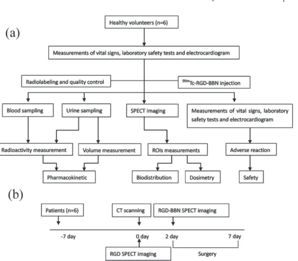
consisted of 3 males and 3 females aged between 25 and 51 years (Table 1). Physical examina-tion and laboratory results from the last 6 months demonstrated no pathologic findings for all volunteers. Informed written consent to participate in the SMM studies was obtained from all subjects. This study was approved by the Ethics Committee of China-Japan Union Hospital of Jilin University. The flow chart of the study protocol for healthy volunteers and patients are shown inFig 1.
All patients were sent to the99mTc-3P4-RGD2and99mTc-RGD-BBN SMM on an individual
basis. The time interval between two imaging procedures was 48–54 hr. Finally,99m Tc-RGD-BBN and99mTc-3P4-RGD2SMM results were compared with each other. To be
Table 1. Demographic and Clinical Characteristics of Volunteers Investigated.
Volunteer No./Sex Age (y) Height (cm) Weight (kg) Body mass index (kg/m2) Injected Activity (MBq)
1/M 51 175 73.5 24.00 1076.7
2/M 30 174 70.0 23.12 952.75
3/M 27 173 77.0 25.73 1061.9
4/F 25 155 41.0 17.07 1128.5
5/F 41 161 58.0 22.38 954.6
6/F 35 158 52.0 20.83 995.3
Mean±SD 34.83±9.81 166±8.99 61.92±13.99 22.19±2.99 1028.29±71.75
doi:10.1371/journal.pone.0123401.t001
Fig 1. Flow chart of the study protocol for healthy volunteers (a) and patients (b), respectively.
included, a patient had to be without any other history of breast disease. Exclusion criteria in-cluded pregnancy, lactation and a body weight greater than 80 kg.
A final diagnosis of the specimens obtained by the surgical procedure was made by histopa-thology. The most representative samples were submitted to immunohistochemical evaluation. For the resected lesions, the largest dimension of the tumor was considered the pathologic size. Results of SPECT/CT were compared with histological findings.
99m
Tc-3P4-RGD2
and
99mTc-RGD-BBN SPECT/CT
The 3P4-RGD2and RGD-BBN were generously provided by the Medical Isotopes Research
Center of Peking University in freeze-dried kits form. Na99mTcO4was obtained from a
com-mercial99Mo/99mTc generator (Beijing Atom High Tech Co., Ltd.). The kit for preparation of
99mTc-3P4-RGD
2was formulated by containing, per millilitre, 20μg of HYNIC-3P4-RGD2,
5 mg of TPPTS, 6.5 mg of tricine, 40 mg of mannitol, 38.5 mg of disodium succinate hexahy-drate and 12.7 mg of succinic acid. Radiolabeling and quality control procedures for99m Tc-3P4-RGD2were performed as described previously4. The kit for preparation of99m
Tc-RGD-BBN was formulated by containing, per millilitre, 20μg of HYNIC-RGD-BBN, 6 mg of
TPPTS, 10 mg of tricine, 40 mg of mannitol, 38.5 mg of disodium succinate hexahydrate and 12.7 mg of succinic acid [9]. Then 1 mL of Na99mTcO4solution in saline was added to each kit
vial followed by 30 min incubation at 100°C. The quality control was conducted by radioactive thin layer chromatography (ITLC), counting the high labeling yield of about 95%. Both99m Tc-3P4-RGD2and99mTc-RGD-BBN with a mean radioactivity of 299 ± 30 MBq and 293 ± 32
MBq were administered via single intravenous bolus injection followed by a 10 mL saline flush.
99mTc-3P4-RGD
2and99mTc-RGD-BBN was performed with SPECT/CT at 1 h after
intrave-nous injection of radiotracers respectively on separate days.
SPECT: Emission images were acquired using a dual-head, large field-of-view scintillation camera (Precedence, Philips Healthcare), equipped with a low-energy, high-resolution and parallel-hole collimator. In all healthy volunteers, planar images in both anterior and posterior views were acquired at 10 min, 30 min, 1 h, 2 h, 4 h, 8 h and 24 h post-injection and were stored digitally, using99mTc with a 20% energy window centered on 140 keV and 128×128 matrix. The velocity of scanning was 15 cm/min. SPECT images in all the patients were acquired over 360° (180°per head) in supine position with raised arms during the imaging. Imaging with both radiotracers was performed using 6°angular steps in a 20 s time frame, with a 64×64 ma-trix size. Distance between the breast and detector was minimized. The position of the patient was recorded in detail and kept consistent to the greatest extent in the second SPECT scan to make sure that the two different SPECT images could be fused to the CT images.
CT images from neck to abdomen were acquired sequentially in a non-dedicated 3rd-gener-ation scanner installed in the SPECT camera gantry, with a 10 mm slice thickness, a maximum current of 2.5 mA and a 140 kV potential. The patients just had one CT scan since99m Tc-3P4-RGD2and99mTc-RGD-BBN SPECT were undertaken on the same device.
Immunohistochemistry of
α
vβ
3and GRPR expression
The immunohistochemistry ofαvβ3and GRPR expression of the breast tissue sample was per-formed as described previously with some modifications [4,10]. The tissues were snap-frozen, sectioned (3μm) and fixed with ice-cold acetone, rinsed with PBS and blocked with 10% goat
serum for 30 min at room temperature. The slices were incubated with goat GRPR anti-body (1:100; Santa Cruz Biotechnology, CA), humanized antihuman integrinαvβ3antibody Abegrin (20μg/mL) (1:100; BD Biosciences, CA) for 1h at room temperature.
Immunohisto-chemical staining was scored according to intensity and distribution in the following way: 1. no staining, 2. weak staining, 3. strong staining; 1. no cells stained, 2. less than 10% cells stained, 3. 10–50% cells stained, 4. 50–90% cells stained, 5. all cells stained. For the purposes of data pre-sentation, tumors were considered positive if the sum score of intensity and distribution was>6—that is, strong staining in at least 10% of cells or weak staining in over half of the
tumor cell population [11].
Blood and urine sampling
The99mTc-RGD-BBN with a range of 786.7 ± 55.8 MBq (19.1–24.2 mCi) was injected into the antecubital vein of all healthy volunteers with a rapid bolus, followed by a 10 mL saline flush. Measurements of vital signs (body temperature, systolic and diastolic blood pressure and pulse rate), laboratory safety tests (renal and liver function chemistry, hematology, and blood coagu-lation parameters) and 12-lead electrocardiogram were recorded before and after
tracer injection.
1.5 mL venous blood sampling was collected via an indwelling catheter throughout the im-aging period specifically at 1, 3, 5, 10, 15, 30, 60 and 120 minutes post-injection. Samples were weighed and counted in aγ-counter (Wallac 1470–002,Perkin Elmer,Finland). Decay corrected time-activity curve was expressed as percentage of injected dose per gram (%ID/g).
A urine sample was collected at the following hourly intervals after tracer injection: 0 to 2, 2 to 4, 4 to 8, 8 to 12 and 12 to 24 hours. Samples had been weighing the volume. Samples’ radio-activity was measured with the gamma counter.
Biodistribution and dosimetry estimation
Visual analysis was applied to determine the integral biodistribution of the tracer and transient and intersubject stability. For each subject, regions of interest (ROIs) were delineated over the identified organs including: lung, heart, liver, kidneys, spleen, intestine, urinary bladder and a background region near the body on the anterior image. The mirror ROIs were applied to the posterior images of each organ. The mean counts of each organ on planar images were measured. The results were expressed as percentage of initial injected activity after decay-correction.
Fitted residence time functions were plotted and multiplied by the exponential decay func-tions for99mTc. These functions were then integrated analytically to determine the area under the curve (AUC) to yield the residence time of each organ. Then, these residence times were input in OLINDA/EXM 1.0 software (Vanderbilt, University, Nashville, TN) to calculate equivalent organ doses and the effective dose (ED) based on the 70-kg reference adult phantom in International Commission on Radiological Protection (ICRP) publication 60 [12].
Statistical analysis
By using SPSS 13.0 statistical analysis software, data are expressed as mean ± SD. A two-tailed Student’sttest for was employed to assess the statistical differences in T/N ratios. APvalue of
Results
No clinically significant abnormalities or abnormal clinical chemistry were reported in any of the patients during the study. The results of measurements of vital signs (body temperature, systolic and diastolic blood pressure and pulse rate), laboratory safety tests (renal and liver function chemistry, hematology, and blood coagulation parameters) and 12-lead electrocardio-gram were normal before and after the study.
The pharmacokinetic results
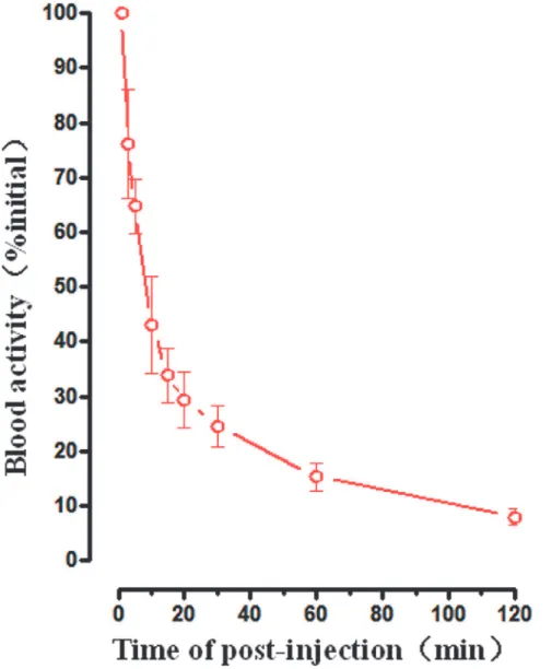
Fig 2gives the time-activity curve reporting the blood clearance over the first 120 min respec-tively. The data showed there was a very sharp decline of radioactivity in the blood. Blood ac-tivity was 43.05%, 24.60% and 15.33% of the initial dosage at 10, 30 and 60 min respectively after injection.
Fig 2. Averaged time-activity curve of99mTc-RGD-BBN in blood for all healthy volunteers.Error bars
indicate standard deviations.
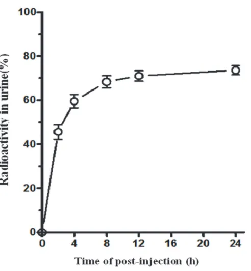
The concentration of radioactivity in the urine was shown inFig 3. The radioactivity in the urine keeps increasing with a total cumulative recovery of (73.56 ± 2.04) % of the original dose at 24h.
Biodistribution of
99mTc-RGD-BBN in normal subjects
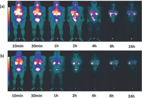
Fig 4shows a representation of a coronal section from whole-body SPECT and the distribution of99mTc-RGD-BBN at 10 min, 30 min, 1 h, 2 h, 4 h, 8 h and 24 h post-injection. The predomi-nant uptake was seen in the bladder, indicating a renal-urinary excretion of the tracer. The kid-neys and liver also showed moderate uptake. Apparent tracer uptake was observed in the nasal cavity and salivary glands in the early time points and almost undetectable at 24 hr
after injection.
By measuring ROIs drawn on both anterior and posterior images, the quantitative tracer up-takes in major organs were presented inFig 5. It shows the highest activity in visceral organs was found in the bladder which ascended from 8.71 ± 2.33% ID/organ (10 min p.i.) to 12.05 ± 8.40% ID/organ (60 min p.i.). It was followed by the kidneys and the liver which declined over time (10 min p.i. to 60 min p.i., 12.40 ± 3.37% to 6.24 ± 1.11% ID/organ, 6.47 ± 0.67% to 4.04 ± 0.63% ID/organ, respectively). Low activities were remained in the spleen and heart till 60 min after injection (1.35 ± 0.49% and 1.17 ± 0.24% ID/organ, respectively).
Fig 3. Averaged time-activity curve of99mTc-RGD-BBN in urine for all healthy volunteers.Error bars
indicate standard deviations.
Dosimetry
A summary of dosimetric parameters for various organs and whole body is given inTable 2. The mean effective dose equivalent for the whole body was 2.17E-03 mSv/MBq.
Scintimammography
All underwent surgery within 1 wk. A total of 6 palpable malignant lesions in 6 patients were described in the standard of truth including 4 invasive ductal carcinomas (IDC) and 2 ductal carcinoma in situ (DCIS) (Table 3). All of them were detected by the prescribed99m Tc-RGD-BBN SPECT/CT imaging with clear uptake. All malignant lesions were also detected
Fig 4. Series of planar whole-body images of a representative subject showing the distribution of
99mTc-RGD-BBN between 10 min and 24 h post-injection.(a) Anterior planar whole-body images. (b)
Posterior planar whole-body images.
doi:10.1371/journal.pone.0123401.g004
Fig 5. The quantified analysis of99mTc-RGD-BBN in major organs of healthy volunteers calculated
from the whole-body images obtained at 10 min, 30 min, 1 h, 2 h, 4 h, 8 h and 24 h after administration.
using99mTc-3P4-RGD2SPECT/CT and the result is five malignant lesions with clear uptake
and the other one with barely an uptake (Fig 6).
Immunohistochemistry of
α
vβ
3expression
For 6 malignant samples, 4 cases were found with dualαvβ3and GRPR expression, 1 case with
only GRPR positive expression (integrinαvβ3negative) (Fig 5) and 1 case with only integrin
αvβ3positive expression (GRPR negative) (Table 3). For the 4 cases with dualαvβ3and GRPR
expression, there was no significant statistical difference in the T/N ratios between99m Tc-RGD-BBN and99mTc-3P4-RGD2imaging (t = 2.6681;P>0.05).
Discussion
The SMM is less widespread than XMM and ultrasound US, but this does not mean that it has to remain in the background, as it is of great diagnostic value in breast tumor, especially after the emergence of SPECT/CT and the constant development of new tracers offers great poten-tial for SMM. The combination of SMM and CT implies a huge progress due to its high diag-nostic performance and ability to detect tumor pathological conditions. As non-invasive and sensitive imaging methods they have been widely used for diagnosing diseases in the clinic [13].
Table 2. Dosimetric data of99mTc-RGD-BBN in all healthy volunteers (n = 6).
Target Organ Dosimetric data(×10–3mSv/MBq)
Male(n = 3) Female(n = 3) Total(n = 6)
Adrenal glands 3.53±0.40 4.16±0.92 3.85±0.66
Brain 1.11±0.19 0.86±0.26 0.99±0.22
Breasts 1.08±0.12 1.17±0.27 1.12±0.18
Gallbladder wall 5.09±0.79 5.22±1.13 5.15±0.80
Lower region of colon 19.40±2.18 27.37±7.33 23.38±5.95
Small intestine 3.08±0.28 3.86±0.76 3.47±0.61
Stomach wall 2.66±0.21 2.93±0.56 2.80±0.37
Upper colon 2.50±0.24 3.04±0.64 2.77±0.48
Heart wall 3.57±0.49 3.77±0.54 3.67±0.43
Kidneys 23.60±4.24 27.50±7.78 25.55±5.48
Liver 5.06±0.51 5.45±1.29 5.26±0.82
Lungs 3.96±0.41 4.48±1.05 4.22±0.70
Muscle 1.65±0.14 1.88±0.40 1.77±0.27
Ovaries - 4.54±0.95 4.54±0.95
Pancreas 9.68±3.03 11.43±1.36 10.56±2.11
Red marrow 1.88±0.20 2.19±0.47 2.03±0.33
Osteogenic cells 3.80±0.42 4.28±1.01 4.04±0.68
Skin 0.91±0.10 0.99±0.23 0.95±0.15
Spleen 14.93±1.96 9.59±4.21 12.26±3.78
Testis 1.48±0.05 - 1.48±0.05
Thymus 1.60±0.20 1.71±0.37 1.66±0.25
Thyroid gland 14.16±4.71 14.57±1.17 14.37±2.81
Urinary bladder wall 12.25±2.99 16.50±5.89 14.38±4.37
Uterus - 4.14±0.77 4.14±0.77
Whole body 2.02±0.19 2.31±0.49 2.17±0.34
Table 3. Characteristics of all patients.
Patients Pathology Location Lesion size(cm)
Visual uptake grading
RGD (T/N Ratio)
Visual uptake grading
RGD-BBN (T/N Ratio)
Immunohistochemistry
RGD RGD-BBN αvβ3 GRPR
1 DCIS R 2.3 1 1.04 3 3.31 - +
2 DCIS L 2.0 3 3.09 3 2.72 + +
3 IDC L 5.1 3 3.12 3 3.06 + +
4 IDC R 2.1 3 2.56 3 2.45 +
-5 IDC L 5.8 3 4.43 3 4.17 + +
6 IDC L 6.3 3 3.12 3 3.03 + +
SPECT/CT with99mTc-RGD-BBN and99mTc-RGD: 3-grade scale, where grade 1 = no abnormal increased uptake; grade 2 = mildly increased or heterogeneous uptake; grade
3 = definite focal increased uptake. IDC = infiltrative ductal carcinoma; DCIS = ductal carcinoma in situ.
doi:10.1371/journal.pone.0123401.t003
Comparis
on
of
Dual
to
Single
Receptor
Imaging
in
Breast
|DOI:10.137
1/journal.p
one.0123401
April
7,
The emergence of various probes has become an indispensable factor for SMM. In previous studies, several one-target based radiotracers have been developed, such as a series of arginine-glycine-aspartic acid (RGD) containing peptides and radiolabeled BBN analogue which can specifically bind to integrinαvβ3and GRPR respectively[14–19]. Some have been successfully
applied to human clinical trials [20]. But monomeric RGD or BBN based radiotracers have some drawbacks for tumor imaging. First, not all tumors highly express cell-surface targets during progression of tumor initiation, growth, invasion and relapse. For instance, GRPR ex-pression is greatly reduced during the dedifferentiation of prostate cancer cells from androgen-controlled to androgen-independent transformation [21]. Second, the relatively feeble binding affinity and unsatisfactory in vivo pharmacokinetics of the monomeric radiotracers may lead to rapid dissociation from the tumor targets and the fuzzy imaging. For these reasons, dual tar-geted peptide provides a new avenue for SMM. It could be with more tumor binding affinity and could identify tumor in its various pathological process. Recently, a dual integrinαvβ3and
GRPR targeted peptide99mTc-RGD-BBN was developed and investigated. Data from previous studies in xenografts mouse models showed99mTc-RGD-BBN has excellent behaviors in vivo and in vitro and exhibited specific tumor imaging with high contrast to the contralateral back-ground [8,9]. Based on these favorable study results and our continuous interest in dual-tar-geted molecular probes for cancer imaging, we tentatively applied this new radiotracer in 6 healthy volunteers and 6 patients with breast cancer for the first time.
No clinically significant abnormalities or abnormal clinical chemistry were reported in any of the patients during our study. This showed that99mTc-RGD-BBN had good security and sta-bility for human use. The pharmacokinetic results were also satisfactory. There was a very sharp decline of radioactivity in the circulation and the radioactivity in the urine kept in-creasing with a total cumulative recovery of (73.56 ± 2.04) % of original dose at 24 h. The bio-distribution of99mTc-RGD-BBN in normal subjects indicated a renal-urinary excretion of the tracer.
Fig 6. A 51-y-old patient with a breast malignant tumor in the right side as Patient 1 inTable 3; (a)
99mTc-3P4-RGD
2SMM demonstrates no tracer uptake observed in the lesion.(b) CT scan demonstrates
a mass in the right breast. (c)99mTc-RGD-BN SMM demonstrates high uptake observed in the lesion. (d) Histopathology staining indicated a ductal carcinoma in situ. ×40 (e) Immunohistochemistry demonstrates barelyαvβ3expression in tumor vessels and tumor cells. ×400. (f) Immunohistochemistry demonstrates intense GRPR expression in tumor vessels and tumor cells. ×400.
The effective radiation dose to the body of99mTc-3P4-RGD2and99mTc-RGD-BBN were
1.15 ± 0.13 mSv and 0.65 ± 0.07 mSv respectively9. According to the 2007 Recommendations of the International Commission on Radiological Protection (ICRP) [22–24], radiation equiva-lent dose which occupational workers’vital organs were exposed to should be less than 500mSv and radiosensitive organs should be less than 150mSv. In our study, the mean absorbed dose equivalent for the whole body was 2.02E-03 mSv / MBq in healthy males and 2.31E-03 mSv / MBq in healthy females. Compared with other clinical markers of99mTc,99mTc-RGD-BBN is significantly lower than the conventional nuclear medical imaging agent, such as99mTc—MDP bone check (5.92 mSv) and99mTc—MIBI heart examination (7.05 mSv) [25].
All of the 6 malignant lesions were clearly detected by99mTc-RGD-BBN SPECT/CT imag-ing with intense uptake, which is consistent with the recent report of18F,64Cu and68Ga labeled RGD-BBN PET imaging [8].99mTc-3P4-RGD2SPECT/CT imaging showed five lesions with
clear uptake and the other one with barely uptake. This case was found with only GRPR posi-tive expression (integrinαvβ3negative), which could be the reason for the different imaging re-sult by99mTc-3P4-RGD2and99mTc-RGD-BBN. The advantage of dual integrinαvβ3and
GRPR targeted peptide is obvious when only one receptor type is overexpressed. For example, in the lesion of patient 1, which expressed GRPR but no integrinαvβ3,99mTc-3P4-RGD2
imag-ing was unable to detect the tumor because it only recognized integrinαvβ3. In contrast,99m Tc-RGD-BBN imaging had a clear tumor uptake due to the function of GRPR. Therefore,99m Tc-RGD-BBN may have the potential to make up for the deficiency of99mTc-3P4-RGD2in the
de-tection of breast malignant tumors with integrinαvβ3negative expression but GRPR positive
expression. There was one case with only integrinαvβ3positive expression (GRPR negative), the T/N ratio of99mTc-3P4-RGD2imaging was higher than that of99mTc-RGD-BBN imaging
(2.56 vs. 2.45), which might have resulted from the improved in tumor affinity and pharmaco-kinetic of dimeric RGD peptide over monomeric RGD. The immunohistochemistry revealed 4 cases with both integrinαvβ3 and GRPR positive expression, the T/N ratio of99m
Tc-3P4-RGD2imaging was higher than that of99mTc-RGD-BBN imaging but there was no
signifi-cant statistical difference (P>0.05). The reasons for this may be as follows:99mTc-3P4-RGD2
is a well-designed, dimeric RGD peptide which can bind to two integrinαvβ3 motifs simulta-neously. Some studies found that99mTc-RGD-BBN is impossible to bind to both GRPR and integrin simultaneously in vivo since the glutamate linker between the RGD and BBN motifs is not long enough. The total number of binding sites (the sum of GRPR and integrin) for99m Tc-RGD-BBN would significantly increase as compared to the monomeric counterparts in the dual-receptor positive tumor models, which would lead to improve in vivo tumor targeting effi-cacy. Since the expression of cell-surface receptors by cancer cells can be heterogeneous and in-homogeneous, it is very hard to distinguish the expression level of each receptor individually when using dual-targeted molecular probes such as99mTc-RGD-BBN. The uptake of99m Tc-3P4-RGD2by tumor with both integrinαvβ3and GRPR positive expression may be higher
than99mTc-RGD-BBN if more integrinαvβ3are expressed. However, due to the small sample
size of this study, there was no significant statistical difference in T/N ratios between99m Tc-3P4-RGD2imaging and99mTc-RGD-BBN imaging.
It is well-known that a realistic strategy for the reduction of breast cancer mortality rates and timely treatment is to detect the disease as early as possible. The most common screening method for early breast cancer is mammography. Our previous research of99mTc-3P4-RGD2
or GRPR expression patterns [9]. The promising imaging results of99mTc-RGD-BBN in pa-tients with breast cancer may give rise to the possibility of extending applications in breast can-cer screening to avoid unnecessary biopsies.
In this study, our findings are of a preliminary nature and need to be further corroborated. It is necessary to investigate the effects of linkers of different lengths, solubility, lipophilicity, and flexibility on the in vitro and in vivo behaviors of the dual integrinαvβ3and GRPR targeted peptide to make it possible to bind to both receptors simultaneously, which may result in en-hanced targeting efficacy and higher uptake of99mTc-RGD-BBN by the tumor. This may help to provide new idea in the development of tumor targeted therapy. The design of heteromulti-meric tracers that recognize other tumor targets is also worth further investigation for tumor-targeted imaging and therapy.
There are several limitations to this study that call for further discussion. First, as a com-bined probe, the sensitivity of99mTc-RGD-BBN SPECT/CT in detecting breast malignant tumor should be improved, but the specificity may be also affected. In our study, there was no false positive or false negative case by99mTc-RGD-BBN SPECT/CT imaging. Such a high accu-racy might be related to the small numbers of patients included in this study and all of them were with breast malignant tumor, which was chosen with a subjective wish. Further research is needed to include a larger patient population. Second, although the immunohistochemistry of integrinαvβ3and GRPR expression were conducted in this study, quantification and
correla-tion with tracer uptake was not performed due to the heterogeneous and inhomogeneous ex-pression of cell-surface receptors by cancer cells.
Conclusions
The dual integrinαvβ3and GRPR targeting99mTc-RGD-BBN showed an excellent ability to de-tect breast cancer without clinically safety problems.99mTc-RGD-BBN may have the potential to make up for the deficiency of99mTc-3P4-RGD2in the detection of breast cancer with only
GRPR positive expression (integrinαvβ3negative). The preliminary application of99m
Tc-RGD-BBN has demonstrated its powerful potential in breast cancer diagnosis and therapy.
Author Contributions
Conceived and designed the experiments: QQC QJM SG. Performed the experiments: QQC MLC BC QW. Analyzed the data: BC QW. Contributed reagents/materials/analysis tools: BJ FW BTS. Wrote the paper: QJM MLC SG.
References
1. Siegel R, Naishadham D, Jemal A. Cancer statistics, 2012. CA: a cancer journal for clinicians. 2012; 62: 10–29. doi:10.3322/caac.20138PMID:22237781
2. DeSantis C, Siegel R, Bandi P, Jemal A. Breast cancer statistics, 2011. CA: a cancer journal for clini-cians. 2011; 61: 408–418. doi:10.3322/caac.20125PMID:22086728
3. Okarvi S. Peptide‐based radiopharmaceuticals: Future tools for diagnostic imaging of cancers and other diseases. Medicinal research reviews. 2004; 24: 357–397. PMID:14994368
4. Ma Q, Ji B, Jia B, Gao S, Ji T, Wang X, et al. Differential diagnosis of solitary pulmonary nodules using 99mTc-3P4-RGD2 scintigraphy. European journal of nuclear medicine and molecular imaging. 2011; 38: 2145–2152. doi:10.1007/s00259-011-1901-2PMID:21874323
5. Guanghui C, Shi G, Tiefeng J, Qingjie M, Bing J, Zuowei C, et al. Pharmacokinetics and radiation do-simetry of~(99m) Tc-3PRGD_2 in healthy individuals: A pilot study. Nuclear Science and Techniques. 2012; 23: 349-349-354.
7. Ji S, Zheng Y, Shao G, Zhou Y, Liu S. Integrinαvβ3-Targeted Radiotracer 99mTc-3P-RGD2 Useful for Noninvasive Monitoring of Breast Tumor Response to Antiangiogenic Linifanib Therapy but not Anti-Integrinαvβ3 RGD2 Therapy. Theranostics. 2013; 3: 816. doi:10.7150/thno.6989PMID:24312152
8. Liu Z, Yan Y, Liu S, Wang F, Chen X. 18F, 64Cu, and 68Ga labeled RGD-bombesin heterodimeric pep-tides for PET imaging of breast cancer. Bioconjugate chemistry. 2009; 20: 1016–1025. doi:10.1021/ bc9000245PMID:20540537
9. Liu Z, Huang J, Dong C, Cui L, Jin X, Jia B, et al. 99mTc-labeled RGD-BBN peptide for small-animal SPECT/CT of lung carcinoma. Molecular pharmaceutics. 2012; 9: 1409–1417. doi:10.1021/ mp200661tPMID:22452411
10. Liu Z, Niu G, Wang F, Chen X. 68Ga-labeled NOTA-RGD-BBN peptide for dual integrin and GRPR-tar-geted tumor imaging. European journal of nuclear medicine and molecular imaging. 2009; 36: 1483– 1494. doi:10.1007/s00259-009-1123-zPMID:19360404
11. Scott N, Millward E, Cartwright E, Preston S, Coletta P. Gastrin releasing peptide and gastrin releasing peptide receptor expression in gastrointestinal carcinoid tumours. Journal of clinical pathology. 2004; 57: 189–192. PMID:14747448
12. Wiegrebe L. An autocorrelation model of bat sonar. Biol. Cybern. 2008; 98: 587–595. doi:10.1007/ s00422-008-0216-2PMID:18491168
13. Yu Z, Ananias HJ, Carlucci G, Hoving HD, Helfrich W, Dierckx RA, et al. An update of radiolabeled bom-besin analogs for gastrin-releasing peptide receptor targeting. Current pharmaceutical design. 2013; 19: 3329–3341. PMID:23431995
14. Liu S, Hsieh W, Jiang Y, Kim Y, Sreerama SG, Chen X, et al. Evaluation of a 99mTc-labeled cyclic RGD tetramer for noninvasive imaging integrinαvβ3-positive breast cancer. Bioconjugate chemistry. 2007; 18: 438–446. PMID:17341108
15. Beer AJ, Grosu A, Carlsen J, Kolk A, Sarbia M, Stangier I, et al. [18F] galacto-RGD positron emission tomography for imaging ofαvβ3 expression on the neovasculature in patients with squamous cell carci-noma of the head and neck. Clinical Cancer Research. 2007; 13: 6610–6616. PMID:18006761
16. Beer AJ, Haubner R, Sarbia M, Goebel M, Luderschmidt S, Grosu AL, et al. Positron emission tomogra-phy using [18F] Galacto-RGD identifies the level of integrinαvβ3 expression in man. Clinical Cancer Research. 2006; 12: 3942–3949. PMID:16818691
17. Zhang X, Cai W, Cao F, Schreibmann E, Wu Y, Wu JC, et al. 18F-labeled bombesin analogs for target-ing GRP receptor-expresstarget-ing prostate cancer. Journal of Nuclear Medicine. 2006; 47: 492–501. PMID:
16513619
18. Yang Y, Zhang X, Xiong Z, Chen X. Comparative in vitro and in vivo evaluation of two64Cu-labeled bombesin analogs in a mouse model of human prostate adenocarcinoma. Nuclear medicine and biolo-gy. 2006; 33: 371–380. PMID:16631086
19. Aitmohand S, Fournier P, Dumulonperreault V, Kiefer GE, Jurek P, Ferreira CL, et al. Evaluation of 64Cu-labeled bifunctional chelate–bombesin conjugates. Bioconjugate chemistry. 2011; 22: 1729– 1735. doi:10.1021/bc2002665PMID:21761921
20. Marrone B, Meurer L, Moretto A, Kleina W, Schwartsmann G. Expression of gastrin‐releasing peptide receptor in patients with cutaneous malignant melanoma. Clinical and experimental dermatology. 2013; 38: 707–712. doi:10.1111/ced.12058PMID:23581973
21. de Visser M, van Weerden WM, de Ridder CM, Reneman S, Melis M, Krenning EP, et al. Androgen-de-pendent expression of the gastrin-releasing peptide receptor in human prostate tumor xenografts. Jour-nal of nuclear medicine: official publication, Society of Nuclear Medicine. 2007; 48: 88–93.
22. Protection TRotICoR. ICRP publication 103. Annals of the ICRP. 2007; 37: 1–332. doi:10.1016/j.icrp.
2008.08.001PMID:18762065
23. Kehoe T. International Commission on Radiological Protection publications 97 and 98: radiation protec-tion can be fun! Clinical Oncology. 2007; 19: 539–541. PMID:17604971
24. Pradhan AS. Evolution of dosimetric quantities of International Commission on Radiological Protection (ICRP): Impact of the forthcoming recommendations. Journal of medical physics / Association of Medi-cal Physicists of India. 2007; 32: 89–91. doi:10.4103/0971-6203.35719PMID:21157526
25. Kubo A, Nakamura K, Sanmiya T, Shimizu S, Hashimoto S, Iwanaga S, et al. [Phase I clinical study on 99mTc-MIBI]. Kaku igaku. The Japanese journal of nuclear medicine. 1991; 28: 1133–1142. PMID:
1839318
26. Liu L, Song Y, Gao S, Ji T, Zhang H, Ji B, et al. (99)mTc-3PRGD2 scintimammography in palpable and nonpalpable breast lesions. Molecular imaging. 2014; 13.