Extracellular Adenosine Protects against
Streptococcus pneumoniae
Lung Infection by
Regulating Pulmonary Neutrophil
Recruitment
Elsa N. Bou Ghanem1, Stacie Clark1, Sara E. Roggensack2, Sally R. McIver3, Pilar Alcaide4, Philip G. Haydon3, John M. Leong1*
1Department of Molecular Biology and Microbiology, Tufts University School of Medicine, Boston,
Massachusetts, United States of America,2Program in Molecular Microbiology, Sackler School of Graduate Biomedical Sciences, Tufts University, Boston, Massachusetts, United States of America,3Department of Neuroscience, Tufts University School of Medicine, Boston, Massachusetts, United States of America, 4Sackler School of Graduate Biomedical Sciences, Tufts University School of Medicine and Molecular Cardiology Research Institute, Tufts Medical Center, Boston, Massachusetts, United States of America
*John.Leong@tufts.edu
Abstract
An important determinant of disease followingStreptococcus pneumoniae (pneumococ-cus) lung infection is pulmonary inflammation mediated by polymorphonuclear leukocytes (PMNs). We found that upon intratracheal challenge of mice, recruitment of PMNs into the lungs within the first 3 hours coincided with decreased pulmonary pneumococci, whereas large numbers of pulmonary PMNs beyond 12 hours correlated with a greater bacterial bur-den. Indeed, mice that survived infection largely resolved inflammation by 72 hours, and PMN depletion at peak infiltration, i.e. 18 hours post-infection, lowered bacterial numbers and enhanced survival. We investigated host signaling pathways that influence both pneu-mococcus clearance and pulmonary inflammation. Pharmacologic inhibition and/or genetic ablation of enzymes that generate extracellular adenosine (EAD) (e.g. the ectoenzyme CD73) or degrade EAD (e.g. adenosine deaminase) revealed that EAD dramatically increases murine resistance toS.pneumoniaelung infection. Moreover, adenosine dimin-ished PMN movement across endothelial monolayersin vitro, and although inhibition or deficiency of CD73 had no discernible impact on PMN recruitment within the first 6 hours after intratracheal inoculation of mice, these measures enhanced PMN numbers in the pul-monary interstitium after 18 hours of infection, culminating in dramatically elevated numbers of pulmonary PMNs at three days post-infection. When assessed at this time point,CD73
-/-mice displayed increased levels of cellular factors that promote leukocyte migration, such as CXCL2 chemokine in the murine lung, as well as CXCR2 andβ-2 integrin on the surface of pulmonary PMNs. The enhanced pneumococcal susceptibility ofCD73-/-mice was signif-icantly reversed by PMN depletion following infection, suggesting that EAD-mediated resis-tance is largely mediated by its effects on PMNs. Finally, CD73-inhibition diminished the ability of PMNs to kill pneumococciin vitro, suggesting that EAD alters both the recruitment
OPEN ACCESS
Citation:Bou Ghanem EN, Clark S, Roggensack SE,
McIver SR, Alcaide P, Haydon PG, et al. (2015) Extracellular Adenosine Protects against Streptococcus pneumoniaeLung Infection by Regulating Pulmonary Neutrophil Recruitment. PLoS Pathog 11(8): e1005126. doi:10.1371/journal. ppat.1005126
Editor:Timothy J. Mitchell, University of Birmingham,
UNITED KINGDOM
Received:April 2, 2015
Accepted:August 4, 2015
Published:August 27, 2015
Copyright:© 2015 Bou Ghanem et al. This is an
open access article distributed under the terms of the
Creative Commons Attribution License, which permits unrestricted use, distribution, and reproduction in any medium, provided the original author and source are credited.
Data Availability Statement:All relevant data are
within the paper and its Supporting Information files.
Funding:Elsa N. Bou Ghanem is a Howard Hughes
Medical Institute Fellow of the Life Sciences Research Foundation that supported this work. The funders had no role in study design, data collection and analysis, decision to publish, or preparation of the manuscript.
Competing Interests:I have read the journal's policy
and bacteriocidal function of PMNs. The EAD-pathway may provide a therapeutic target for regulating potentially harmful inflammatory host responses during Gram-positive bacterial pneumonia.
Author Summary
Despite the presence of vaccines and antibiotic therapies, invasiveStreptococcus pneumo-niae(pneumococcus) infections, such as pneumonia, bacteremia and meningitis, remain a leading cause of mortality and morbidity worldwide. Understanding the host factors that influence the outcome ofS.pneumoniaeinfection will allow us to design better therapies. Here, we elucidate the role of rapidly responding innate immune cells termed neutrophils, or PMNs (polymorphonuclear leukocytes), whose role inS.pneumoniaeinfection has long been controversial. We found that PMNs are initially required for controlling bacte-rial numbers, but their extended presence in the lungs leads to significant damage and poor control of infection. The signals that control the movement of PMNs into the infected lungs are not well understood. Here, we identified extracellular adenosine (EAD), a molecule produced by the host in response to cellular damage, as important in limiting PMN movement into the lungs upon pneumococcal challenge. Importantly, EAD-medi-ated control of PMNs was crucial for fighting lung infection byS.pneumoniae. This study may lead to the potential use of clinically available adenosine-based therapies to combat pneumococcal pneumonia in the future.
Introduction
Despite vaccines and antibiotic therapies, invasiveStreptococcus pneumoniae(pneumococcus) infections such as pneumonia, meningitis and bacteremia remain a considerable health and economic burden [1,2]. A major determinant of disease followingS.pneumoniaeinfection is pulmonary inflammation, which, if excessive, can result in tissue destruction, compromised gas exchange, and/or acute respiratory distress syndrome [3]. Many conditions associated with enhanced inflammation, including influenza infection [4–6] and aging [7,8], lead to increased susceptibility to pneumococcal pneumonia.
Effective inflammatory responses to infection balance host defense with the potentially competing demand of a rapid return to homeostasis. Indeed, pneumococcal pneumonia trig-gers a massive neutrophil, or polymorphonuclear leukocyte (PMN), influx into the alveolar spaces [9,10], but the role of these innate immune cells during infection is complex. Several findings suggest that PMNs are needed to control the infection: neutropenic patients are at increased risk for pneumonia [11], and in several mouse studies, depletion of PMNs prior toS. pneumoniaeinfection [12,13] or delay in PMN recruitment into the lungs [14,15] resulted in higher pulmonary bacterial loads and lethal septicemia. Paradoxically however, conditions associated with increased numbers of PMNs in the lungs several days afterS.pneumoniae infection of mice, such as advanced age [8,16], deficiency in regulatory T cells [17], or influenza infection [18], result in more severe systemic infection and reduced survival. Conversely, reducing PMN influx into mouse airways dramatically decreases bacteremia, resulting in uni-form survival to a normally lethal pneumococcal pulmonary challenge [9]. These findings sug-gest that host survival may require an initial acute PMN response that is rapidly resolved later in the course ofS.pneumoniaeinfection.
To reachS.pneumoniaein alveolar spaces, circulating PMNs cross the endothelium, enter into the interstitial space, then breach the lung epithelium to access the airway spaces [19]. This complex process involves multiple pathways of chemotaxis, including those mediated by eicosanoids [9] or chemokines [19] [20], as well as a network of ligand-receptor interactions, including those mediated by lectins or integrins [15]. Although many studies have focused on positive regulators of PMN recruitment into the lungs following pneumococcal challenge [9,14,15], signals that negatively regulate this process and ultimately promote resolution of this response are poorly understood.
Extracellular adenosine (EAD) is a potentially crucial regulator of PMN-mediated pulmo-nary inflammation. Basal EAD levels in tissues are typically low (<1μM) [21], but can increase more than ten-fold during pathological conditions [22]. Upon cellular insult, such as infection [23], ATP is released from cells and metabolized to adenosine by the sequential action of two extracellular enzymes, CD39, which converts ATP to AMP, and CD73, an ecto-5’-nucleotidase that de-phosphorylates AMP to EAD [22]. EAD is recognized by four G-protein coupled receptors, A1, A2A, A2B and A3 [23] leading to enhanced or diminished acute inflammation, depending on the target receptor, cell type, and/or EAD concentration [23]. Thus, the EAD pathway may provide a means for complex regulation of PMN movement [22].
Several non-infectious acute pulmonary injury models indicate that EAD generated by endothelial cell CD73 binds to cognate adenosine receptors on PMNs, leading to reduced PMN-endothelial cell adhesion, inflammation, and tissue damage [24–26]. Lung epithelial cells are both an important EAD source [25] and, given that they produce all four adenosine recep-tors [21], a potential EAD target.CD73-/-mice show impaired clearance of bacteremia and enhanced pulmonary inflammation in a cecal puncture model [27], whereas deficiency of adenosine A2B or A1 receptors was protective againstKlebsiella pneumoniae[28] or influenza lung infection [29], respectively. Thus, the role of EAD in pathogen lung burden, inflamma-tion, and injury during bacterial infection is not fully characterized.
In this study, we characterized the kinetics of PMN entry into the lung during murine pneu-mococcal challenge with an invasiveS.pneumoniaestrain, and addressed potential beneficial and detrimental roles of PMNs in disease. We found that PMNs promoted microbial control early, but inhibited bacterial clearance later in infection. We identified the EAD pathway as a regulator of endothelial transmigration and PMN recruitment into the lung at later time points after pneumococcal infection, as well as PMNs anti- pneumococci function. This study is a first step in elucidating the potentially complex role of the EAD-pathway in regulating pulmonary inflammation and host defense against Gram-positive bacterial pneumonia.
Results
Kinetic analysis of PMN influx into the lungs and bacterial infection
following
S
.
pneumoniae
intratracheal challenge
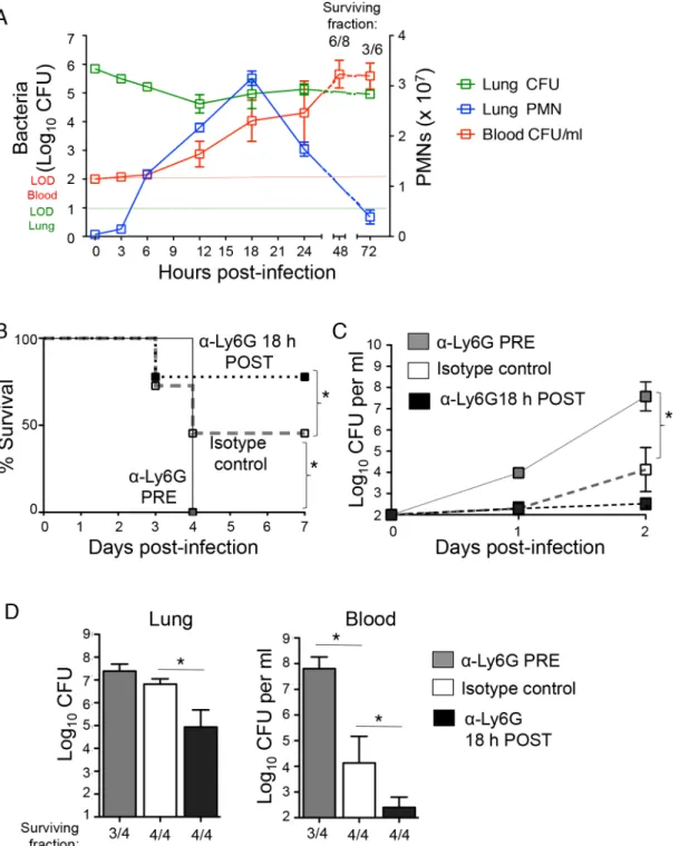
Fig 1. PMNs promote pulmonary and systemic disease during later stages ofS.pneumoniaelung infection.(A) C57BL/6J mice were inoculated I.T with 5x10⁵CFU ofS.pneumoniaeTIGR4 and pulmonary (green) and bloodstream (red) bacterial loads, as well as pulmonary PMNs (blue) were monitored through 72 hours post-infection. Shown are representative data from one of two separate experiments (using 3 to 4 mice per time point). The numbers above the graph represent the fraction of surviving mice within that group at the corresponding time. (B-D) C57BL/6J mice were treated i.p with PMN depleting antibodies (anti-ly6G) or isotype control either 18 hours pre or post pulmonary challenge with 5x10⁵CFU ofS.pneumoniaeTIGR4. Survival (B) and bacterial burdens in the blood (C) were monitored over time and shown are pooled data from two separate experiments. (D) Pneumococcal burdens in the lungs and blood were determined 3 days post-infection. The numbers below the graph represent the fraction of surviving mice within that group. Representative data from one of 4 separate experiments with 3 to 4 mice per group are shown. Means +/- SEM are given in Panels A, C and D, and values significantly (p<0.05) different from isotype control-treated group by Student’s t-test are indicated by asterisk. In Panel B, asterisk indicates survival rate was significantly (p<0.05) different from isotype control-treated controls by Log-rank (Mantel-Cox) test.
Quantitation of bacterial numbers in the lung revealed two phases of infection control. In spite of the fact thatS.pneumoniaeTIGR4 is a virulent strain capable of replication in the murine lung [30], bacterial numbers in the lung decreased ~30-fold in the first 12 hours of infection, a period in which PMN numbers increased dramatically (Fig 1A). However, between 12 and 18 hours, during which mice continued to experience a striking increase in pulmonary PMNs, bacterial lung burden increased 5-fold to approximately 2 x 10⁵, and this level of infec-tion or higher was maintained for the remainder of the 72-hour experiment. Moreover, the three-fold increase in pulmonary PMNs, peaking at 18 hours post-challenge correlated with a large increase in bacterial numbers in the circulation, with titers of more than 104/ml, consis-tent with our previous findings that PMN entry into the lung facilitates bacterial spread [9]. Over the next 30 hours, a majority of infected mice succumbed to infection (Fig 1A) and even among survivors, bacterial titers in the blood increased 100-fold to over a million CFU/ml. Thus, although the initial increase in PMN influx into the lungs corresponded to a transient control of infection during the first 12 hours, the further accumulation of PMNs after this time point, peaking at 18 hours post-infection, coincided with the development of serious systemic infection.
PMNs are required for protection at the beginning of infection, but are
detrimental at later times
To experimentally address the role of PMNs during lung infection byS.pneumoniae, we depleted PMNs with intra-peritoneal (i.p.) injections of the anti-Ly6G antibody (IA8) either one day before I.T. infection with ~5x10⁵colony forming units (CFU) ofS.pneumoniaeTIGR4 strain, or 18 hours post-infection (seeMethods), a time point that corresponded to peak pul-monary infiltration by PMNs (Fig 1A). At both time points, treatment with the Ly6G anti-body resulted in>90% depletion of lung and circulating PMNs compared to isotype-treated controls (seeMethods). Survival and bacteremia, as well bacterial burdens in the lungs and blood at day three following infection were compared between the groups (Fig 1). Consistent with previous reports [12,13], mice depleted of PMNs pre-infection were extremely susceptible toS.pneumoniae(Fig 1B). In comparison to the matched isotype-treated control group, the pre-depleted mice suffered more than a thousand-fold greater bacterial load in the bloodstream (Fig 1C and 1D), and failed to survive the infection (Fig 1B).
In contrast, depletion of PMNs at 18 hours post-infection significantly increased the sur-vival rate (Fig 1B) and lowered bacterial burdens a hundred-fold in both the lungs and blood (Fig 1C and 1D). Our findings strongly support the hypothesis that while PMNs are required for bacterial control at the beginning of pneumococcal infection, their persistence following infection is detrimental to the host.
Inhibition of adenosine breakdown promotes host defense against
S
.
pneumoniae
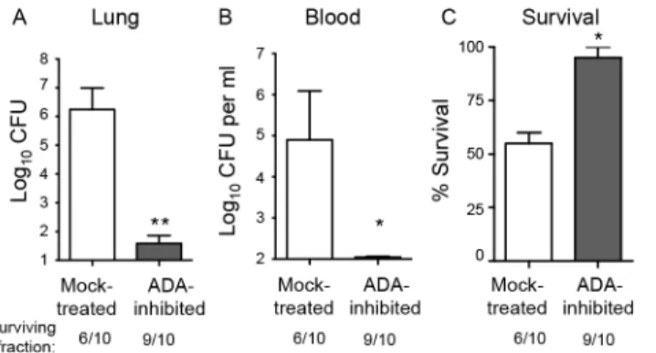
detectable bacteremia (Fig 2B). By day 3 post-infection, 40% of the mock-treated mice had suc-cumbed to the infection, compared to 10% of the ADA-inhibited mice (Fig 2C). Although in some infection models, adenosine-mediated protection is due to the direct effects of adenosine on the infectious agent [34], we found that adenosine concentrations typically present in inflamed tissues [22] had no effect on bacterial growthin vitro(S1 Fig).
Since the ADA inhibitor EHNA hydrochloride may also target other enzymes [35], we tested whether the protective phenotype was dependent on adenosine signaling. Adenosine receptor blockade partially reversed the protective effect of inhibition of adenosine breakdown by ADA (S2 Fig), consistent with the hypothesis that the protection we observed upon treat-ment with EHNA hydrochloride is at least partly mediated via the interaction between adeno-sine and its receptors.
Inhibition of CD73 increases susceptibility of mice to infection after
S
.
pneumoniae
lung challenge
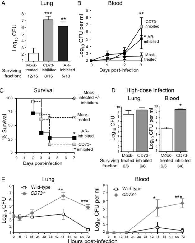
A prediction of the hypothesis that EAD is responsible for the protective effect of ADA inhibi-tion is that inhibiinhibi-tion of adenosine producinhibi-tion should enhance susceptibility to infecinhibi-tion. To test that, wild-type C57BL/6 mice were either mock-treated or injected intra-peritoneally with the CD73 inhibitorα,βmethylene ADP which was shown to drastically lower adenosine levels in mice [24]. The mice were then challenged I.T. with 5x10³S.pneumoniaeTIGR4, a dose ~2-fold below the LD50. Neither the addition of adenosine orα,βmethylene ADP had any
effect onS.pneumoniaegrowthin vitro(S1 Fig). However, at day 3 post-infection, mice treated with the inhibitor suffered approximately a million-fold higher bacterial burden in their lungs compared to mock-treated controls (Fig 3A). In addition, whereas mock-treated mice suffered low-level bacteremia that eventually resolved, CD73-inhibited mice suffered bacteremia that reached a million CFU per milliliter of blood at day 3 post-infection (Fig 3B). To determine if CD73 inhibition resulted in enhanced bacteremia in a mouse strain more resistant toS. pneu-moniaeinfection [36], we I.T. challenged either mock-treated or CD73-inhibited BALB/c mice and found that CD73 inhibition was associated with a 100- to 1000-fold increase in bacteremia after day 3 post-infection (S3 Fig). In C57BL/6 mice, CD73 inhibition was also associated with apparent neurological dysfunction, such as hind limb twitching, weakness, paralysis and an
Fig 2. Inhibition of adenosine breakdown promotes resistance toS.pneumoniaelung challenge. C57BL/6J mice mock-treated or treated with EHNA-hydrochloride, an adenosine deaminase inhibitor, were inoculated I.T with 5x10⁵CFU ofS.pneumoniaeTIGR4. Bacterial burdens in the lungs (A) or blood (B), as well as survival (C), was determined 3 days post-infection. Data pooled from 2 separate experiments (n = 6 mice per group) are shown. Data represent means +/- SEM.**=p<0.001 and*=p<0.05 indicate means
significantly different from mock-treated group by Student’s t-test. Below the graphs are indicated the fraction of surviving mice within each group.
Fig 3. Inhibition of EAD production or signaling enhances susceptibility of mice toS.pneumoniaelung challenge.(A-D) Wild-type C57BL/6J mice were either treated with the CD73 inhibitor (α,β- methylene ADP), the pan adenosine receptor (AR) inhibitor (CGS 15943) or mock-treated with a vehicle control. (A) Bacterial loads in the lungs of a group of mice were determined 3 days after I.T. inoculation with 5x10³CFU ofS.pneumoniaeTIGR4. (B-C) Bacteremia (B) and survival (C) were monitored over time for another group of mice. Pooled data from 3 separate experiments (n = 6–12 mice per group) are shown. (D) Mice were challenged I.T. with a high dose, ~1x10⁷CFU ofS.pneumoniaeTIGR4 and bacterial burdens in the lungs and blood were assessed at two days post-infection. Pooled data from two separate experiments (n = 6 mice per group) are shown. Below figures A and D are indicated the fraction of surviving mice within each group. (E)CD73
-/-mice and wild-type B6 controls were inoculated I.T. with 5x10³CFU ofS.pneumoniaeTIGR4 and bacterial burdens in the lungs and blood were measured at the indicated time points post-infection. Data pooled from 3 separate experiments (n = 6–9 mice per group) are shown. None of the mice died within the time frame of these (E) experiments. Data represent means +/- SEM. Means that are significantly different from mock-treated group (A-B) or wild-type control group (E) by student t-test are indicated by asterisks (***=p<0.0001;**=p<0.001;*=p<0.05). In Panel C,
a survival rate significantly (p<0.05) different from mock-treated controls by Log-rank (Mantel-Cox) test is indicated by asterisk.
inability to walk normally. By day 4 post-infection, 85% of CD73-inhibited C57BL/6 mice suc-cumbed to the infection (Fig 3C).
To test whether the increased systemic spread of pneumococci was simply a reflection of increased bacterial loads in the lungs, we challenged mice with a high dose ofS.pneumoniae, i.e. 2x10⁷CFU. CD73-inhibited mice suffered only a 1.1-fold higher (and statistically indistin-guishable) bacterial lung burden than mock-treated mice (Fig 3D). Despite similar numbers of bacteria in the lung, CD73-inhibition resulted in 1000-fold higher levels of bacteremia. Our findings suggest that in addition to impacting the ability of the host to control lung infection, CD73 inhibition also promotes systemic spread ofS.pneumoniaefrom the lungs.
To test the role of CD73 during pneumococcal infection using genetic rather than pharma-cological means, and to determine whether CD73 activity alters bacterial load early in infection, we inoculatedCD73-/-mice I.T with 5x 10³S.pneumoniaeand followed lung and blood CFU over time. CD73-deficiency had no significant effect on bacterial burden at either site at 6 or 18 hours post-infection (Fig 3E), suggesting that EAD does not play a major role in controlling bacterial numbers at the early stages of infection. Beyond 18 hours post-infection,CD73-/-mice were incapable of controlling pneumococcal burdens, reflected in a 100- to 1000-fold increase in both infection sites (Fig 3E). In contrast, bacterial numbers in the lung and blood of wild-type mice increased only slightly in the first 48 hours of infection and were largely cleared by 72 hours. Thus, pharmacological inhibition or genetic ablation of CD73, an enzyme required for EAD production [24], drastically increased theS.pneumoniaelung burden and susceptibil-ity to systemic disease.
The effect of EAD on host susceptibility to pneumococcal challenge is
dependent on adenosine receptor signaling
To test whether EAD-mediated protection upon pneumococcal infection was dependent on signaling via adenosine receptors in the host, mice were treated with the pan-adenosine recep-tor antagonist CGS-15943 [37] prior to challenge withS.pneumoniae. This inhibitor targets all four adenosine receptors, with Ki values of 3.5, 4.2, 16 and 51 nM for human A1, A2A, A2B and A3 receptors respectively [37,38]. Although CGS-15943 had no effect on the viability ofS. pneumoniae in vitro(S1 Fig), treatment of mice with this inhibitor resulted in increased suscep-tibility toS.pneumoniaelung challenge that was virtually identical to that observed upon inhi-bition of CD73-mediated EAD production (Fig 3). Compared to mock-treated controls, mice treated with the adenosine receptors antagonist suffered ten thousand-fold higher bacterial loads in their lungs (Fig 3A) as well as bacteremia exceeding 103CFU/ml (Fig 3B). The mice treated with the adenosine receptors antagonist also displayed a significant survival defect com-pared to mock-treated mice following pneumococcal lung challenge (Fig 3C). These findings clearly show that inhibition of adenosine receptors signaling render mice highly susceptible to pneumococcal challenge.
Extracellular adenosine signaling diminishes recruitment of PMNs
across endothelial but not epithelial monolayers
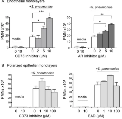
endothelial cell monolayer byS.pneumoniae(Fig 4A), suggesting that, in this model, pneumo-coccal infection activates the endothelium to trigger PMN transmigration.
To test the role of EAD production on PMN migration in this system, we added the phar-macological inhibitor of CD73,α,βmethylene ADP, to the media during the migration process, and found that it resulted in a significant dose-dependent increase in PMN migration in response to pneumococcal infection (Fig 4A, left panel). Importantly, a similar increase in PMN migration was observed when adenosine receptor signaling was blocked using the pan-adenosine receptor inhibitor CGS-15943 (Fig 4A, right panel).
We previously showed that blocking the movement of PMNs across the lung epithelium and into the airways protected mice against an otherwise lethalS.pneumoniaeinfection [9]. To test whether EAD also regulates PMN movement across this barrier, we utilized a well-establishedin vitrohuman PMN trans-epithelial migration assay [9]. As previously observed, apical pneumococcal infection of confluent polarized lung epithelial cells grown on filter mem-branes elicited robust basolateral to apical migration of PMNs (Fig 4B). Addition of the CD73 inhibitor, or exogenous adenosine to this assay had no significant effect on migration (Fig 4B). Together with our studies on endothelium, these results indicate that EAD negatively regulates PMN transmigration across endothelial but not epithelial monolayers in response toS. pneu-moniaeinfection.
Fig 4. EAD negatively regulates PMN migration across the endothelium but not the epithelium.(A) Media alone orS.pneumoniae-containing media was added to the lower chamber of HUVEC-seeded Transwell dishes for 3 hours. Transmigration of PMNs added to the upper chamber in media containing vehicle control or increasing concentrations of the CD73 inhibitorα,β- methylene ADP (left panel) or pan-adenosine receptor (AR) inhibitor CGS 15943 (right panel) was measured using a hemocytometer. The means +/- SEM from one representative of three experiments are shown and values significantly different from media control, determined by student’s t-test, are indicated by asterisk (***=p<0.0001;**=p<0.001;
*=p<0.05). (B) Polarized H292 epithelial cells, pre-treated with increasing concentrations CD73 inhibitorα,
β- methylene ADP (left panel) or just adenosine (right panel), were left uninfected (“media”) or infected apically with pneumococcus (“+S.pneumoniae”). The transmigration of PMNs, added to the basolateral side in media alone or the indicated concentrations of CD73 inhibitor or adenosine was measured by
myeloperoxidase ELISA. Data represent means +/- SEM, and shown are one of three separate experiments.
Blocking EAD production or signaling increases interstitial pulmonary
PMNs
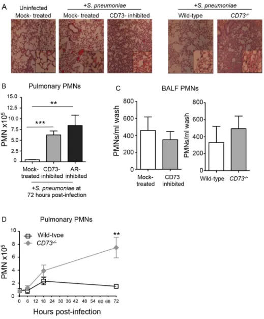
To test whether EAD regulates transmigration specifically across pulmonary endothelium dur-ing infection, we administered 5x10³CFU ofS.pneumoniaeI.T to mock-treated or CD73-in-hibited mice, as well as toCD73-/-or wild type C57BL/6 mice. We assessed cellular recruitment into the lungs at day three post-infection, a time point that coincides with the resolution of pul-monary inflammation following a sub-lethal pneumococcal infection [40]. Consistent with the hypothesis that blocking EAD synthesis results in enhanced egress of PMNs from the vascula-ture, histological analysis of H&E stained lung sections at 3 days post-infection, revealed an increase in cellular infiltrates into the lungs of bothCD73-/-mice and wild type C57BL/6 mice treated with the CD73 inhibitor (Fig 5A).
To quantify the apparent increase in pulmonary PMNs upon inhibition of EAD production or signaling, we measured pulmonary PMNs of mice that had been treated with the CD73 or the pan-adenosine receptor inhibitors and previously analyzed for lung and blood CFU inFig 3A and 3B. Single-cell suspensions of lung tissue at day 3 post-infection were analyzed by flow cytometry after staining with antibody directed against the PMN marker Ly6G. Genetic abla-tion of CD73 (Fig 5D), as well as inhibition of CD73 or adenosine receptors (Fig 5B), resulted in a 6- to 8-fold increase in pulmonary PMNs, respectively, compared to mock-treated controls.
The failure of EAD to regulate PMN transmigration across human epithelial monolayersin vitropredicts that migration of PMNs into the airway spaces should be unaltered by manipula-tion of EAD signaling. To estimate the number of airway PMN, the number of PMNs in bronchoalveolar lavage fluid (BALF) of mock-treated and CD73-inhibited mice, orCD73 -/-and wildtype control mice, three days after I.T. infection was determined by flow cytometry. In spite of the large increase in total pulmonary PMNs, no significant increase in the number of PMNs in BALF was observed upon pharmacological inhibition or genetic ablation of CD73 compared to control mice (Fig 5C). Importantly, the increase in pulmonary PMNs in the absence of CD73 was not simply a reflection of an increase in circulating PMNs, because both control andCD73-/-mice had comparable numbers of PMNs in the blood at 72 hours post-infection (S4 Fig). Our findings suggest that during pneumococcoal infection, EAD production and signaling are crucial for regulating the movement of PMNs specifically from the blood-stream across the endothelium, highlighting the differences in the regulation of PMN traffick-ing across the distinct endothelial and epithelial barriers.
EAD regulates the expression of several molecules critical for PMN
transmigration during pulmonary challenge by
S
.
pneumoniae
PMN recruitment from the vasculature into the lung interstitial space involves a complex com-bination of chemotaxis signaling and cell adhesion molecule interactions [15,20]. In assessing whether EAD regulated some of the key molecules implicated in PMN recruitment into the
Fig 5. Inhibition of EAD production or signaling significantly increases PMN numbers in the pulmonary tissues.Wild-type untreated, mock-treated or CD73-inhibited C57BL/6 mice and CD73-ablated (CD73
-/-) mice were inoculated I.T. with 5x 10³CFU ofS.pneumoniaeTIGR4. (A) H&E-stained lung sections examined by light microscopy at 3 days post-infection (10× magnification; inset at 40x magnification). (B) The mean +/- SEM of pulmonary PMNs (Ly6G+cells) in mock-treated, CD73-inhibited, or adenosine receptor (AR)-inhibited mice, measured by flow cytometry at 72 hours post-infection. Uninfected mice of the different treatment groups had comparable low numbers (less than 105) of PMNs in the lungs. (C) The number of PMNs in the bronchio-alveolar lavage fluid (BALF) was also determined by flow cytometry. (D) The mean +/-SEM of pulmonary PMNs of wild-type orCD73-/-mice was determined over time at the indicated times post-infection. Pooled data from three separate experiments (Panels B and C n = 8–9 mice per group; Panel D n = 6–9 mice per group) are shown. Statistically significant differences determined by student’s t-test are indicated by asterisks (***=p<0.0001;**=p<0.001;*=p<0.05).
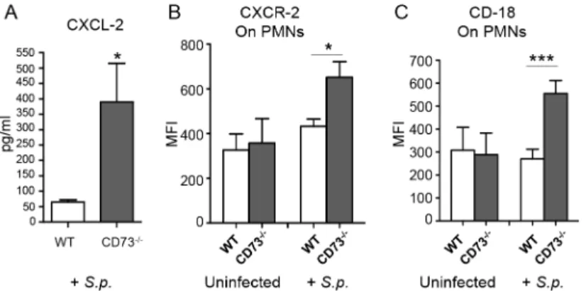
lungs during pneumococcal infection, we found by ELISA that upon infection,CD73-/-mice had 7-fold higher levels of the chemokine CXCL2 in their lungs than did wild type mice (Fig 6A). Flow cytometric analysis revealed that the expression of the cognate receptor, CXCR2, was increased by 1.5-fold on the surface of PMNs fromCD73-/-compared to wild type mice (Fig 6B). Similarly, levels of surface expressedβ2integrin (CD18), an adhesion molecule critical
for PMN transmigration across endothelial barriers, was more than 2-fold higher on PMNs isolated fromCD73-/-mice compared to wild type mice (Fig 6C). These findings suggest that EAD may be involved in regulating both chemotactic and cell adhesion steps during endothe-lial transmigration by PMNs.
CD73-inhibition impairs the ability of PMNs to kill
S
.
pneumoniae
in vitro
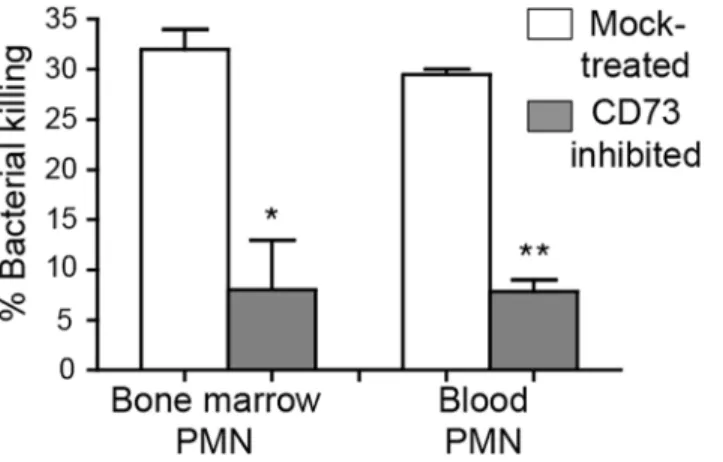
Although CD73 inhibition resulted in enhanced recruitment of PMNs into the lungs, these PMNs failed to control infection. Indeed, by day 3 post-infection, CD73-inhibition was associ-ated with ~100,000-fold more pulmonary pneumococci compared to mock-treatment (Fig 3). Thus, the absence of CD73 activity appeared to diminish the ability of PMNs recruited to the site of infection to clear the infection. We compared opsonophagocytic killing of pneumococci by PMNs isolated from the blood and bone marrow of CD73-inhibited or mock-treated mice. PMNs isolated from both the blood and bone marrow of CD73-inhibited mice displayed a ~5-fold defect in bacterial killing as compared to PMNs from mock-treated mice (Fig 7). These data are consistent with the suggestion that, in addition to a role for EAD in modulating trans-endothelial migration by PMNS, EAD may enhance bacteriocidal functions of PMNs.
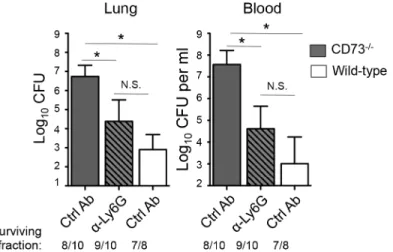
Depleting PMNs post-infection in the absence of EAD production
restores resistance to pneumococcal challenge
To determine whether the heightenedS.pneumoniaesusceptibility of mice inhibited for EAD signaling was due to a dysregulated recruitment and function of PMN,CD73-/-or wildtype control mice were treated with the Ly6G antibody 18 hours after I.T. infection with 5x10³CFU ofS.pneumoniaeand bacterial burdens in the lungs, as well as spread to the blood were assessed. Treatment with the Ly6G antibodies resulted in ~80% PMN reduction inCD73
-/-Fig 6. CD73 modulates the induction of leukocyte recruitment signals upon I.T. challenge byS. pneumoniae.Wild-type C57BL/6 orCD73
-/-mice were mock-infected or I.T. challenged with 5 x 10³CFU of
S.pneumoniaeTIGR4 (+S.p). Three days after challenge, levels of CXCL-2 in the lung homogenates were determined by ELISA (A) and the mean florescent intensities (MFI) of CXCR2 (B) or CD18 (C) on PMNs (Ly6G+) recruited into the lungs were determined by flow cytometry (seeMaterials and Methods). Pooled data from two separate experiments (n = 6 infected and n = 4 uninfected mice per group) are shown. Data represent means +/- SEM, and significant differences determined by Student’s t-test are indicated by asterisks (***=p<0.0001 and*=p<0.05).
mice at day 3 post-infection compared to isotype-treated controls. In comparison to the untreatedCD73-/-mice, PMN depletedCD73-/-mice had significantly lower bacterial burdens in both the lung and the blood, statistically indistinguishable (albeit slightly higher) from those of wildtype control mice (Fig 8). Our data suggest that the increased susceptibility of mice diminished for EAD signaling during pneumococcal infection is at least in part mediated by PMNs.
Discussion
Acute inflammation following microbial infection may have either beneficial or detrimental effects. We investigated the role of PMNs in shaping the course of disease caused by the global pathogenS.pneumoniae. We first showed that within the first 12 hours after I.T. inoculation of mice, PMN entry into the lungs correlates with initial control of pulmonary bacterial burdens and that depletion of PMNs prior to pulmonary challenge withS.pneumoniaeresults in increased susceptibility and lethal septicemia. AlthoughS.pneumoniaestrains are quite hetero-geneous, and PMN depletion enhances survival during murine infection by a serotype 8 pneu-mococcal strain [41], our current findings with strain TIGR4, a serotype 4 strain, are consistent with previous work indicating that PMNs, which phagocytose and kill pneumococci [42], are crucial for host defense against many serotypes ofS.pneumoniae[13–15,43].
We also found that in the next phase of infection, beginning at approximately twelve hours after inoculation, PMN influx into the lungs corresponded with increased bacterial lung bur-dens and pneumoccocal spread into the systemic circulation. Depletion of PMNs 18 h after pulmonary challenge resulted in lower bacterial loads and enhanced survival, suggesting that timely resolution of inflammation may diminish deleterious effects of an over-exuberant host response. Indeed, mice that survived infection had drastically fewer pulmonary PMNs at day 3 post-infection, and many studies have shown that conditions that result in increased numbers of PMNs in the lungs several days afterS.pneumoniaelung infection, such as influenza virus infection [18], aging [8,16,44] or deficiency in regulatory T cells [17], suffer more severe sys-temic spread and reduced survival. Conversely, reducing chemotaxis of PMNs into airways after I.T. pneumococcal challenge of mice resulted in uniform survival after an otherwise lethal pneumococcal pulmonary challenge [9]. Mice protected fromS.pneumoniaechallenge by
Fig 7. CD73 inhibition impairs the ability of PMNs to kill pneumococci.The viability ofS.pneumoniae
TIGR4 after 45 minute incubation with PMNs isolated from the blood and bone marrow of mock-treated C57BL/6 mice or mice treated with the CD73 inhibitor was determined by plating. The mean +/- SEM of % bacterial killing compared to a no PMN control was determined. Significant difference, determined by student’s t test, are indicated by asterisks (**= p<0.001 and*= p<0.05). Data representative of one of two
separate experiments performed are shown (n = 4 mice per group).
treatment with anti-capsular antibody experience only transient influx in PMNs into the lung followed by resolution by 24 hours post-infection [45]. Thus, although PMNs are initially needed to clearS.pneumoniaeinfection, later in infection they function in ways that are detri-mental to the host, suggesting that regulation of PMN influx is crucial to protect against disease.
Although EAD is a crucial regulator of acute pulmonary inflammation in several sterile lung injury models [24,25,46,47], its role in infection-induced inflammation remains relatively unexplored. EAD is recognized by four distinct adenosine receptors, termed A1, A2A, A2B and A3, and stimulation of a particular adenosine receptor may have a positive or negative effect on pulmonary inflammation depending on the type of lung injury [23]. A1 receptor stimulation diminished PMN infiltration and tissue damage in murine lung injury models [48–50] but pro-moted damaging lung inflammation during influenza infection [29]. Stimulation of the A2B adenosine receptor blocked LPS-mediated PMN recruitment into the lungs in mice [46,47,51], but had no effect on leukocyte recruitment following pulmonary infection by the Gram-nega-tive bacteriumK.pneumoniae[28]. In the context of the Gram-positive pathogen,S. pneumo-niae, we found here that EAD negatively regulates trans-endothelial migrationin vitro, and inhibition of EAD signaling by pan-adenosine receptor blockade, or by genetic ablation or chemical inhibition of CD73, resulted in a four- to 20-fold fold increase in pulmonary PMNs three days following I.T. pneumococcal challenge. Adenosine enhanced basolateral-to-apical transmigration of PMNs across endothelial monolayersin vitro, but did not regulate PMN migration across epithelial monolayers. Correspondingly, the increase in pulmonary PMNs during murine infection was not reflected in an increase in airway PMNs, as sampled by bronchoalveolar lavage. Thus, similar to previous findings after A2B receptor inhibition in an LPS-induced lung injury model [51], upon disruption of EAD signaling, PMNs accumulated predominantly in the interstitium.
The mechanism by which EAD modulates PMN transendothelial migration during pneu-mococcal infection could involve chemotactic signals or molecules that directly mediate PMN-endothelial cell interactions, or both.In vitro, the production of the chemokine CXCL-8 (IL-8)
Fig 8. Post-infection depletion of PMNs partially reverses the susceptibility ofCD73-/-mice to
pneumococcal challenge.CD73
-/-mice were treated with anti-Ly6G antibodies 18 hours after I.T inoculation of 5x10³CFU ofS.pneumoniaeTIGR4. Three days after infection, bacterial burdens in the lungs and blood were compared to those of isotype control antibody-treatedCD73-/-and wildtype mice. Pooled data from three separate experiments (n = 7 mice per group) are shown. Data represent means +/- SEM and significant (p<0.05) differences, determined by student’s t-test, are indicated by asterisk. Below the graphs
are indicated the fraction of surviving mice within each group.
by endothelial monolayers is diminished by adenosine [52], and in a murine LPS-induced lung injury model, the level of CXCL2/3 (i.e. the murine paralog of IL-8), a chemokine that pro-motes PMN and macrophage recruitment during murine pneumococcal infection [20], is diminished by A1 receptor stimulation [48]. On activated PMNs, adenosine inhibits up-regula-tion of theβ2 integrin CD11b/CD18 [53], which has been implicated in pulmonary PMN recruitment during pneumococcal murine infection[15]. We found that, following pneumo-coccal infection, the level of pulmonary CXCL-2 was significantly elevated inCD73-/-mice compared to wild-type mice. In addition, levels of CXCR-2 (i.e. the CXCL-2 receptor) and the integrin CD18 were elevated onCD73-/-PMNs. Thus, EAD likely regulates multiple signals involved in pulmonary recruitment of PMNs in response toS.pneumoniaeinfection.
A striking finding was that disruption of EAD production or signaling resulted in an increase of many orders of magnitude in bacterial numbers in the lung and blood, as well as significantly higher mortality rates. Conversely, inhibition of EAD breakdown decreased bacte-rial loads and diminished lethality. Although we cannot rule out that altering extracellular ATP or adenosine levels in the host may have direct effects onS.pneumoniae, especially given their far ranging metabolic and/or regulatory effects on pneumococcus [5], neither adenosine nor the ADA or CD73 inhibitors alteredS.pneumoniaeviabilityin vitro. EAD can regulate PMN phagocytosis and degranulationin vitro[22], features that are crucial for anti-pneumococcal activity of PMNs [42]. Interestingly, several Gram-positive pathogens (although likely notS. pneumoniae) express ectonucleotidases that produce EAD that inhibits PMN-mediated phago-cytosis [54] and oxidative killing [55]. A2B-deficient PMNs form neutrophil extracellular traps (NETs) and clearK.pneumoniaemore efficiently than wild type PMNs [28]. In contrast, here we found that pharmacologic blockade of CD73 impaired opsonophagocytic killing ofS. pneu-moniaeby PMNsex vivo. Phagocytic killing of pneumococci by PMNs requires serine prote-ases but is independent of oxidative burst [13,42], raising the possibility that the effect of EAD on a given infection may depend on the specific mechanism(s) by which PMNs kill the particu-lar infecting microbe.
Importantly, however, the effects of CD73 ablation or inhibition and adenosine signaling blockade on lung infection cannot be fully explained by the loss of a putative PMN defense function, because depletion of these cells 18 hours after inoculation significantly mitigated the susceptibility ofCD73-/-mice. Thus, EAD appears to limit disease by blunting the detrimental effect of PMNs later in infection. The nature of this PMN-mediated harmful effect on immune control is unknown, but it is possible that once bacterial burden reaches a threshold beyond which PMNs can no longer control the infection, they instead contribute to an environment permissive for bacterial persistence and growth. Some pathogens, such asSalmonella enterica, harbor metabolic capacities well adapted to the inflamed environment [56], and given that sugar utilization and other metabolic pathways have been shown to be critical determinants of pneumococcal virulencein vivo[57], PMN-derived products in inflamed tissue might make available growth-limiting nutrients utilized by this organism [58]. PMNs are also known to modulate other arms of the host immune response, such as the recruitment and function of T cells [59] and monocytes [60], and may influence pneumococcal persistence indirectly. Finally, although PMN depletion following infection significantly mitigated the susceptibility of CD73-/-mice, these mice still suffered somewhat (albeit not statistical significant) higher bacte-rial burdens than wild-type mice. Thus, EAD, which regulates the function of immune cells such as macrophages [61] and regulatory T- cells [62,63] that promote pneumococcal defense [17,64], may also enhance resistance by PMN-independent mechanisms.
depletion 18 hours post-infection or chemical inhibition of adenosine breakdown reduced bac-terial spread. Although the reduced spread may partially reflect lower bacbac-terial burden in the lung, CD73-inhibited mice challenged with a high (107) dose of pneumococci harbored num-bers of bacteria in the lung equivalent to untreated controls, yet suffered greater bloodstream spread. In other infection models, PMN influx into infected tissues was associated with tissue damage and poor infection outcome, without altering pathogen numbers [65]. In addition, we previously showed that transmigration of PMN across a respiratory epithelial monolayer dis-rupted its barrier functionin vitroand inhibition of PMN influx into the airways prevented lethal septicemia in mice [9]. Given that we found here that EAD signaling controls transmi-gration across endothelium but not epithelium, inflammation may promote disseminated pneumococcal disease by multiple mechanisms.
All four EAD receptors are produced in the lung [21] and on PMNs [22], and in future stud-ies it will be essential to characterize the adenosine receptor(s) that influence the course of pneumococcal infection. Adenosine receptors vary in both their effect on pulmonary inflam-mation and their affinity for adenosine, with EC50‘s varying from<0.5 to 64μM, raising the possibility that EAD could be pro- or anti-inflammatory depending on EAD tissue concentra-tion. Previous studies indicate that administration of the ADA inhibitor EHNA-hydrochloride and the CD73 inhibitorα,βmethylene ADP to mice results in the predicted effects on adeno-sine [24,33] concentration, but we did not directly measure changes in EAD levels during pneumococcal infection. Changes in the expression of adenosine receptors [61] could also raise another dynamic variable that may influence EAD signaling. Adenosine receptor signaling resulted in either a pro-, or anti-inflammatory T-cell response during autoimmune uveitis depending on the phase of the disease [66], and one might imagine that the effect of EAD sig-naling may differ with phase of pneumococcal infection, providing a rationale for the lack of discernable effect of CD73 inhibition or ablation soon after I.T. inoculation, but a dramatic effect on the resolution of pulmonary inflammation later in infection. The use of receptor-spe-cific agonists and antagonist or mice that are genetically ablated for a spereceptor-spe-cific adenosine recep-tor provide future avenues to better define specific pathways that control inflammation and disease during pneumococcal infection, potentially revealing new therapeutic strategies to combat this important disease.
Material and Methods
Ethics statement
This work was performed in accordance with the recommendations in the Guide for the Care and Use of Laboratory Animals published by the National Institutes of Health. All procedures were reviewed and approved by Tufts University Institutional Animal Care and Use Commit-tee (IACUC) and are under protocol # B2014-86.
Mice
Wild type BALB/c/By/J (BALB/c), C57BL/6J (B6) and CD73 knockout (CD73-/-) mice on a B6 background were purchased from The Jackson Laboratory (Bar Harbor, ME) and bred at Tufts University. Mice were matched for age and sex and maintained in a specific-pathogen free facility at Tufts University.
Murine infections
Mice were challenged I.T. withS.pneumoniaeTIGR4 grown at 37°C in 5% CO2in
previously described [9]. For every experiment, the inoculum was plated on blood agar plates for CFU enumeration. If the bacterial inoculae differed by less than 20%, the data from separate experiments were pooled. If the titers varied by more 20% between individual experiments, then representative data are shown.
EAD-pathway inhibitors
The effect of EAD on infection was assessed using the following: The selective and competitive inhibitor of CD73,α,βmethylene ADP; the pan-adenosine receptor antagonist CGS-15943; and the adenosine deaminase inhibitor EHNA hydrochloride. All chemicals were purchased from Sigma Aldrich, dissolved in DMSO and filter sterilized by passing through a 0.22μm filter.
The mice were then given intraperitoneal (i.p.) injections of 10mg/kg daily at days 0 (immedi-ately before I.T. infection), 1 and 2 post-infection. Control mice were mock-treated with the vehicle control.
Assessment of bacterial burden and survival
For enumeration of bacterial numbers, lung and blood samples were harvested from the live mice and plated on TSA plates supplemented with 5% sheep blood agar (Northeast Laboratory Services). The limit of detection was 20 CFU (1.3 Log10) per lung and 200 CFU (2.3 Log10) per
ml blood. When no colonies were detected on the plates, the numbers of bacteria were assumed to be slightly under our limit of detection (2.0 Log10bacteria per 1ml of blood and 1.0 Log10
bacteria in the lungs). For survival analysis, the mice were monitored for 7 days following infec-tion. At a dose of 5x10⁵CFUS.pneumoniaeTIGR4, we typically observed ~ 60% total mortal-ity rate over the course of a week, with all deaths occurring on days 2, 3 or 4. However, the specific kinetics of death over this three-day period varied between experiments. We consis-tently observed an ~70% survival rate in mice inoculated with 5x10³CFU ofS.pneumoniae TIGR4. Despite slight variation in kinetics between experiments, the differences between experimental groups were consistent from experiment to experiment.
Bacterial growth assays
Frozen aliquots of log-phaseS.pneumoniaeTIGR4 strain (serotype 4) were thawed, washed and diluted to an OD600~ 0.1 in THY liquid media supplemented with oxyrase. To measure
the effect of the CD73 inhibitor on bacterial growth, 40μg/ml of the drug was added to the
media. To measure the effect of adenosine on growth, the chemical (Sigma Aldrich) was added exogenously to a final concentration of 10μM or 100μM. The compounds were added at 0 h
and bacterial growth in 37°C / 5% CO2was monitored overtime by measuring OD600and
com-pared to growth in media alone. To measure the effect of the adenosine deaminase inhibitor and the pan-adenosine receptor inhibitor on bacterial viability, 40μg/ml of the drugs was
added to the media at 37°C / 5% CO2and two hours later bacterial viability was measured by
plating on blood agar plates for CFU enumeration.
PMN depletions
Mice were injected i.p. with 100μg of the Ly6G-depleting antibody IA8 or isotype IgG control
Isolation of cells from alveolar spaces and lung tissues
Mice were euthanized at the indicated times post-infection and the bronchio-alveolar lavage fluid (BALF) obtained by washing the lungs with PBS. The lungs were then digested with Type II collagenase (Worthington) and DNase (Worthington) and single-cell suspensions obtained as previously described [8].
Flow cytometry
Cells were stained with anti-mouse Ly6G (clone 1A8, BD Biosciences), CD18 (Clone M18/2, Biolegend) and CXCR2 (Clone SA045E1, Biolegend) antibodies. Fluorescence intensities were measured on a FACSCalibur and at least 25,000 events for lung tissue and 10,000 events for BALF were analyzed using FlowJo.
CXCL-2 ELISA
Three days post-infection, the lungs were harvested, homogenized in sterile PBS and the result-ing supernatants were used CXCL-2 concentrations usresult-ing the mouse MIP2/CXCL-2 ELISA kit (Sigma-Aldrich) following the manufacturer’s protocol.
Histology
For histological analysis mice were euthanized 3 days post-infection and whole lungs were fixed in 10% buffered formalin (Sigma-Aldrich). Lungs were then embedded in paraffin, sec-tioned at 5μm, stained with hematoxylin and eosin (H&E) and analyzed using a Nikon eclipse
TE2000-U microscope.
Isolation of human PMNs
Healthy human volunteers were recruited in accordance to IRB protocols and signed informed consent forms. Whole blood was obtained and anticoagulated with acid citrate/dextrose. PMNs were isolated using a 2% gelatin sedimentation technique as previously described [9].
Maintenance of epithelial cells
Human pulmonary mucoepidermoid carcinoma-derived NCI-H292 (H292) cells were grown on the underside of collagen-coated Transwell filters (0.33-cm2, Corning Life Sciences) in RPMI 1640 medium (ATCC) with 2 mM L-glutamine, 10% FBS, and 100 U penicillin/strepto-mycin following a previously described protocol [9].
PMN migration assay across epithelium
Maintenance of endothelial cells
Human umbilical vascular endothelial cells (HUVECs) were seeded on the inner chamber of collagen-coated Transwell filters (0.33-cm2, Corning Life Sciences) in M199 medium (Biowhit-taker) supplemented with 2 mM L-glutamine, 10% FBS, 10μg/ml endothelium mitogen
(Fisher), 20μg/ml heparin sodium salt (Sigma) and 100 U penicillin/streptomycin following a
previously described protocol [68]. The cell monolayer was allowed to form over 4–5 days.
PMN migration assay across endothelium
The PMN migration assay across the endothelium was performed as previously described [39] with the following modifications. The endothelial cells seeded on Transwells were infected for 3 h byS.pneumoniaeadded to the lower chamber. 5x10⁵PMNs were added to the upper cham-ber and migration was allowed to occur +/- CD73 or adenosine receptors inhibitors for 3 h at 37°C/ 5% CO2. Since this assay utilizes standard RPMI with phenol red, which precludes
color-imetric assays such as MPO, the number of PMNs that migrated was determined by counting in a hemacytometer in triplicate, as previously described [39]. The inhibitors had no significant affect on cell viability within the timeframe of the assay as measured by trypan-blue exclusion.
Isolation of murine PMNs
Bone marrow PMNs were isolated from the femurs of mice as previously described [43] and enriched using Percoll (Sigma) density gradient centrifugation. For isolation of PMNs from the circulation, blood was collected by cardiac puncture using acid citrate/dextrose as an anticoag-ulant. PMNs were then enriched by Ficoll density gradient centrifugation in Mono-poly (MP-Biomedicals) resolving medium based on the manufacturer’s instructions. The enriched cells were ~ 85–90% Ly6G+by flowcytometry.
Opsonophagocytic killing assay
The ability of PMNs to kill pneumococci was assessedex vivoas previously described. Briefly, 200μl reactions in Hank’s buffer/0.1% gelatin consisted of 1x10⁵PMNs incubated with 1x10²
bacteria grown to mid log phase and pre-opsonized with 20μl mouse sera. The reactions were
incubated rotating for 45 minutes at 37°C. Percent killing relative to parallel incubations with-out PMNs was determined by plating serial dilutions on blood agar plates.
Statistics
All statistical analysis was performed using Prism4 for Macintosh (Graph Pad). For analysis of survival curves, Log-rank (Mantel-Cox) test was performed. CFU data were log-transformed to normalize distribution. Student t-test was used for comparison between groups.pvalues less than 0.05 were considered significant. For all graphs, the mean values +/- SEM are shown.
Supporting Information
S1 Fig. Extracellular adenosine and the CD73 inhibitor do not affect bacterial growthin
vitro.Growth ofS.pneumoniaeTIGR4 was measured in THY media after the addition of (A) increasing concentrations of exogenously added adenosine or (B) the CD73 inhibitor (40μg/ml). (C) Viability ofS.pneumoniaeTIGR4 was measured after 2 h incubation in THY
media +/- the addition of the ADA-inhibitor EHNA-hydrochloride or the adenosine receptors (AR)-inhibitor CGS 15943 (40μg/ml). Representative data from (A) three and (B and C) two
S2 Fig. Adenosine mediated-protection againstS.pneumoniaelung infection is in part
dependent on adenosine receptors.Wild type C57BL/6 mice were either given the adenosine
deaminase (ADA) inhibitor (EHNA hydrochloride) alone or in conjunction with a pan adeno-sine receptor (AR) inhibitor (CGS 15943). Survival (D) as well as bacterial numbers in the lung (A) and blood (B) at 3 days post lung infection with 5x10⁵CFU ofS.pneumoniaeTIGR4 were assessed. Representative data from one of two separate experiments are shown.=p<0.001; =p<0.05 indicate that the means are significantly different by student’s t-test.
(TIF)
S3 Fig. Inhibition of EAD production increases systemic spread of the pneumococci
follow-ing high dose lung challenge.Mock-treated and CD73-inhibited BALB/c mice were
chal-lenged I.T. with ~1x10⁶CFU ofS.pneumoniaeTIGR4. Bacteremia was monitored overtime. Data represent means +/- SEM. Significant (p<0.05) differences are indicated by asterisk. Pooled data from two separate experiments (n = 5 mice per group) are shown. None of the mice succumbed to infection within the monitored time.
(TIF)
S4 Fig. The effect of CD73 ablation on immune cell numbers in the blood Wildtype control
andCD73-/-mice were either inoculated intratracheally with 5× 10³CFU ofS.pneumoniae
TIGR4 (+Sp) or left uninfected.At day 3 post-infection, the number of PMNs (Ly6G+) that
were circulating in the blood was determined by flow cytometry. Data shown are pooled from three separate experiments performed (n = 9 mice per group).= p<0.05 indicate that the means are significantly different by student’s t-test.
(TIF)
Acknowledgments
We would like to thank Mihalis Lionakis and Beth McCormick for invaluable discussion and critical revision of the paper as well as Guillermo Arroyo for technical assistance.
Author Contributions
Conceived and designed the experiments: ENBG JML. Performed the experiments: ENBG SC SER. Analyzed the data: ENBG. Contributed reagents/materials/analysis tools: SRM PA PGH. Wrote the paper: ENBG JML.
References
1. Kadioglu A, Weiser JN, Paton JC, Andrew PW (2008) The role of Streptococcus pneumoniae virulence factors in host respiratory colonization and disease. Nat Rev Microbiol 6: 288–301. doi:10.1038/ nrmicro1871PMID:18340341
2. Chong CP, Street PR (2008) Pneumonia in the elderly: a review of the epidemiology, pathogenesis, microbiology, and clinical features. South Med J 101: 1141–1145; quiz 1132, 1179. doi:10.1097/SMJ. 0b013e318181d5b5PMID:19088525
3. Fruchtman SM, Gombert ME, Lyons HA (1983) Adult respiratory distress syndrome as a cause of death in pneumococcal pneumonia. Report of ten cases. Chest 83: 598–601. PMID:6831945 4. Sun K, Metzger DW (2014) Influenza infection suppresses NADPH oxidase-dependent phagocytic
bac-terial clearance and enhances susceptibility to secondary methicillin-resistant Staphylococcus aureus infection. J Immunol 192: 3301–3307. doi:10.4049/jimmunol.1303049PMID:24563256
7. Hinojosa E, Boyd AR, Orihuela CJ (2009) Age-associated inflammation and toll-like receptor dysfunc-tion prime the lungs for pneumococcal pneumonia. J Infect Dis 200: 546–554. doi:10.1086/600870 PMID:19586419
8. Bou Ghanem EN CS, Du X, Wu D, Camilli A, Leong JM, Meydani SN Theα-Tocopherol Form of Vitamin E Reverses Age-Associated Susceptibility to Streptococcus pneumoniae Lung Infection by Modulating Pulmonary Neutrophil Recruitment. [published online head of print December 15, 2014] J immunol doi: 10.4049/jimmunol1402401
9. Bhowmick R, Tin Maung NH, Hurley BP, Ghanem EB, Gronert K, et al. (2013) Systemic disease during Streptococcus pneumoniae acute lung infection requires 12-lipoxygenase-dependent inflammation. J Immunol 191: 5115–5123. doi:10.4049/jimmunol.1300522PMID:24089193
10. Doerschuk CM, Markos J, Coxson HO, English D, Hogg JC (1994) Quantitation of neutrophil migration in acute bacterial pneumonia in rabbits. J Appl Physiol 77: 2593–2599. PMID:7896596
11. Rolston KV (2001) The spectrum of pulmonary infections in cancer patients. Curr Opin Oncol 13: 218–
223. PMID:11429477
12. Garvy BA, Harmsen AG (1996) The importance of neutrophils in resistance to pneumococcal pneumo-nia in adult and neonatal mice. Inflammation 20: 499–512. PMID:8894714
13. Hahn I, Klaus A, Janze AK, Steinwede K, Ding N, et al. (2011) Cathepsin G and neutrophil elastase play critical and nonredundant roles in lung-protective immunity against Streptococcus pneumoniae in mice. Infect Immun 79: 4893–4901. doi:10.1128/IAI.05593-11PMID:21911460
14. Nieminen J, St-Pierre C, Bhaumik P, Poirier F, Sato S (2008) Role of galectin-3 in leukocyte recruitment in a murine model of lung infection by Streptococcus pneumoniae. J Immunol 180: 2466–2473. PMID: 18250456
15. Kadioglu A, De Filippo K, Bangert M, Fernandes VE, Richards L, et al. (2011) The integrins Mac-1 and alpha4beta1 perform crucial roles in neutrophil and T cell recruitment to lungs during Streptococcus pneumoniae infection. J Immunol 186: 5907–5915. doi:10.4049/jimmunol.1001533PMID:21460207 16. Boyd AR, Shivshankar P, Jiang S, Berton MT, Orihuela CJ (2012) Age-related defects in TLR2 signal-ing diminish the cytokine response by alveolar macrophages dursignal-ing murine pneumococcal pneumonia. Exp Gerontol 47: 507–518. doi:10.1016/j.exger.2012.04.004PMID:22548913
17. Neill DR, Fernandes VE, Wisby L, Haynes AR, Ferreira DM, et al. (2012) T regulatory cells control sus-ceptibility to invasive pneumococcal pneumonia in mice. PLoS Pathog 8: e1002660. doi:10.1371/ journal.ppat.1002660PMID:22563306
18. Narayana Moorthy A, Narasaraju T, Rai P, Perumalsamy R, Tan KB, et al. (2013) In vivo and in vitro studies on the roles of neutrophil extracellular traps during secondary pneumococcal pneumonia after primary pulmonary influenza infection. Front Immunol 4: 56. doi:10.3389/fimmu.2013.00056PMID: 23467809
19. Craig A, Mai J, Cai S, Jeyaseelan S (2009) Neutrophil recruitment to the lungs during bacterial pneumo-nia. Infect Immun 77: 568–575. doi:10.1128/IAI.00832-08PMID:19015252
20. Herbold W, Maus R, Hahn I, Ding N, Srivastava M, et al. (2010) Importance of CXC chemokine receptor 2 in alveolar neutrophil and exudate macrophage recruitment in response to pneumococcal lung infec-tion. Infect Immun 78: 2620–2630. doi:10.1128/IAI.01169-09PMID:20368349
21. Factor P, Mutlu GM, Chen L, Mohameed J, Akhmedov AT, et al. (2007) Adenosine regulation of alveo-lar fluid clearance. Proc Natl Acad Sci U S A 104: 4083–4088. PMID:17360481
22. Barletta KE, Ley K, Mehrad B (2012) Regulation of neutrophil function by adenosine. Arterioscler Thromb Vasc Biol 32: 856–864. doi:10.1161/ATVBAHA.111.226845PMID:22423037
23. Hasko G, Linden J, Cronstein B, Pacher P (2008) Adenosine receptors: therapeutic aspects for inflam-matory and immune diseases. Nat Rev Drug Discov 7: 759–770. doi:10.1038/nrd2638PMID: 18758473
24. Thompson LF, Eltzschig HK, Ibla JC, Van De Wiele CJ, Resta R, et al. (2004) Crucial role for ecto-5'-nucleotidase (CD73) in vascular leakage during hypoxia. J Exp Med 200: 1395–1405. PMID: 15583013
25. Eckle T, Fullbier L, Wehrmann M, Khoury J, Mittelbronn M, et al. (2007) Identification of ectonucleoti-dases CD39 and CD73 in innate protection during acute lung injury. J Immunol 178: 8127–8137. PMID:17548651
26. Eltzschig HK, Thompson LF, Karhausen J, Cotta RJ, Ibla JC, et al. (2004) Endogenous adenosine pro-duced during hypoxia attenuates neutrophil accumulation: coordination by extracellular nucleotide metabolism. Blood 104: 3986–3992. PMID:15319286
28. Barletta KE, Cagnina RE, Burdick MD, Linden J, Mehrad B (2012) Adenosine A(2B) receptor deficiency promotes host defenses against gram-negative bacterial pneumonia. Am J Respir Crit Care Med 186: 1044–1050. doi:10.1164/rccm.201204-0622OCPMID:22997203
29. Aeffner F, Woods PS, Davis IC (2014) Activation of A1-adenosine receptors promotes leukocyte recruitment to the lung and attenuates acute lung injury in mice infected with influenza A/WSN/33 (H1N1) virus. J Virol 88: 10214–10227. doi:10.1128/JVI.01068-14PMID:24965449
30. van Opijnen T, Camilli A (2012) A fine scale phenotype-genotype virulence map of a bacterial patho-gen. Genome Res 22: 2541–2551. doi:10.1101/gr.137430.112PMID:22826510
31. Kumar V, Sharma A (2009) Adenosine: an endogenous modulator of innate immune system with thera-peutic potential. Eur J Pharmacol 616: 7–15. doi:10.1016/j.ejphar.2009.05.005PMID:19464286 32. Bessodes M, Bastian G, Abushanab E, Panzica RP, Berman SF, et al. (1982) Effect of chirality in
ery-thro-9-(2-hydroxy-3-nonyl) adenine (EHNA) on adenosine deaminase inhibition. Biochem Pharmacol 31: 879–882. PMID:7082355
33. Lambe CU, Nelson DJ (1982) Pharmacokinetics of inhibition of adenosine deaminase by erythro-9-(2-hydroxy-3-nonyl)adenine in CBA mice. Biochem Pharmacol 31: 535–539. PMID:7066021
34. Mahamed DA, Mills JH, Egan CE, Denkers EY, Bynoe MS (2012) CD73-generated adenosine facili-tates Toxoplasma gondii differentiation to long-lived tissue cysts in the central nervous system. Proc Natl Acad Sci U S A 109: 16312–16317. doi:10.1073/pnas.1205589109PMID:22988118
35. Podzuweit T, Nennstiel P, Muller A (1995) Isozyme selective inhibition of cGMP-stimulated cyclic nucle-otide phosphodiesterases by erythro-9-(2-hydroxy-3-nonyl) adenine. Cell Signal 7: 733–738. PMID: 8519602
36. Gingles NA, Alexander JE, Kadioglu A, Andrew PW, Kerr A, et al. (2001) Role of genetic resistance in invasive pneumococcal infection: identification and study of susceptibility and resistance in inbred mouse strains. Infect Immun 69: 426–434. PMID:11119534
37. Williams M, Francis J, Ghai G, Braunwalder A, Psychoyos S, et al. (1987) Biochemical characterization of the triazoloquinazoline, CGS 15943, a novel, non-xanthine adenosine antagonist. J Pharmacol Exp Ther 241: 415–420. PMID:2883298
38. Klotz KN (2000) Adenosine receptors and their ligands. Naunyn Schmiedebergs Arch Pharmacol 362: 382–391. PMID:11111832
39. Moreland JG, Bailey G (2006) Neutrophil transendothelial migration in vitro to Streptococcus pneumo-niae is pneumolysin dependent. Am J Physiol Lung Cell Mol Physiol 290: L833–840. PMID:16339781 40. Preston JA, Beagley KW, Gibson PG, Hansbro PM (2004) Genetic background affects susceptibility in
nonfatal pneumococcal bronchopneumonia. Eur Respir J 23: 224–231. PMID:14979496
41. Marks M, Burns T, Abadi M, Seyoum B, Thornton J, et al. (2007) Influence of neutropenia on the course of serotype 8 pneumococcal pneumonia in mice. Infect Immun 75: 1586–1597. PMID:17296760 42. Standish AJ, Weiser JN (2009) Human neutrophils kill Streptococcus pneumoniae via serine
prote-ases. J Immunol 183: 2602–2609. doi:10.4049/jimmunol.0900688PMID:19620298
43. Lysenko ES, Clarke TB, Shchepetov M, Ratner AJ, Roper DI, et al. (2007) Nod1 signaling overcomes resistance of S. pneumoniae to opsonophagocytic killing. PLoS Pathog 3: e118. PMID:17722978 44. Krone CL, Trzcinski K, Zborowski T, Sanders EA, Bogaert D (2013) Impaired innate mucosal immunity
in aged mice permits prolonged Streptococcus pneumoniae colonization. Infect Immun 81: 4615–
4625. doi:10.1128/IAI.00618-13PMID:24082075
45. Burns T, Abadi M, Pirofski LA (2005) Modulation of the lung inflammatory response to serotype 8 pneu-mococcal infection by a human immunoglobulin m monoclonal antibody to serotype 8 capsular polysac-charide. Infect Immun 73: 4530–4538. PMID:16040964
46. Schingnitz U, Hartmann K, Macmanus CF, Eckle T, Zug S, et al. (2010) Signaling through the A2B adenosine receptor dampens endotoxin-induced acute lung injury. J Immunol 184: 5271–5279. doi: 10.4049/jimmunol.0903035PMID:20348420
47. Yang D, Zhang Y, Nguyen HG, Koupenova M, Chauhan AK, et al. (2006) The A2B adenosine receptor protects against inflammation and excessive vascular adhesion. J Clin Invest 116: 1913–1923. PMID: 16823489
48. Ngamsri KC, Wagner R, Vollmer I, Stark S, Reutershan J (2010) Adenosine receptor A1 regulates poly-morphonuclear cell trafficking and microvascular permeability in lipopolysaccharide-induced lung injury. J Immunol 185: 4374–4384. doi:10.4049/jimmunol.1000433PMID:20729330
50. Fernandez LG, Sharma AK, LaPar DJ, Kron IL, Laubach VE (2013) Adenosine A1 receptor activation attenuates lung ischemia-reperfusion injury. J Thorac Cardiovasc Surg 145: 1654–1659. doi:10.1016/ j.jtcvs.2013.01.006PMID:23398646
51. Konrad FM, Witte E, Vollmer I, Stark S, Reutershan J (2012) Adenosine receptor A2b on hematopoietic cells mediates LPS-induced migration of PMNs into the lung interstitium. Am J Physiol Lung Cell Mol Physiol 303: L425–438. doi:10.1152/ajplung.00387.2011PMID:22707616
52. Bouma MG, van den Wildenberg FA, Buurman WA (1996) Adenosine inhibits cytokine release and expression of adhesion molecules by activated human endothelial cells. Am J Physiol 270: C522–529. PMID:8779915
53. Wollner A, Wollner S, Smith JB (1993) Acting via A2 receptors, adenosine inhibits the upregulation of Mac-1 (Cd11b/CD18) expression on FMLP-stimulated neutrophils. Am J Respir Cell Mol Biol 9: 179–
185. PMID:8393328
54. Thammavongsa V, Kern JW, Missiakas DM, Schneewind O (2009) Staphylococcus aureus synthe-sizes adenosine to escape host immune responses. J Exp Med 206: 2417–2427. doi:10.1084/jem. 20090097PMID:19808256
55. Liu P, Pian Y, Li X, Liu R, Xie W, et al. (2014) Streptococcus suis adenosine synthase functions as an effector in evasion of PMN-mediated innate immunit. J Infect Dis 210: 35–45. PMID:24446521 56. Winter SE, Thiennimitr P, Winter MG, Butler BP, Huseby DL, et al. (2010) Gut inflammation provides a
respiratory electron acceptor for Salmonella. Nature 467: 426–429. doi:10.1038/nature09415PMID: 20864996
57. Shelburne SA, Davenport MT, Keith DB, Musser JM (2008) The role of complex carbohydrate catabo-lism in the pathogenesis of invasive streptococci. Trends Microbiol 16: 318–325. doi:10.1016/j.tim. 2008.04.002PMID:18508271
58. Burnaugh AM, Frantz LJ, King SJ (2008) Growth of Streptococcus pneumoniae on human glycoconju-gates is dependent upon the sequential activity of bacterial exoglycosidases. J Bacteriol 190: 221–
230. PMID:17981977
59. Muller I, Munder M, Kropf P, Hansch GM (2009) Polymorphonuclear neutrophils and T lymphocytes: strange bedfellows or brothers in arms? Trends Immunol 30: 522–530. doi:10.1016/j.it.2009.07.007 PMID:19775938
60. Soehnlein O, Weber C, Lindbom L (2009) Neutrophil granule proteins tune monocytic cell function. Trends Immunol 30: 538–546. doi:10.1016/j.it.2009.06.006PMID:19699683
61. Cohen HB, Briggs KT, Marino JP, Ravid K, Robson SC, et al. (2013) TLR stimulation initiates a CD39-based autoregulatory mechanism that limits macrophage inflammatory responses. Blood 122: 1935–
1945. doi:10.1182/blood-2013-04-496216PMID:23908469
62. Ohta A, Kini R, Ohta A, Subramanian M, Madasu M, et al. (2012) The development and immunosup-pressive functions of CD4(+) CD25(+) FoxP3(+) regulatory T cells are under influence of the adeno-sine-A2A adenosine receptor pathway. Front Immunol 3: 190. doi:10.3389/fimmu.2012.00190PMID: 22783261
63. Romio M, Reinbeck B, Bongardt S, Huls S, Burghoff S, et al. (2011) Extracellular purine metabolism and signaling of CD73-derived adenosine in murine Treg and Teff cells. Am J Physiol Cell Physiol 301: C530–539. doi:10.1152/ajpcell.00385.2010PMID:21593451
64. Dockrell DH, Marriott HM, Prince LR, Ridger VC, Ince PG, et al. (2003) Alveolar macrophage apoptosis contributes to pneumococcal clearance in a resolving model of pulmonary infection. J Immunol 171: 5380–5388. PMID:14607941
65. Lionakis MS, Fischer BG, Lim JK, Swamydas M, Wan W, et al. (2012) Chemokine receptor Ccr1 drives neutrophil-mediated kidney immunopathology and mortality in invasive candidiasis. PLoS Pathog 8: e1002865. doi:10.1371/journal.ppat.1002865PMID:22916017
66. Liang D, Zuo A, Shao H, Chen M, Kaplan HJ, et al. (2014) Anti-inflammatory or proinflammatory effect of an adenosine receptor agonist on the th17 autoimmune response is inflammatory environment-dependent. J Immunol 193: 5498–5505. doi:10.4049/jimmunol.1401959PMID:25367119 67. McCormick BA, Hofman PM, Kim J, Carnes DK, Miller SI, et al. (1995) Surface attachment of
Salmo-nella typhimurium to intestinal epithelia imprints the subepithelial matrix with gradients chemotactic for neutrophils. J Cell Biol 131: 1599–1608. PMID:8522615