Rev. bras. ortop. vol.46 número6 en a04v46n6
Texto
Imagem
Documentos relacionados
Conditioned medium from bone marrow- derived mesenchymal stem cells improves recovery after spinal cord injury in rats: an original strategy to avoid cell
economy since 1980 to 2010, such as: (i) rising skill premium; (ii) increase in income and wealth Gini coefficient; (iii) decrease in the wealth share owned by the bottom 90%
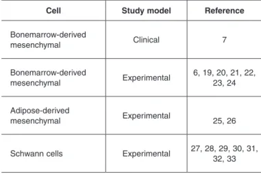
Bone marrow -derived mesenchymal stem cells remain host-derived despite suc- cessful hematopoietic engraftment after allogeneic transplantation in patients w ith lysosomal
We determined the effects of these epigenetic mechanisms on adipocyte differentiation in mesenchymal stem cells (MSCs) derived from bone marrow (BM-MSCs) and adipose tissue (ADSCs)
Transplantation of Bone Marrow – Derived Mesenchymal Stem Cells Improves Diabetic Polyneuropathy in Rats.. Transplantation of Bone Marrow-Derived Mononuclear Cells Improves
Intracerebral transplantation of bone marrow stromal cells in a 1-methyl-4-phenyl-1,2,3,6-tetrahydropyridine mouse model of Parkinson’s disease. Human mesenchymal stem cells
A feasibility of useful cell-based therapy by bone regeneration with deciduous tooth stem cells, dental pulp stem cells, or bone marrow-derived mesenchymal stem cells for
