Protective Effects of Inactivated EV71 Vaccines in Mice
Qunying Mao1, Chenghong Dong2, Xiuling Li3, Qiang Gao4, Zengbing Guo5, Xin Yao1, Yiping Wang1, Fan Gao1, Fengxiang Li1, Miao Xu1, Weidong Yin4, Qihan Li5, Xinliang Shen3, Zhenglun Liang1*, Junzhi Wang1*
1National Institutes for Food and Drug Control, Beijing, China,2Institute of Medical Biology, Chinese Academy of Medical Sciences, Kunming, China,3National Vaccine and Serum Institute, Beijing, China,4Sinovac Biotech Co., Ltd., Beijing, China,5Hualan Biological Engineering Inc, Henan, China
Abstract
Background:Enterovirus 71 (EV71) is the major causative agent of hand, foot, and mouth disease (HFMD). Three inactivated EV71 whole-virus vaccines of different strains developed by different manufacturers in mainland China have recently entered clinical trials. Although several studies on these vaccines have been published, a study directly comparing the immunogenicity and protective effects among them has not been carried out, which makes evaluating their relative effectiveness difficult. Thus, properly comparing newly developed vaccines has become a priority, especially in China.
Methods and Findings: This comparative immunogenicity study was carried out on vaccine strains (both live and inactivated), final container products (FCPs) without adjuvant, and corresponding FCPs containing adjuvant (FCP-As) produced by three manufacturers. These vaccines were evaluated by neutralizing antibody (NAb) responses induced by the same or different dosages at one or multiple time points post-immunization. The protective efficacy of the three vaccines was also determined in one-day-old ICR mice born to immunized female mice. Survival rates were observed in these
suckling mice after challenge with 20 LD50of EV71/048M3C2. Three FCP-As, in a dose of 200 U, generated nearly 100% NAb
positivity rates and similar geometric mean titers (GMTs), especially at 14–21 days post-inoculation. However, the dynamic NAb responses were different among three vaccine strains or three FCPs. The FCP-As at the lowest dose used in clinical trials
(162 U) showed good protective effects in suckling mice against lethal challenge (90–100% survival), while the ED50of NAb
responses and protective effects varied among three FCP-As.
Conclusions:These studies establish a standard method for measuring the immunogenicity of EV71 vaccines in mice. The data generated from our mouse model study indicated a clear dose-response relationship, which is important for vaccine quality control and assessment, especially for predicting protective efficacy in humans when combined with future clinical trial results.
Citation:Mao Q, Dong C, Li X, Gao Q, Guo Z, et al. (2012) Comparative Analysis of the Immunogenicity and Protective Effects of Inactivated EV71 Vaccines in Mice. PLoS ONE 7(9): e46043. doi:10.1371/journal.pone.0046043
Editor:Lijun Rong, University of Illinois at Chicago, United States of America
ReceivedJune 3, 2012;AcceptedAugust 28, 2012;PublishedSeptember 28, 2012
Copyright:ß2012 Mao et al. This is an open-access article distributed under the terms of the Creative Commons Attribution License, which permits unrestricted use, distribution, and reproduction in any medium, provided the original author and source are credited.
Funding:The current study was sponsored by the National Science Project (No. 2008BAI69B01) and the National 11th Five Major Special Projects Funding Program (No. 2009ZX10004-804) from the Ministry of Science and Technology of the People’s Republic of China. The funders had no role in study design, data collection and analysis, decision to publish, or preparation of the manuscript.
Competing Interests:Q Gao and WD Yin are affiliated to Sinovac Biotech Co., Ltd. ZB Guo is affiliated to Hualan Biological Engineering Inc.; XL Li and XL Shen are affiliated to National Vaccine and Serum Institute; CH Dong and QH Li are affiliated to Institute of Medical Biology, Chinese Academy of Medical Sciences. As a Board member, WD Yin holds shares of Sinovac Biotech Co., Ltd. There are EV71 vaccines in development to declare. There are no consultancies, patents, or marketed products to declare. This does not alter the authors’ adherence to all the PLOS ONE policies concerning sharing data and materials as detailed online in the guide for authors.
* E-mail: [email protected] (ZL); [email protected] (JW)
Introduction
Enterovirus 71 (EV71) is a small RNA virus belonging to the
Enterovirusgenus. It is a spherical particle with icosahedral (cubic)
symmetry and contains a positive-sense single-stranded RNA approximately 7.4 kb long. Each subunit of the viral capsid contains a copy of the four structural viral proteins (VP1–VP4); VP1, VP2, and VP3 are external, while VP4 is completely within the interior of the viral particle and is not, therefore, exposed to the host antibody response [1]. VP1 displays the predominant neutralizing epitope [2]. EV71 infection mainly leads to hand, foot, and mouth disease (HFMD) and EV71-associated
research community’s extensive experience in developing other enterovirus vaccines, such as the polio and hepatitis A vaccines, development of inactivated virus vaccines has proceeded faster than the others and exhibits the highest apparent immunogenicity [18,20,22]. In mainland China [23], Taiwan [24] and Singapore [25], these inactivated virus vaccines have been tested in clinical trials and are expected to be the first class of vaccines to be employed to prevent EV71-associated diseases worldwide [23].
In mainland China, three inactivated EV71 vaccines have been developed by different manufacturers. Although the three vaccines are all inactivated virus vaccines, differences in their manufactur-ing processes exist, includmanufactur-ing the strains (though all three are the C4 genotype), cell substrate (Vero or diploid cells), cell culture system (roller bottles, cell factories or microcarrier bioreactor system), production process, and vaccine dose (Table 1). All these factors may lead to differences in immunogenicity [15,26]. Although good immunogenicity and protective effects have been reported at particular time points after immunization, the antigen content of these vaccines was reported in different units (mg/ml,
KU/ml, EU/ml), and different animal models were empolyed by the different manufacturers to test these vaccines [27,28]. These differences make it difficult to compare the immunogenicity and protective effects among the different EV71 vaccines, which will be important for testing in clinical trials. A prior collaborative effort was carried out to standardize the EV71 antigen content of three aqueous bulk and three final container products (FCPs) without adjuvant from three manufacturers (unpublished data). Based on the standardized results of the collaborative study, experiments were carried out to compare the immunogenicity and protective effects of EV71 vaccine antigens from the three different manufacturers at different production stages, including the vaccine strains themselves, FCP, and FCP with alum adjuvant (FCP-A). Additionally, the relationship between NAb response and protec-tive effect was determined. These studies provide a basis for the design of clinical trials to confirm dosage and evaluate the protective effects of EV71 vaccines.
Materials and Methods
1. Collaborative laboratories
The following laboratories were involved in this collaborative study comparing the EV71 vaccines: the National Institutes for Food and Drug Control of China (lab 1), the National Vaccine & Serum Institute (lab 2), and Sinovac Biotech Co., Ltd., Beijing (lab 3).
2. Testing for antigen content
The EV71 antigen content of three aqueous bulks and three FCPs from three manufacturers (A, B, C; Table 1) was assayed in a previous collaborative study in the four labs (the same three labs in this study and the lab of the Institute of Medical Biology, Chinese Academy of Medical Sciences). A quantitative ELISA assay kit [29] was used to detect the EV71 antigen content using the reference standard (1600 U/ml) provided by the National Institutes for Food and Drug Control [30]. The samples and EV71 antigen standard (1600 U/ml) were serially diluted two-fold and tested in duplicate wells. Variance analysis (F-test) was performed to determine the linearity and parallelism of the samples and the EV71 antigen standard. Only when bothPvalues
were greater than 0.05, were the EV71 antigen reference and the samples considered to have a parallel linear relationship. The parallel-line method was used to calculate the antigen content of the samples. Results are expressed in standard national EV71
antigen units/ml (U/ml). Table
3. EV71 vaccine strains
The three vaccine strains (labeled M1, M2, and M3: C4 genotype) came from three different vaccine manufacturers in mainland China herein termed A, B and C (because the vaccines are still in clinical trials, we have kept the manufacturers’ names anonymous). M1, M2, and M3 were isolated from EV71 viruses in HFMD epidemic areas in mainland China since 2008 (Table 1). The sequence homologies between the VP1 region of the three vaccine strains (M1, M2, and M3) and the reference strain BJ08 (GenBank accession no: FJ828519) were 97.4%, 96.6%, and 97.9%, respectively [26]. The virus titers of M1, M2, and M3 were 7.19, 6.98 and 6.58 lg PFU/ml [26], and the antigen concentra-tions were 626, 127, and 332 U/ml (unpublished data), respec-tively.
The three inactivated EV71 strains (labeled IM1, IM2, and IM3) were treated with formalin (0.25% wt/vol) at 37uC for 3 d. All EV71 strains and inactivated EV71 strains were stored at 280uC before use. Appropriate amounts of Minimum Essential Media (MEM) were used to dilute each to an equivalent EV71 virus titer of 6.50 lg PFU/ml. Samples were blinded and distributed by lab 1 to the collaborative labs for mouse immunization and testing of the NAb response.
4. EV71 FCPs
Three EV71 aqueous bulks (Q1, Q2 and Q3) were produced by manufacturers A, B, and C and derived from M1, M2, and M3 vaccine strains, respectively, using their own processing techniques (Table 1). The purity of all three aqueous bulks was verified by HPLC to be above 95%. Three EV71 FCPs (B1, B2, and B3) were diluted by the manufacturers (A, B, and C) from aqueous bulks in PBS containing the protective agent (Table 1). The EV71 antigen content of the three EV71 FCPs and three aqueous bulks was assayed three consecutive times in a previous collaborative study using the quantitative ELISA kit [29] mentioned above with the EV71 antigen reference standard (1600 U/ml, from the National Institutes for Food and Drug Control) [26]. The EV71 antigen content of the three FCPs ranged from 506 to 1047 U/ml (CV: 4.8%–12.7% by collaborative labs), as measured by the parallel-line method. EV71 FCPs were stored at 4uC before use. Appropriate amounts of PBS were used to dilute each to an equivalent EV71 antigen content of 500 U/ml. Samples were blinded and distributed by lab 1 to the collaborative labs.
5. EV71 FCP-As
Three FCP-As (V1, V2, and V3) were produced through the absorption of aqueous bulks onto alum adjuvant by manufacturers A, B, and C (Table 1). The EV71 antigen content of the three FCP-As varied between 324 and 1012 U/ml (162–506 U/dose/ 0.5 ml) which was calculated according to the previous collabo-rative study on EV71 antigen content of the three aqueous bulks. EV71 FCP-As were stored at 4uC before use. Appropriate amounts of alum adjuvant were used to dilute each vaccine to an equivalent EV71 antigen concentration of 200 U/ml (for comparative studies of NAbs responses) or 324 U/ml (for protection studies). Samples were blinded and distributed by lab 1 to the collaborative labs.
6. Comparative studies of NAbs induced in mice by EV71 viral strains, FCPs, and FCP-As from three different manufacturers
The mice were immunized with the blinded samples at the three labs according to the same protocol. Female BALB/c mice (4–6 weeks old) were obtained from Vital River Laboratories, Ltd.
Beijing, China. Samples of the three vaccine strains (M1, M2, and M3), inactivated EV71 strains (IM1, IM2, and IM3), FCPs (B1, B2, and B3) and EV71 FCP-As (V1, V2, and V3) containing equivalent virus titer or antigen content in a volume of 1.0 ml were used to immunize mice intraperitoneally (i.p.). Each group receiving EV71 vaccine strains or inactivated EV71 strains contained 20 mice; the sera were collected 14 d and 28 d post-inoculation (10 mice/time point). The groups receiving FCPs each included 60 mice, and the sera were collected at six time points: 7, 14, 21, 28, 56 and 84 d post-inoculation (10 mice/time point). The sera were isolated and tested by each lab with the identical standard operating procedure (SOP). In all experiments, the corresponding diluent was used as a negative control (Figure 1a, Figure S1 and Table S1).
7. ED50of three FCP-As at 7 d after inoculation
The blinded samples were used to immunize mice according to the same protocol in the three labs. Female BALB/c mice (4–6 weeks old) were obtained from Vital River Laboratories, Ltd. Beijing, China. The alum adjuvant was used to serially dilute the three FCP-As (V1, V2, and V3) four times from 200 U/ml to 0.8 U/ml. Each diluted FCP-A was used to i.p. immunize mice with an injection volume of 1.0 ml. Each group contained 10 mice, from which the sera were collected at 7 d after inoculation. The sera were isolated and tested by each lab with the same SOP. In all experiments, the corresponding diluent was used as a negative control.
Figure 1. Dynamic trend of neutralizing antibody GMTs for EV71 strains.Female BALB/c mice (4–6 weeks old) were i.p. inoculated with three vaccine strains (M1, M2, and M3) and three inactivated EV71
strains (IM1, IM2, and IM3) containing 6.50 lg CCID50per dose. NAb
were detected at 14 d and 28 d after inoculation. NAb titers equal to and above 1:1536 were assigned a value of 1:1536. Common logarithmic transformation of the NAb titer raw data was used to
calculate the GMT and CV. SPSS10.0software was used for statistical
analyses. * With the exception of the NAb induced by IM1 at 28 d, NAbs induced by inactivated EV71 were significantly higher than those of live
EV71 (P,0.05).
8. EV71 NAb assay
The titer of NAb against EV71 was measured in all samples with the cytopathogenic effect (CPE) assay performed with the same SOP at each lab [31,32]. The protocol utilized in this experiment was a modified version of the one used for polioviruses (WHO 1997) [33]. Briefly, blood samples were diluted 1:8, and the serum was inactivated at 5660.5uC for 30 min. Fifty microliters of each serum dilution (ranging from 1:8 to 1:1024) was mixed with 100 TCID50 EV71 (EV71/523-07T, C4 genotype) per well in a
96-well microplate (Thermo Fisher Scientific, NUNC, Denmark) and incubated at 3760.5uC for 2 h. Next, a 100ml suspension of
rhabdomyosarcoma cells (RD cells: ATCC, CCL-136, a gift from the National Vaccine & Serum Institute) (16105 cells/ml) was added per well. Each assay set a cell control, a virus control (no serum) and EV71 NAb standards (one quantitative standard and three reference sera). The plates were placed in a CO2incubator
at 3560.5uC for 7 days after which CPEs were observed by microscopy [26]. NAb titers were defined as the highest dilution capable of inhibiting 50% of the CPEs. Only the results of assays in which each control fit the specifications were considered valid. NAb titers against EV71 were defined as positive if equal to or greater than 1:8. NAb titers equal to and above 1:1536 were assigned a value of 1:1536. All samples were assayed in a blinded manner and were double-checked by a second investigator. The results of experiments from all labs were collected and analyzed by lab 1.
9. Evaluation of protective efficacy in suckling mice Female ICR mice (9–10 weeks old) were obtained from Vital River Laboratories, Ltd. Beijing, China. As shown in Figure 2a, for each vaccine (V1, V2, or V3) female mice were divided into three groups and immunized at three different doses (162 U/ 0.5 ml/mouse, 54 U/0.5 ml/mouse, and 18 U/0.5 ml/mouse) by i.p. injection. Aluminum salt adjuvant and inactivated CA16 virus solution (G-10, titer: 107.5TCID50/ml) were used as a negative
control and a CA16 control, respectively. One hour after immunization, the female mice were caged and mated with naı¨ve males. Pregnant dams delivered pups 21–28 d post first immuni-zation. On the first postnatal day, EV71/048M3C2 (mouse-adapted strain, C4 genotype, provided by the National Vaccine & Serum Institute) was administered intracerebrally to all newborn suckling mice at 20 times the median lethal dose (LD50). The
suckling mice were then observed for 14 days, recording their health, disease onset, and death rate. The median effective dose (ED50) was calculated for each experimental group based on the
survival rates of the newborn suckling mice. Results were only considered valid if the death rate in the negative control group reached 90% within 14 days. Two independent experiments were performed; due to good repeatability, the results were combined for statistical analysis.
10. Statistical methods
EV71 antigen content was measured and calculated using the parallel-line method with the EV71 antigen reference standard (1600 U/ml). Variance analysis (F-test) was performed to
deter-mine the linearity and parallelism of the sample and standard EV71 antigen. Only when bothPvalues were greater than 0.05,
were the antigen reference and the sample considered to have a parallel linear relationship. Results are expressed in standard national EV71 antigen units/ml (U/ml) [30]. ED50was calculated
using the Spearman-Karber method [34]. A comparison of EV71 NAb positive rates was performed with the x2 test. Common
logarithmic transformation of the NAb titer raw data was used to calculate the geometric mean titer (GMT) and coefficient of
variance (CV). SPSS10.0software was used for statistical analyses.
Data were considered significant at P,0.05. The results were
highly repeatable in the three labs and were combined for statistical analysis.
Results
1. NAb responses to EV71 vaccine strains
As shown in Table 2, the NAb seroconversion rates of mice inoculated with the three EV71 vaccine strains and inactivated EV71 (6.50 lg PFU/ml/mouse) ranged from 90.0% to 100.0% at 14 and 28 d after immunization. However, the NAb GMTs were significantly different at each time point with the same order: M2.M3.M1 and IM2.IM3.IM1 (Figure 1b and 1c). The anti-EV71 NAb GMTs elicited by M1, M2, and M3 were 1:14.9, 1:54.5, and 1:33.2, respectively, at 14 d post-inoculation. The differences in GMTs among the three groups varied between 1.6-to 3.7-fold. The respective Nab GMTs were 1:15.3, 1:67.1, and 1:23.4 (1.5- to 4.4-fold difference among the three virus strains) at 28 d after inoculation, and no sharp decreases were observed from 14 to 28 d.
For the inactivated EV71 strains, the NAb GMTs induced by IM1, IM2, and IM3 were 1:38.4, 1:114.1, and 1:107.9 (a 1.0- to Figure 2. Protection of EV71 inoculated suckling mice by three vaccines administered to dams.Female ICR mice (9–10 weeks old) were divided into three groups and immunized with V1, V2 or V3 at doses of 162 U/0.5 ml/mouse, 54 U/0.5 ml/mouse, and 18 U/0.5 ml/ mouse by i.p. injection. Aluminum salt adjuvant and inactivated CA16
virus solution (G-10, titer: 107.5TCID50/ml) were used as negative and
CA16 controls, respectively. After immunization of the female mice with
vaccines V1, V2, or V3 at the dose of 162 U/0.5 ml/mouse (b), 54 U/
0.5 ml/mouse (c), or 18 U/0.5 ml/mouse (d), they were caged and
mated with naı¨ve males. On the first postnatal day, EV71/048M3C2 was
administered to the ICR suckling mice intracerebrally at 20 LD50. After
infection, the suckling mice were observed daily to determine the protective effect of maternal antibody transfer against EV71 challenge, and the survival rates were calculated. Data shown are representative of three independent experiments.
3.0-fold difference) and 1:23.0, 1:143.7, and 1:52.6 (a 2.3- to 6.2-fold difference) at 14 d and 28 d post-inoculation, respectively. With the exception of NAbs induced by M1 at 28 d, NAbs induced by inactivated EV71 at both 14 and 28 d post-inoculation were higher than that of live EV71 (P,0.05), indicating that
formalin inactivation increased the immunogenicity of EV71. Comparing the immune response to the three EV71 strains, both live and inactive, the lowest NAb GMTs were observed with the M1 and IM1 strains among three EV71 groups at 14 and 28 d post-inoculation. Thus, even though the vaccine strains were of the same genotype (C4a) and were administered at the equivalent dosage of 6.50 lg PFU/mouse, different levels of immunogenicity were observed.
2. NAbs induced by EV71 FCP
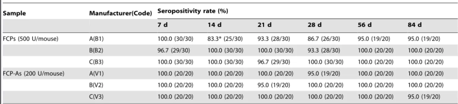
The anti-EV71 NAb seropositivity rates of groups immunized with the three FCPs ranged from 96.7–100% at 7 d, and were higher than 83.3% from 7 d to 84 d. The seroconversion rates induced by B2 and B3 (93.3–100%) were higher than that of B1 (83.3–100%) from 7 to 84 d post-inoculation with a dose of 500 U/mouse; the differences were not significant except at 14 d post-inoculation (P,0.01, Table 3).
There were differences in NAb GMTs and dynamic changes induced by the three FCPs (Table 4). Over time after immuni-zation, the NAb GMTs of B2 slowly rose from 118.0 at 7 d to 259.9 at 84 d, while those of B3 gradually reduced (from 138.3 at 7 d to 23.5 at 84 d), and those of B1 remained stable (from 37.4 at 7 d to 26.5 at 84 d) with the lowest titers. The NAb GMTs of B1 were significantly lower than those of B2 and B3 from 7–21 d; however, the NAb GMTs of B2 were significantly higher than those of B1 and B3 from 21–84 d (Table 4). On the whole, the relative order of the three FCPs by NAb GMT was B2.B3.B1, which was similar to the NAb responses to the EV71 strains reported above.
3. NAb induced by EV1 FCP-A
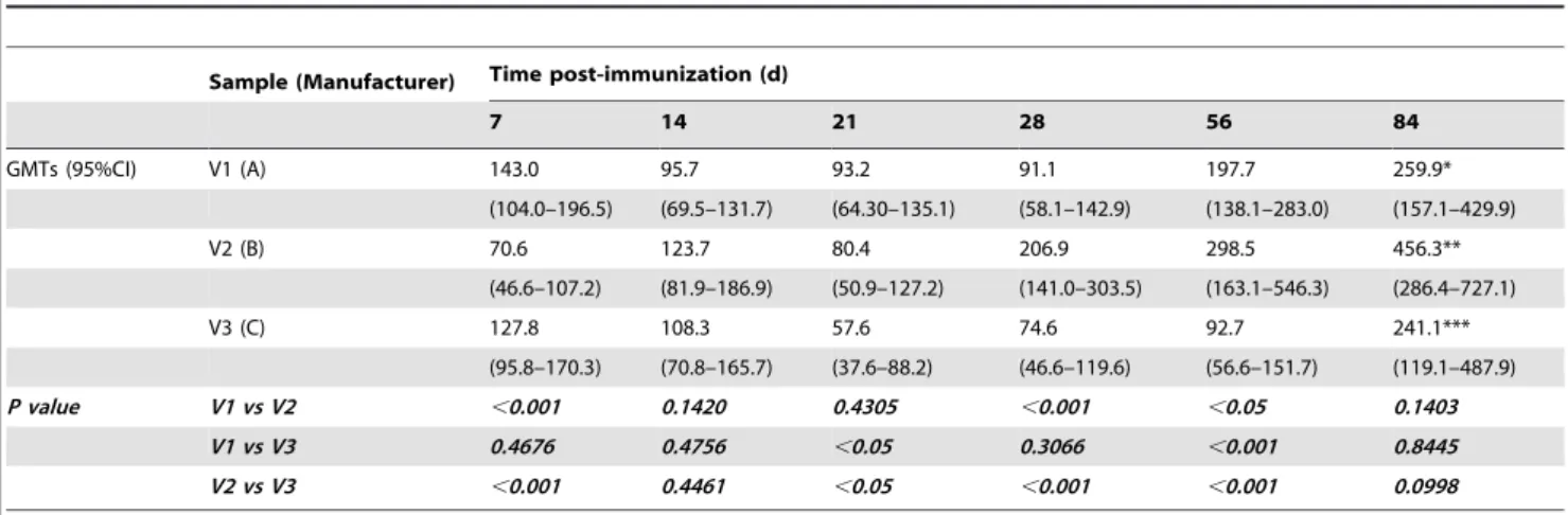
Unlike the EV71 FCPs, the NAb seroconversion rates of groups inoculated with one of the three EV71 FCP-As (200 U/mouse) ranged from 95.0–100.0% at each time point after immunization, and no significant differences in seropositivity were observed among the three FCP-As (Table 3), while the NAb GMTs were different (Table 5). The NAb GMTs induced by V1 (143.0, 7 d; 91.1, 28 d; and 259.9, 84 d) and V3 (127.8, 7 d; 57.6, 21 d; and 241.1, 84 d) were low with similar titers at 21 d or 28 d, but they were high at 7 d and 84 d. The highest NAb GMTs appeared at
84 d, with 2.9-fold (V1) and 4.2-fold (V3) increases compared with the GMTs at 21 d and 28 d, respectively. Meanwhile, the NAb GMTs elicited by V2 increased gradually (70.6, 7 d; 206.9, 28 d; and 456.3, 84 d) and reached the highest titer at 84 d, which was 6.5-fold higher than that observed at 7 d.
In comparing the three FCP-As at 200 U/mouse, similar NAb seroconversion rates were found (with slight variations) and the same trend of GMTs over time was observed (Table 3 and 5). The GMTs of the mice inoculated with the three FCP-As had the smallest differences (95.7, 123.7, and 108.3 with 1.3-fold difference) at 14 d, and the largest differences (197.7, 298.5, and 92.7 with 3.2-fold difference) at 56 d. The NAb GMT induced by V2 was significantly lower than that of V1 and V3 at 7 d, but significantly higher than that of V1 and V3 at 28 d and 56 d (P,0.05).
4. ED50of EV1 FCP-A at 7 d after inoculation
In addition to studying the dynamic NAb response, the NAb response to four serial dilutions of FCP-As was studied at 7 d after inoculation. Similar to the NAb response at 7 d after inoculation reported above, 100% seroconversion rate was observed in the 200 U dose groups (Table 6), and the lowest NAb GMT was induced by V2 at 7 d after inoculation (NAb GMTs induced by V1, V2, and V3 were 106.3, 37.4, and 52.0, respectively).
Every final product showed a good dose-response relationship. In response to a reduction in vaccine dose, the NAb-positive rate of the V3 group showed the smallest decrease among the three vaccines and V3 had the lowest ED50value (Table 6). The positive
rate of V3 group was still 33.3% when the dose was dropped to 0.8 U/mouse, while that of V1 group and V2 group were 3.3% and 0%, respectively. The NAb-positive rates and GMTs of the V2-inoculated mice all decreased faster than those immunized with V1 and V3, with significant differences in seropositive rates observed among three vaccines at doses of 12.5 U, 3.1 U, and 0.8 U (P,0.01). The ED
50values of V1, V2, and V3 were 6.1 U,
25.3 U, and 2.5 U, respectively. Based on seroconversion rates at 7 d post-inoculation, V2 had the highest ED50value; however, this
vaccine also induced the highest NAb GMTs from 21 d to 84 d post-inoculation. These results indicated that the NAb response of any one dose will not fully reflect the immunogenicity of the EV71 FCP-A at any time point post-inoculation.
5. Protective efficacy of EV71 FCP-As in suckling mice As shown in Figure 2a, in the protective efficacy evaluation by maternal antibodies, V1, V2, and V3 at doses of 162, 54, and
Table 2.NAb seropositivity rate (dilution$1:8) of mice inoculated with live and inactivated EV71 vaccine strains from three difference manufacturers.
Sample Manufacturer (Code) Seropositive rate (%)
14 d 28 d
EV71 strains (6.50 lg PFU/mouse) A(M1) 100.0 (10/10) 100.0 (10/10)
B(M2) 90.0 (9/10) 100.0 (10/10)
C(M3) 100.0 (10/10) 100.0 (10/10)
Inactivated EV71 strains (6.50 lg PFU/mouse) A(IM1) 96.7 (29/30) 90.0 (27/30)
B(IM2) 100.0 (30/30) 100.0 (30/30)
C(IM3) 100.0 (30/30) 96.7 (29/30)
Immunization and NAb detection were carried out in three collaborative labs using the same SOP. Ten to thirty mice were immunized in each dose group. The results were highly repeatable between the three labs and were combined for statistical analysis.
18 U/0.5 ml/dam were used to immunize adult ICR female mice. EV71/048M3C2 was administered intracerebrally to the newborn suckling mice at 20 LD50. At 2–3 days after inoculation, suckling
mice born to negative control dams began to show symptoms of EV71 infection, including slow movement and limb paralysis, eventually leading to death (Figures 2b, c, d and Figure S2). In the negative control and CA16 control groups, all suckling mice died 5 days after inoculation (of all two independent experiments, only one pup in the negative control group survived, yielding a survival rate of 3.7%). In comparison, only a few suckling mice died in the EV71 experimental groups. When dams were immunized with a dose of 162 U/mouse, the pup survival rates were 96.2–97.0% in all three FCP-A groups. When dams were immunized with a dose of 54 U/mouse, the pup survival rate in the V1 group was 0%, but those in the V2 and V3 groups were as high as 93.9–95.8%. At 18 U/dam, 92.0% of the suckling mice survived in the V3 group. The ED50of V1, V2, and V3 were calculated to be 96.9, 34.8, and
12.3 U, a 2.79- to 7.89-fold difference in dose. These results indicate that V3 has better protective effects against the 048M3C2 virus strain than V1 or V2.
Discussion
Immunogenicity and protective efficacy in animal models are two key indexes required for the approval of a new vaccine [35,36]. Several animal models have been developed for testing EV71 vaccines [16–18,20,22,35], including the maternal–NAb mouse model, which has been used to evaluate the protective efficacy of these vaccines [16,18,20]. However, the lack of standard methods for measuring EV71 antigen content and NAb titer have been major limitations in the evaluation and comparison of vaccine immunogenicity [37,38]. Two recent reports from Chinese Taiwan and mainland China addressed this problem by establishing assays to measure antigen content using a VP2 monoclonal antibody (MAb) and a VP1 MAb with high neutralizing activity [29,39]. Prior to these recent studies, the antigenicity of EV71 bulks from three manufacturers was compared with the EV71 antigen standard by kits using the VP1 MAb. The EV71 antigen reference standard demonstrated good parallelism and linearity with the different vaccine antigens, and the R2values were all higher than 0.999 [30]. These findings suggested that the vaccine antigens produced by the three
Table 3.NAb seropositivity rates (dilution$1:8) of mice inoculated with EV71 FCPs and FCP-As from three different manufacturers.
Sample Manufacturer(Code) Seropositivity rate (%)
7 d 14 d 21 d 28 d 56 d 84 d
FCPs (500 U/mouse) A(B1) 100.0 (30/30) 83.3* (25/30) 93.3 (28/30) 86.7 (26/30) 95.0 (19/20) 95.0 (19/20)
B(B2) 96.7 (29/30) 100.0 (30/30) 100.0 (30/30) 93.3 (28/30) 100.0 (20/20) 100.0 (20/20)
C(B3) 100.0 (30/30) 100.0 (30/30) 96.7 (29/30) 100.0 (30/30) 100.0 (20/20) 100.0 (20/20)
FCP-As (200 U/mouse) A(V1) 100.0 (20/20) 100.0 (20/20) 100.0 (20/20) 95.0 (19/20) 100.0 (20/20) 100.0 (20/20)
B(V2) 100.0 (20/20) 100.0 (20/20) 95.0 (19/20) 100.0 (20/20) 100.0 (20/20) 100.0 (20/20)
C(V3) 100.0 (20/20) 100.0 (20/20) 100.0 (20/20) 100.0 (20/20) 100.0 (20/20) 95.0 (19/20)
Immunization and NAb detection were carried out in three collaborative labs according to the same SOP. Twenty to thirty mice were immunized in each dose group. Results were highly repeatable in the three labs and were combined for statistical analysis.
*Anti-EV71 seropositive rate induced by B1 was significantly lower than that of B2 and B3 at 14 d post-inoculation,x2= 10.61,P,0.01. doi:10.1371/journal.pone.0046043.t003
Table 4.Dynamic trend of neutralizing antibody GMTs (95%CI) for EV71 FCPs.
Sample
(Manufacturer) Time post-immunization (d)
7 14 21 28 56 84
GMTs (95% CI) B1 (A) 37.4 24.6 25.7 36.4 35.2 26.5
(27.8–50.4) (17.2–35.2) (18.9–35.0) (24.5–50.6) (24.5–50.6) (17.5–40.3)
B2 (B) 118.0 100.5 135.2 129.1 182.7 259.9*
(91.2–152.8) (78.4–128.9) (98.4–185.8) (95.2–175.2) (102.7–324.8) (137.9–489.6)
B3 (C) 138.3 117.5 51.9 48.2 55.9 23.5**
(101.7–188.2) (85.0–162.3) (39.9–67.6) (37.0–62.8) (32.1–97.5) (17.0–32.6)
P value B1 vs B2 ,0.001 ,0.001 ,0.001 ,0.001 ,0.001 ,0.001
B1 vs B3 ,0.001 ,0.001 ,0.05 0.2083 0.1878 0.7115
B2 vs B3 0.4311 0.4535 ,0.001 ,0.001 ,0.05 ,0.001
Female BALB/c mice (4–6 weeks old) were i.p. inoculated with three EV71 FCPs (B1, B2, and B3) containing 500 U antigen. The sera were isolated and tested at 7, 14, 21, 28, 56 and 84 d post-inoculation. NAb titers equal to and above 1:1536 were assigned a value of 1:1536. Common logarithmic transformation of the NAb titer raw data was used to calculate the GMT and CV. SPSS10.0 software was used for statistical analyses.
*Denotes a significantly higher NAb GMT for B2 at 84 d post-inoculation than that of B2 at 7 d and 14 d post-inoculation(P,0.05).
manufacturers could be accurately quantitated using the same units of measure (U/ml) when using this kit and standard. In addition, the CPE assays performed in each collaborative lab followed the same SOP and employed the national reference standards for neutralizing antibody as the quality control. Only when the results of three reference standards and virus back-titration all met the detection range allowed, were the data of CPE assays accepted. To further confirm the results of CPE assays, when all CPE assays were completed, 120 samples were randomly selected and tested again; the differences among all 120 samples between two tests were within 4-fold of each other, indicating that the CPE assays have good repeatability. These data suggest that the use of a standardin vitroneutralization test is critical to confirm
the repeatability, comparability, and accuracy of NAb titer measurements, which are essential for the proper comparison of the immunogenicity and protective effects of vaccines from different manufacturers.
The immunogenicity of antigens in inactivated virus vaccines is crucial for successful vaccine development [15,26]. We previously compared the genomes of 11 EV71 vaccine strains in China that
have differences in nucleotide and amino acids in VP1–VP3 regions [26]. To compare the immunogenicity of the vaccines in this study, the three vaccine strains were diluted to an equivalent virus titer (6.50 lg PFU/mouse) before immunization. Although they are all of the C4a genotype,different NAb GMTs were induced by the vaccine strains, which was similar to the results obtained by Wang et al. and Chang et al. [40,41]. In the study by Liu et al [42], two peptides (211–220aa in VP1 and 136–150aa in VP2) were considered to be potentially of greater importance for influencing NAb responses. In our studies, only one residue of these peptides was different (M1, M2, M3 is T, S, T at 144aa in VP2) among the three vaccine strains. Whether this differential residue has an effect on the immunogenicity of the vaccine strains is an important area of future investigation. Chang et al. reported that formaldehyde inactivation may influence the immunogenicity of the C4 strain [41]. However, our research showed that the EV71 immunogenicity of all three vaccine strains to induce NAbs did not decrease after inactivation, indicating that the key conformational epitopes were not affected by formaldehyde treatment.
The seroconversion rate induced by FCPs (500 U) and FCP-As (200 U) were higher than 80% and 95%, respectively, and the NAb titers induced by FCP-As (200 U) were all higher than those induced by FCPs (500 U). The seroconversion rates and the NAb titers produced by vaccines from the three manufacturers were relatively close. These results suggest that the aluminum hydroxide adjuvant used at similar concentrations (1.00–1.16 mg/ml) in the EV71 vaccines from different manufacturers had different immunological enhancing effects (Figure S3). Combined with the comparisons in a recent report [41] of an EV71 vaccine absorbed with either aluminium phosphate adjuvant or aluminium hydrox-ide adjuvant, we can conclude from our results that different types of alum adjuvant and different manufacturing processes may influence the degree of immunogenicity enhancement [43,44]. Compared with the FCPs, the differences in immunogenicity among the FCP-As produced by the three manufacturers were reduced, especially at 14 d and 21 d after immunization. These results confirmed that the levels of immunogenicity of FCP-A produced by the three manufacturers were similar, and may
Table 5.Dynamic trend of neutralizing antibody GMTs (95%CI) for EV71 FCP-As.
Sample (Manufacturer) Time post-immunization (d)
7 14 21 28 56 84
GMTs (95%CI) V1 (A) 143.0 95.7 93.2 91.1 197.7 259.9*
(104.0–196.5) (69.5–131.7) (64.30–135.1) (58.1–142.9) (138.1–283.0) (157.1–429.9)
V2 (B) 70.6 123.7 80.4 206.9 298.5 456.3**
(46.6–107.2) (81.9–186.9) (50.9–127.2) (141.0–303.5) (163.1–546.3) (286.4–727.1)
V3 (C) 127.8 108.3 57.6 74.6 92.7 241.1***
(95.8–170.3) (70.8–165.7) (37.6–88.2) (46.6–119.6) (56.6–151.7) (119.1–487.9)
P value V1 vs V2 ,0.001 0.1420 0.4305 ,0.001 ,0.05 0.1403
V1 vs V3 0.4676 0.4756 ,0.05 0.3066 ,0.001 0.8445
V2 vs V3 ,0.001 0.4461 ,0.05 ,0.001 ,0.001 0.0998
Female BALB/c mice (4–6 weeks old) were i.p. inoculated with three EV71 FCP-As (V1, V2, and V3) containing 200 U antigen. The sera were isolated and tested at 7, 14, 21, 28, 56 and 84 d post-inoculation. NAb titers equal to and above 1:1536 were assigned a value of 1:1536. Common logarithmic transformation of the NAb titer raw data was used to calculate the GMT and CV. SPSS10.0 software was used for statistical analyses.
*Denotes a significantly higher NAb GMT for V1 at 84 d post- inoculation than that of V1 at 14 d, 21 d and 28 d post-inoculation(P,0.05). **Denotes a significantly higher NAb GMT for V2 at 84 d post- inoculation than that of V2 at 7 d, 14 d and 21 d post-inoculation(P,0.05). ***Denotes a significantly higher NAb GMT for V3 at 84 d post- inoculation than that of V3 at 21 d, 28 d and 56 d post-inoculation(P,0.05). doi:10.1371/journal.pone.0046043.t005
Table 6.Seroconversion rates and ED50values of mice inoculated with EV71 FCP-As from three different manufacturers.
Sample Seroconversion rate at 7 d pos-inoculation (%) ED50(U)
200 (U) 50 (U) 12.5 (U)* 3.1 (U)* 0.8 (U)*
V1 100 86.7 80.0 30.0 3.3 6.1
V2 100 76.7 16.7 6.7 0.0 25.3
V3 100 93.3 83.3 53.3 33.3 2.5
Immunization and NAb detection were carried out in three collaborative labs according to the same SOP. Thirty mice were immunized in each dose group. Results were highly repeatable in the three labs and were combined for statistical analysis. ED50was calculated using the Spearman-Karber method [34].
*There were significant differences in seropositive rates induced by the three vaccines at doses of 12.5 U, 3.1 U and 0.8 U (x2= 35.22, 15.51 and 18.86,
P,0.01).
provide the basis for establishing a uniform antigen unit (U) for EV71 vaccine dosages in clinical trials.
However, the ED50 for the NAb response and the in vivo
protection of the three vaccines were different. The V2 vaccine elicited the most potent NAb response at 28–84 d after inoculation; however, its ED50 with regards to both in vivo
protection and NAb response were higher than V3. There are several possible reasons for this observation: 1) the efficacy of the V2 vaccine may be most affected by dilution, as its protein content was the lowest among the three vaccines; 2) differences in the cell substrates and processes used during production may lead to different ratio of E-type particles (empty) and F-type particles (full) in the vaccines;3) the effects of different aluminium adjuvant adsorption technologies used by the three different manufacturers may contribute to the range of antibody responses. This result demonstrated that evaluating NAb responses at a single time point or using a single index is not adequate for evaluating the immunogenicity of the EV71 vaccines. However, the index of ED50 was suitable for monitoring the vaccine production from
different manufacturers.
The pup EV71 challenge model for evaluating protective efficacy by maternally transferred antibodies was applied in this study as an indirect method for comparing the protective efficacy of the inactivated virus vaccines [16,18,20]. In an earlier study carried out by Bek et al., all mice immunized with more than 50 U of vaccine antigen could survive when challenged with a mouse adapted strain (B3 genotype) of EV71 [27]. Additionally, in a report by Dong et al., another inactivated vaccine (with dose of 40 EU or 160 EU) exhibited nearly 100% immunoprotective efficacy in mice and monkeys that were challenged by the FY-23 virus (C4 genotype of EV71) [28]. However, the immunoprotective effects of the vaccines from different manufacturers are not comparable because of differences in animal experimental design and challenge virus.
By titrating the vaccines in this study, we found that every final product showed a good dose-response relationship. After only one immunization at a dose of 162 U/0.5 ml/dam, the protection rate passed 90%, consistent with the NAb seropositivity rate and GMTs of the three final products at 21 d post-inoculation. In particular, V3, produced by manufacturer C, showed good protective effects at a dose of only 18 U/0.5 ml/dam, which was better than that observed with V1 or V2. These results were consistent with the NAb responses in the vaccine titration experiment, in which the NAb-positive rate elicited by V3 showed the best ED50among the three vaccines. These results were also
consistent with a study reported by Bek et al. [27], which showed that the higher protection level conferred by the inactivated virus vaccine mainly correlated with the NAb titer elicited, although the ED50value for protecting suckling mice was not absolutely related
with ED50 value for NAb. However, some researchers have
speculated thatin vitroassays do not provide a full representation of
the in vivo activity and functions of pathogen-specific
immuno-globulin, as in vivo IgG can activate complement cascades,
participate in opsonization, and mediate antibody-dependent cytotoxicity, all of which play an important role in the protective process [22].
EV71 has only one serotype but 11 subtypes including A, B (B1–B5), and C (C1–C5). C4 has been the predominant subtype in mainland China in recent years, and therefore three C4 genotype strains were selected as vaccine strains. Whether these EV71 vaccines of C4 genotype can protect against the EV71 infection caused by other genotypes is another key indicator in the evaluation of vaccine immunogenicity. However, there are few studies addressing this matter. Among all subtypes, the genetic
homology between the C4 subtype and A genotype is the lowest. In previous studies conducted in our lab, we investigated the cross-neutralizing reaction capacity between 10 strains of C4 subtype (including 3 vaccine strains) and one A genotype strain. The results indicated that all 10 strains of C4 subtype had a cross-neutralizing capacity towards the A genotype strain, but the magnitude varied [26]. Bek et al. reported that EV71 vaccine of the C4 subtype can effectively protect mice from the lethal challenge of B3 subtype EV71 [27]. However, the cross-protective capacity of these EV71 vaccines (C4 subtype) against other subtypes of EV71 needs further study.
In summary, using uniform antigen units (U/ml) and a standardized NAb assay, protective effects and NAb responses induced by three EV71 inactivated vaccines were studied at the vaccine strain (activated and inactivated) stage as well as the final product stage without adjuvant (FCP) or with adjuvant (FCP-A). Although the three products had differences in the sequences of the vaccine strains as well as manufacturing process, the three FCP-As (200 U) showed good immunogenicity, inducing NAbs at a rate of greater than 95% in mice at 7 d after one immunization, with the NAb titer gradually increasing thereafter. The antigen content of the three EV71 vaccines, which are being used in current clinical trials, ranged from 162 U/dose to 506 U/dose. Our analysis indicated that the lowest dose (162 U/dose) among the three FPC-As showed good protective effects, with 90–100% of suckling mice surviving lethal challenge. This study compared the immunogenicity of different inactivated EV71 vaccines, and provided a basis for evaluating immunogenicity during clinical trials, indicating that the comparison of vaccine effects at one time point may not be representative of other time points. Finally, the standardized EV71 antigen unit (U/ml) should be used to evaluate the antibody responses and protective efficacy of EV71 vaccines in future clinical trials.
Supporting Information
Figure S1 Diagrammatic drawing of comparative stud-ies for NAbs induced in mice by EV71 FCPs and FCP-As from three different manufacturers.
(TIF)
Figure S2 Immune protection experiment–symptoms displayed by suckling mice. (a) Healthy suckling mice. (b) Inoculated suckling mice with disease onset on 5th day (arrows: rear limb paralysis).
(TIF)
Figure S3 Dynamic trend analysis of neutralizing antibody GMTs for EV71 FCPs and FCP-As from different manufacturers. * Denotes significant differences between EV71 FCPs and FCP-As (P,0.05).
(TIF)
Table S1 The targets and projects were researched in this paper.
(DOC)
Acknowledgments
Author Contributions
Conceived and designed the experiments: ZL JW WY QL XS. Performed the experiments: CD XL QG ZG XY YW FG. Analyzed the data: QM FL
MX. Contributed reagents/materials/analysis tools: ZL JW. Wrote the paper: QM ZL.
References
1. Solomon T, Lewthwaite P, Perera D, Cardosa MJ, McMinn P, et al. (2010) Virology, epidemiology, pathogenesis, and control of enterovirus 71. Lancet Infect Dis 10: 778–790.
2. Yi L, Lu J, Kung HF, He ML (2011) The virology and developments toward control of human enterovirus 71. Crit Rev Microbiol 37: 313–327. 3. McMinn PC (2002) An overview of the evolution of enterovirus 71 and its
clinical and public health signifi cance. FEMS Microbiol Rev 26: 91–107. 4. Chen SC, Chang HL, Yan TR, Cheng YT, Chen KT (2007) An eight-year
study of epidemiologic features of enterovirus 71 infection in Taiwan. Am J Trop Med Hyg 77:188–191.
5. Ministry of Health of the People’s Republic of China website. Available: http:// www.moh.gov.cn/publicfiles/business/htmlfiles/wsb/pyqxx/list.htm. Accessed 2012 Aug 30.
6. Chan LG, Parashar UD, Lye MS, Ong FG, Zaki SR, et al. (2000) Deaths of children during an outbreak of hand, foot, and mouth disease in sarawak, malaysia: clinical and pathological characteristics of the disease. For the Outbreak Study Group. Clin Infect Dis 31: 678–683.
7. Tu PV, Thao NT, Perera D, Huu TK, Tien NT, et al. (2007) Epidemiologic and virologic investigation of hand, foot, and mouth disease, Southern Vietnam, 2005. Emerg Infect Dis 13: 1733–1741.
8. Tan X, Huang X, Zhu S, Chen H, Yu Q, et al. (2011) The persistent circulation of enterovirus 71 in People’s Republic of China: causing emerging nationwide epidemics since 2008. PLoS One 6: e25662.
9. Kim KH (2010) Enterovirus 71 infection: An experience in Korea, 2009. Korean J Pediatr 53: 616–622.
10. Ma E, Lam T, Chan KC, Wong C, Chuang SK (2010) Changing epidemiology of hand, foot, and mouth disease in Hong Kong, 2001–2009. Jpn J Infect Dis 63: 422–426.
11. Mizuta K, Abiko C, Murata T, Matsuzaki Y, Itagaki T, et al. (2005) Frequent importation of enterovirus 71 from surrounding countries into the local community of Yamagata, Japan, between1998 and 2003. J Clin Microbiol 43: 6171–6175.
12. Chua KB, Kasri AR (2011) Hand Foot and Mouth Disease Due to Enterovirus 71 in Malaysia. Virol Sin 26: 221–228.
13. Wang SM, Ho TS, Lin HC, Lei HY, Wang JR, et al. (2012) Reemerging of enterovirus 71 in Taiwan: the age impact on disease severity. Eur J Clin Microbiol Infect Dis 31: 1219–1224.
14. Arita M, Nagata N, Iwata N, Ami Y, Suzaki Y, et al. (2007) An attenuated strain of enterovirus 71 belonging to genotype a showed a broad spectrum of antigenicity with attenuated neurovirulence in cynomolgus monkeys. J Virol 81: 9386–9395.
15. Liu CC, Lian WC, Butler M, Wu SC (2007) High immunogenic enterovirus 71 strain and its production using serum-free microcarrier Vero cell culture. Vaccine 25: 19–24.
16. Ong KC, Devi S, Cardosa MJ, Wong KT (2010) Formadehyde-inactivated whole-virus vaccine protects a murine model of enterovirus 71 encephalomyelitis against disease. J Virol 84: 661–665.
17. Chen HF, Chang MH, Chiang BL, Jeng ST (2006) Oral immunization of mice using transgenic tomato fruit expressing VP1 protein from enterovirus 71. Vaccine 24: 2944–2951.
18. Chung YC, Ho MS, Wu JC, Chen WJ, Huang JH, et al. (2008) Immunization with virus-like particles of enterovirus 71 elicits potent immune responses and protects mice against lethal challenge. Vaccine 26: 1855–1862.
19. Tung W, Bakar SA, Sekawi Z, Rosli R (2007) DNA vaccine constructs against enterovirus 71 elicit immune response in mice. Genetic vaccines and therapy 5: 6.
20. Chiu CH, Chu C, He CC, Lin TY (2006) Protection of neonatal mice from lethal enterovirus 71 infection by maternal immunization with attenuated Salmonella enterica serovar Typhimurium expressing VP1 of enterovirus 71. Microbes and Infection 8: 1671–1678.
21. Sivasamugham LA, Cardosa MJ, Tan WS, Yusoff K (2006) Recombinant Newcastle Disease virus capsids displaying enterovirus 71 VP1 fragment induce a strong immune response in rabbits. Journal of Medical Virology 78:1096– 1104.
22. Wu CN, Lin YC, Fann C, Liao NS, Shih SR, et al. (2001) Protection against lethal enterovirus 71 infection in newborn mice by passive immunization with subunit VP1 vaccines and inactivated virus. Vaccine 20: 895–904.
23. Li YP, Liang ZL, Gao Q, Huang LR, Mao QY, et al. (2012) Safety and immunogenicity of a novel human Enterovirus 71 (EV71) vaccine: A randomized, placebo-controlled, double-blind, Phase I clinical trial.Vaccine 30(22):3295–303.
24. Clinical Trials website. Available: http://clinicaltrials.gov/ct2/show/ NCT01268787?term=NCT01268787&rank=1. Accessed 2012 Aug 30. 25. Clinical Trials website. Available: http://clinicaltrials.gov/ct2/show/
NCT01376479?term=NCT01376479&rank=1. Accessed 2012 Aug 30. 26. Mao QY, Li N, Yu X, Yao X, Li FX, et al. (2011) Antigenicity, animal
protective effect and genetic characteristics of candidate vaccine strains of enterovirus 71. Arch Virol 157: 37–41.
27. Bek EJ, Hussain KM, Phuektes P, Kok CC, Gao Q et al. (2011) Formalin-inactivated vaccine provokes cross-protective immunity in a mouse model of human enterovirus 71 infection.Vaccine 29: 4829–4838.
28. Dong C, Liu L, Zhao H, Wang J, Liao Y, et al. (2011) Immunoprotection elicited by an enterovirus type 71 experimental inactivated vaccine in mice and rhesus monkeys. Vaccine 29: 6269–6275.
29. Jia H, Cai F, Gao Q, Zhang JS, Jing SR (2010) Development of A Quantitative ELISA Method for EV71 Antigen. Chin J Biologicals 23: 87–90.
30. Liang ZL, Mao QY, Gao Q, Li XL, Dong CH, et al. (2011) Establishing China’s national standards of antigen content and neutralizing antibody responses for evaluation of enterovirus 71 (EV71) vaccines. Vaccine 29: 9668–9674. 31. Grandien M, Fosgren M, Ehrnet A (1995) Enterovirua. In: Lennette EH,
Lennette DA, Lennette ET, editors. Diagnostic procedures for Viral, Rickettsial and Chlamydial Infections. 7th ed. Washington, DC: American Public Health Association. pp. 279–298.
32. Mao QY, He P, Yu X, Li N, Hao CS (2010) Laboratory Evaluation of Method for Determination of Neutralizing Antibody against Human Enterovirus 71. Chin J Biologicals 23: 885–888.
33. Manual for the virological investigation of polio. Available: http://whqlibdoc. who.int/hq/1997/WHO_EPI_GEN_97.01.pdf. Accessed 2007 Nov 25. 34. Martin A.Hamilton (1979) Robust estimates of the ED50. Journal of the
American Statistical Association 74: 344–354.
35. Yu CK, Chen CC, Chen CL, Wang JR, Liu CC, et al. (2000) Neutralizing antibody provided protection against enterovirus type 71 lethal challenge in neonatal mice. J Biomed Sci 7: 523–528.
36. Chang LY, King CC, Hsu KH, Ning HC, Tsao KC, et al. (2002) Risk factors of enterovirus 71 infection and associated hand, foot, and mouth disease/ herpangina in children during an epidemic in Taiwan. Pediatrics 109: e88–e93. 37. Lee MS, Chang LY (2010) Development of enterovirus 71 vaccines. Exerpt Rev.
Vaccines 9: 149–156.
38. Mao QY, Yang ZW, Yu X, Jing Q, He P, et al. (2009) Epidemic Tendency of Neutralizing Antibody against Enterovirus 71 and Coxsackievirus A 16 in Infants in Rural Area of Kaifeng City, Henan Province, China. Chin J Biologicals 22: 911–913.
39. Liu CC, Chang HW, Yang G, Chiang JR, Chow YH, et al. (2011) Development of a quantitative enzyme linked immunosorbent assay for monitoring the Enterovirus 71 vaccine manufacturing process. J Virol Methods 176: 60–68. 40. Wang LC, Tang SQ, Li YM, Zhao HL, Dong CH, et al. (2010) A Comparison
of the Biological Characteristics of EV71 C4 Subtypes from Different Epidemic Strains. Virol Sin 25: 98–106.
41. Chang JY, Chang CP, Tsai HH, Lee CD, Lian WC, et al. (2012) Selection and characterization of vaccine strain for Enterovirus 71 vaccine development.Vac-Vaccine 30: 703–711.
42. Liu CC, Chou AH, Lien SP, Lin HY, Liu SJ, et al. (2011) Identification and characterization of a cross-neutralization epitope of Enterovirus 71. Vaccine 29: 4362–4372.
43. White JL, Hem SL (2000) Characterization of aluminum-containing adjuvants. In Brown F, Corbel M, Griffiths E, eds. Physico-Chemical procedures for the Characterization of vaccines. Basel, karger. pp. 217–228.