Influence of brain lesion and educational
background on language tests in aphasic subjects
Ellen Cristina Siqueira Soares
1, Karin Zazo Ortiz
2Abstract – In language assessment, several socio-demographic variables must be taken into account. Objectives:
To characterize the performance of aphasic patients with different educational background on language tasks and to compare their performance to that of individuals with no language disorders. Methods: Thirty aphasic
patients and 30 healthy individuals were selected. Patients were divided into two groups according to educational level: A (1–4 years) n=15 and B (5–11 years) n=15. Age ranged from 27 to 78 years. All subjects were submitted to the Montreal Toulouse language assessment protocol. The pertinent statistical tests were applied. Results:
Educational level interfered in the linguistic performance of normal subjects but not in that of aphasic subjects, whose performance was influenced more by the lesion. Conclusions: The present study can contribute toward greater understanding of the influence of lesions and educational background on the language performance of aphasic subjects.
Key words: language, aphasia, language test.
Impacto da lesão e da escolaridade na avaliação de linguagem em sujeitos afásicos
Resumo – Para a avaliação de linguagem várias variáveis sócio-demográficas devem ser levadas em conta.
Objetivo: Estudar e caracterizar o desempenho de sujeitos afásicos com diferentes escolaridades em tarefas lingüísticas e comparar o desempenho destes pacientes com indivíduos sem alteração de linguagem. Métodos:
Foram selecionados 30 pacientes afásicos e 30 indivíduos, pareados com mesma idade, sexo e escolaridade. A escolaridade variou em duas faixas de acordo com o nível educacional: A (1–4 anos) n=15 e B (5–11 anos) n=15. A idade variou entre 27 e 78 anos. Todos os sujeitos foram submetidos ao protocolo de avaliação de linguagem Montreal Toulouse. Foram aplicados os testes estatísticos pertinentes. Resultados: A escolaridade interferiu no desempenho lingüístico de sujeitos normais, mas não de sujeitos afásicos, sendo para este grupo a lesão mais impactante e determinante do desempenho. Conclusões: Acreditamos que o nosso estudo contribua para a uma melhor compreensão do impacto da lesão e da escolaridade no desempenho lingüístico de sujeitos afásicos.
Palavras-chave: linguagem, afasia, teste de linguagem.
1Master in Sciences by Universidade Federal de São Paulo (Unifesp), São Paulo, Brazil. 2Post Doctor in Sciences by Universidade Federal de São Paulo
(Unifesp), Departamento de Fonoaudiologia, Universidade Federal de São Paulo, São Paulo, SP, Brazil.
Ellen Cristina Siqueira Soares – Av. Miguel Stefano, 1973/121 - 04301-900 São Paulo SP - Brazil. E-mail: [email protected]
Received September 2, 2008. Accepted in final form October 30, 2008.
Language is a complex cerebral function comprising many linguistic processes. The multiple functional com-ponents of language interact to produce the end function of verbal communication.1
Aphasia is a loss or deficit in language function as a result of brain injury.
Language assessment in aphasic subjects seeks to mea-sure oral and graphical production and comprehension, repetition, naming and fluency, while also assessing prag-matic and discursive functions of the language.
The investigator can devise their own tests to perform
language assessment, or may elect to use previously stan-dardized formal instruments. The tests allow performance comparisons between brain-damaged and normal subjects with the same educational background. Moreover, com-parisons can also be inter-patient when the test is used longitudinally.
patients with healthy individuals of the same age, sex and schooling in order to ascertain:
A) Which language tasks were most compromised in the aphasic group;
B) Which language tasks were most affected by educa-tional background in both aphasic and control groups.
Methods
The present studied was approved by the UNIFESP Re-search Ethics Committee (protocol nº 0151/05).
Thirty subjects diagnosed with aphasia were selected. Inclusion criteria were adult subjects, with single em-bolic ischemic stroke in the left hemisphere, having neu-rologic diagnosis and neuroimaging exam.
All the patients were assessed at the Acquired Neurolog-ic Disturbances of Speech and Language Outpatient Unit of the Speech Therapy Department of UNIFESP-EPM in 2006. Patients were divided into two groups based on their educational background: A (1–4 years of education) n=15 and B (5–11 years of education) n=15. Age ranged from 27 to 78 years.
All subjects were informed regarding the study and un-derwent language assessment using the Montreal Toulouse protocol.
The Montreal Toulouse protocol2 was used in the as-sessment. It has 22 different subtests that characterize oral and graphical production, oral and graphical comprehsion, besides repetition, naming and fluency. The test en-ables the examiner to verify speech and language manifes-tations and to diagnose the type of aphasia.
The control group for the aphasic population comprised 30 subjects matched with the aphasics for age, sex and edu-cational background, with ages ranging from 27 to 78 years. Inclusion criteria were absence of language deficits, where-as exclusion criteria were diagnosis or history of hearing, psychiatric and/or neurological disorders, based on a previ-ously administered questionnaire. For this control group, the same criteria in relation to educational background was applied, one group comprised those with 1 to 4 years of education and a second with 5 to 11 years of education. The Toulouse Montreal Test encompasses the follow-ing tests:
1) Guided interview: Subject must answer 12 open
questions, some subdivided. Subtests assess both oral pro-duction and comprehension. This study scored oral com-prehension.
2) Reciting or automatisms: The task is made up of
natural series (numbers, months of the year and days of the week). One point is given for each series produced correctly.
3) Oral comprehension: This task involves a total of 42
items assessing oral comprehension of words, along with simple and complex phrases. Subjects provide answers through the designation task.
4) Repetition: The subject must repeat a total of 33
items, comprising words, non-words and phrases.
5) Reading: The subject has to read a total of 33
prompts such as words, non-words and phrases.
6) Written comprehension: 16 boards containing 6 to 4
figures are presented, and 16 written cards which the sub-ject must read and match with the corresponding graphical prompt. There are boards representing words or simple and complex phrases.
7) Naming: The subject must name 31 figures.
8) Verbal fluency: The subject must produce the
high-est number of animals they can within a 60-second inter-val, while the investigator records the number of words produced. Each item scores 1 point. Timings are achieved using a stop-watch.
9) Buco-facial praxis: The subject must make 6 praxic
non-verbal gestures, elicited by verbal command.
10) Naming of body parts (oral): The subject must
un-derstand and indicate on themselves, 8 body parts named by the investigators.
11) Handling of objects following verbal instructions:
The subject must understand and carry out 8 commands given by the Investigator, using concrete objects (key, comb, cup, ashtray and paper). Phrase complexity steadily increases.
12) Copying: The subject must copy 3 words and 1 phrase.
13) Dictation: The subject must note down 10 words
and 3 phrases dictated by the investigator.
14) Naming of body parts (written): The subject must
understand and indicate 8 parts of the body.
15) Written naming of actions: The subject must
graphically produce phrases referring to 6 figures arranged on 6 different boards shown by the investigator.
16) Repetition of numbers: The subject must repeat 10
items referring to numbers.
17) Reading of numbers: The subject must read 10
numbers.
All tasks in the test are awarded 1 point for each cor-rect answer.
The data from the 17 subtests, including performance of both aphasics and controls according to educational level, underwent statistical analysis.
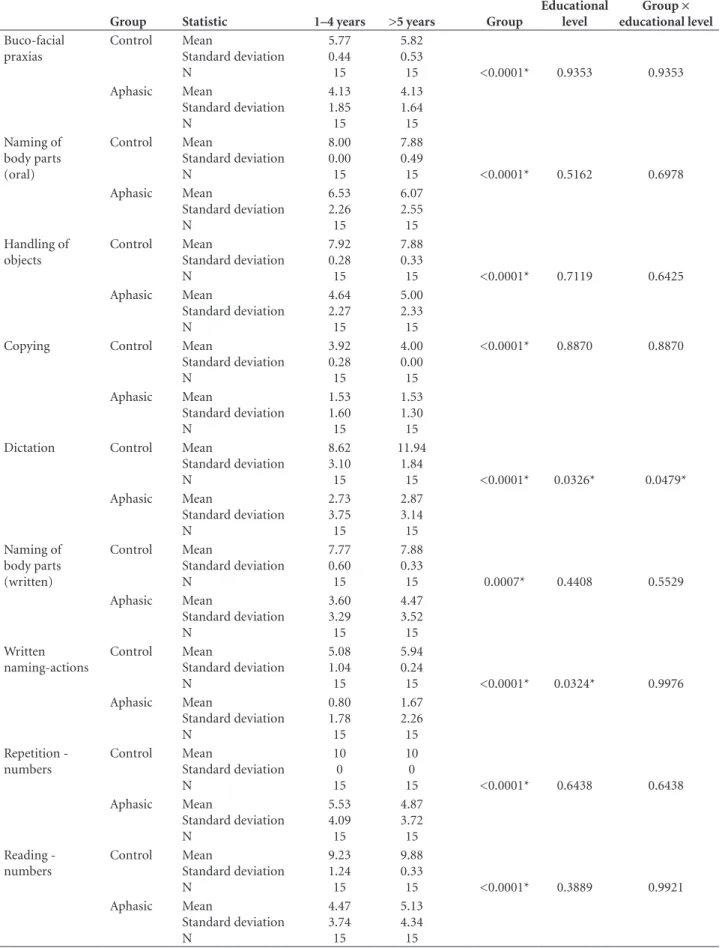
Table 1. Comparison between performance of aphasics and controls by mean and standard deviation on tasks from the Montreal Tou-louse test, and ANOVA values.
Group Statistic
Educational background ANOVA (p) values
1–4 years >5 years Group
Educational level Group × educational level Guided interview Control Mean Standard deviation N 11.85 0.38 15 11.88 0.33
15 <0.0001* 0.2634 0.2906 Aphasic Mean Standard deviation N 8.13 3.29 15 9.40 2.95 15 Automatisms form Control Mean Standard deviation N 29.00 0.00 15 29.00 0.00
15 <0.0001* 0.9517 0.9517 Aphasic Mean Standard deviation N 17.47 12.39 15 17.20 11.47 15 Automatisms content Control Mean Standard deviation N 3.00 0.00 15 3.00 0.00
15 <0.0001* 0.7860 0.7860 Aphasic Mean Standard deviation N 1.80 1.37 15 1.67 1.29 15 Oral comprehension Control Mean Standard deviation N 38.54 2.57 15 39.82 2.35
15 <0.0001* 0.3609 0.7564 Aphasic Mean Standard deviation N 25.93 11.85 15 28.53 10.59 15 Repetition Control Mean
Standard deviation N 32.46 0.97 15 32.12 0.93
15 <0.0001* 0.7508 0.8707 Aphasic Mean Standard deviation N 16.40 12.55 15 15.33 11.46 15 Reading Control Mean
Standard deviation N 31.54 1.90 15 32.41 0.80
15 <0.0001* 0.7789 0.8812 Aphasic Mean Standard deviation N 11.67 11.97 15 11.93 9.78 15 Written comprehension Control Mean Standard deviation N 12.08 1.32 15 12.71 0.59
15 <0.0001* 0.1583 0.5699 Aphasic Mean Standard deviation N 6.60 3.92 15 8.07 3.83 15 Naming Control Mean
Standard deviation N 26.69 3.12 15 28.00 2.37
15 <0.0001* 0.2940 0.7100 Aphasic Mean Standard deviation N 11.07 10.26 15 13.80 9.81 15 Verbal fluency Control Mean Standard deviation N 13.92 6.45 15 21.24 7.66
Table 1. Continuation.
Group Statistic
Educational background ANOVA (p) values
1–4 years >5 years Group
Educational level Group × educational level Buco-facial praxias Control Mean Standard deviation N 5.77 0.44 15 5.82 0.53
15 <0.0001* 0.9353 0.9353 Aphasic Mean Standard deviation N 4.13 1.85 15 4.13 1.64 15 Naming of body parts (oral) Control Mean Standard deviation N 8.00 0.00 15 7.88 0.49
15 <0.0001* 0.5162 0.6978 Aphasic Mean Standard deviation N 6.53 2.26 15 6.07 2.55 15 Handling of objects Control Mean Standard deviation N 7.92 0.28 15 7.88 0.33
15 <0.0001* 0.7119 0.6425 Aphasic Mean Standard deviation N 4.64 2.27 15 5.00 2.33 15 Copying Control Mean
Standard deviation N 3.92 0.28 15 4.00 0.00 15
<0.0001* 0.8870 0.8870
Aphasic Mean Standard deviation N 1.53 1.60 15 1.53 1.30 15 Dictation Control Mean
Standard deviation N 8.62 3.10 15 11.94 1.84
15 <0.0001* 0.0326* 0.0479* Aphasic Mean Standard deviation N 2.73 3.75 15 2.87 3.14 15 Naming of body parts (written) Control Mean Standard deviation N 7.77 0.60 15 7.88 0.33
15 0.0007* 0.4408 0.5529 Aphasic Mean Standard deviation N 3.60 3.29 15 4.47 3.52 15 Written naming-actions Control Mean Standard deviation N 5.08 1.04 15 5.94 0.24
15 <0.0001* 0.0324* 0.9976 Aphasic Mean Standard deviation N 0.80 1.78 15 1.67 2.26 15 Repetition -numbers Control Mean Standard deviation N 10 0 15 10 0
15 <0.0001* 0.6438 0.6438 Aphasic Mean Standard deviation N 5.53 4.09 15 4.87 3.72 15 Reading -numbers Control Mean Standard deviation N 9.23 1.24 15 9.88 0.33
Results
In our casuistic, 8 (26.6%) patients presented expres-sive aphasia, 10 (33.3%) receptive aphasia while 12 (40%) presented mixed aphasia.
Table 1 compares performance of aphasics and controls on tasks from the Montreal Toulouse test.
This table shows that the group effect compared the performance of aphasics and controls whereas the edu-cational effect compared eduedu-cational background in each group – aphasic and control groups. Concerning the Group effect vs. educational background, comparisons were made between groups according to educational profile.
For verbal fluency and dictation tasks, we observed that in terms of group effect, aphasic subjects presented fewer responses than controls at both levels of education. For the educational background effect, the control group presented fewer responses in the 1–4 year education bracket, yet no such difference was seen among aphasics with different educational background.
Concerning written naming of action tasks, we ob-served that in terms of group effect, aphasic subjects always presented fewer responses than controls. For the educa-tional background effect, the control group presented fewer responses in the 1–4 year education bracket, yet no such difference was seen among aphasics with different educa-tional background.
With regard to the remaining tasks in terms of group effect, aphasic subjects presented fewer responses for both educational bands.
Discussion
The aphasic subjects presented lesions at different sites within the left hemisphere, although all had suffered a single embolic ischemic stroke. It is known that there is no strict correlation between the injured area of the central nervous system and the type of aphasia,3,4 a fact corroborated by the present study. Studies in the literature have also indicated that, in the majority of right-handed patients, language pro-cessing takes place predominantly in the left hemisphere.4 In terms of characterizing the sample distribution, we observed that the groups of patients with expressive, recep-tive and mixed aphasia were similar in number.
Table 1 shows no difference comparing performance of aphasic subjects on a range of tasks from the Montreal Tou-louse protocol, with regard to educational background.
First, we can observe that the comparison of the per-formance of aphasic subjects and controls of the same age, sex and schooling, yielded statistically significant differ-ences across all tasks in the Montreal Toulouse protocol, showing this to be an effective tool for assessing language compromise in aphasic patients.
The control group presented statistically significant dif-ference in performance with increased educational-level on verbal fluency, dictation and written naming of action tasks. Drawing on these results, we may infer that educa-tional level modifies the performance profile in subjects on these tasks, most likely due to the development of a meta-language and skills acquired over the course of the knowledge-building process during school and academic learning. These differences were not observed in aphasic patients. This data will be discussed below.
Several models in cognitive neuropsychology explain some of the oral and graphical language processes.5-7 These models are commonly used for studying single clinical cas-es, in which specific changes to some linguistic processing components can be better identified. The models can ensure greater understanding of the disorder presented by the pa-tient, while also guaranteeing improved treatment planning. Population studies provide a profile of the most prevalent manifestations but do not specify which language processes are impaired. This can only be achieved using individual case studies. In the present study, statistically significant differences were observed between aphasic and control groups for all language tasks of the Montreal Toulouse test. Language tasks are known to entail complex involve-ment of many areas of the brain. Our results allow us to hypothesize that a brain lesion in an aphasic patient may correlate with poorer performance in language tasks.
Our study demonstrated that all oral comprehension tasks in the Montreal Toulouse Test, word comprehension, simple phrases (subject, verb and complement) and com-plex phrases (coordinated, subordinated and passive voice utterances), allowed us to differentiate aphasic subjects from those without brain injury.
Aphasic subjects can present a range of errors in oral production tasks, whereas educated subjects without brain injury are able to correctly repeat words, pseudowords and non-words.8,9 The statistically significant difference found between the group of aphasics and controls can be explained upon verifying the manifestations presented by aphasic subjects. Examination of errors committed by aphasic subjects reveals that these errors must have oc-curred due to failure in phonologic access or failure in mo-tor programming of speech, followed by, to a lesser extent, failures in lexical and semantic access.
most frequent manifestation in aphasias.1 It is known that large anterior and posterior areas seem to be involved in lexical access10 making aphasic subjects vulnerable to this manifestation.
Concerning writing, cognitive writing models assume the existence of different storage and procedure memories involved in this function. These models assist in analyz-ing performance of patients followanalyz-ing neurologic impair-ment. Paragraphies are manifestations in writing which are seen in aphasic pictures. This type of manifestation was observed in all tasks of the present study such as copying, graphical naming and writing by dictation. The writing process, including copying, writing by dictation and nam-ing, involves the cognitive domains of attention, cognitive flexibility, memory, visual processing and praxias, render-ing it more vulnerable in the event of a neurologic injury. In the dictation task, after hearing a prompt the subject should recognize the words as a sequence of syllables of the language, or as a whole word in the lexical recognition. The stimulus may undergo three types of processing: lexical, syllabic or phonological. Information can be processed by lexical semantic access or can undergo phonological-ortho-graphic conversion, with information flowing to the gra-phosyllabic output register, having two options of process-ing: information can proceed to the orthographical output and on to the letter-alphabetic output, or can go directly to the latter (syllabic segmentation processing). Another information processing mechanism is whereby phonosyl-labic input register information goes to the literal input register, and from here letters are matched in the alphabetic output register (literal segmentation processing). After any of the three processes outlined above, the information is processed in the graphemic buffer, proceeding to the allo-graphic buffer where programming and execution of chi-roarticulatory hand movements finally takes place resulting in writing.11 Regardless of the kind of processing used by aphasic subjects in the dictation task, their performance was statistically lower when compared to controls (Table 1). This appears to indicate distinct alterations in language processing of writing compared with performance on other tasks of the test in the aphasic population. Taken together, the data highlight that the aphasic group indeed presented specific alteration in writing.
In reading tasks, graphical symbols are processed within the occipital, inferior-temporal and parietal lobes.12-14 Sev-eral cortical and subcortical areas in the left hemisphere are involved in reading processing explaining the frequent oc-currence of central dyslexias co-occurring with aphasias.15,16 The reading aloud tasks performed in this study from the Montreal Toulouse test are composed of stimuli able to as-sess the lexical and phonological routes.11 Aphasic subjects
presented errors in all cues, demonstrating lower perfor-mance than controls.
Written comprehension tasks entail matching written content with a given figure. In this case, the subject has to perform visual analysis both of written content and the cor-responding figure. In the event that the patient presents vi-sual difficulty or when the figure is unclear, the subject may fail on the subtest due to a failure in visual analysis prior to graphical processing of the written information. Although this possibility existed in the present study, we observed that aphasic subjects presented difficulties in matching the written content with the appropriate figure, most likely ow-ing to failure in graphical comprehension or arisow-ing from difficulties in carrying out integration of the written con-tent with the pictograph. Aphasic subjects presented defi-cits in graphical comprehension ranging from mild to se-vere, leading to worse performances compared to controls.
Concerning buco-facial praxias, a statistically signifi-cant difference was evidenced between aphasic and con-trol groups. In fact, aphasic patients presenting verbal and non-verbal praxic alterations are frequently seen in clinical practice. Dronkers17 found that 48% of patients presenting brain injury also had verbal or non-verbal apraxia. More-over, phonemic paraphasias are manifestations which may stem from verbal apraxia. The high rate of phonemic para-phasias in expressive and mixed aphasia groups, in con-junction with the non-verbal praxic alterations in these two groups, lead us to hypothesize on the co-occurrence of ver-bal and non-verver-bal apraxia in some patients of this study.
We can argue that no difference was evident since the factor determining performance was language impairment resulting from brain injury and not schooling. Thus, le-sions were the strongest determinant of language behavior in aphasics, preventing the stronger language performance in more highly educated individuals commonly seen in healthy populations.
In fact, several studies18-20 have reported no perfor-mance difference in brain-damaged subjects on neurop-sychological tests based on level of schooling. Our findings revealed the same type of effect, albeit in relation to lan-guage, that is, higher-education in brain-damaged subjects was not associated with better performance in language tests. This is most likely because the injury compromised language abilities thereby causing aphasic groups with dif-ferent educational level to perform similarly in terms of language processing.
occur in the group of aphasic patients, in which the le-sion itself appeared to have greater influence. Finally, worse performance was observed in aphasics on verbal fluency, dictation and written naming of action tasks.
The authors believe the present study contributes toward understanding the role played by lesions and schooling in aphasic patients, proving valuable to professionals involved in assessment and rehabilitation of this patient population, especially in countries with large social cultural diversity.
References
1. Rogers MA, Off CA, Silkes J, Potts A, Kenny A, Pompon RH. The effect of repetition priming on picture naming accuracy and latency in individuals with aphasia. Brain Lang 2006;199-200. 2. Nespoulous JL, Lecours AR, Lafond D, et al. Protocole Mon-tréal-Toulouse d’examen linguistique de l’aphasie. MT 86. Module standard initial: MIB. Laboratoire Théophile-Alaj-ouanine, Montréal; 1986.
3. Basso A. The aphasias: fall and renaissance of the neurological model? Brain Lang 2000;71:15-17.
4. Marsh EB, Hillis AE. Recovery from aphasia following brain injury: the role of reorganization. Prog Brain Res2006;157: 143-156.
5. Berndt RS, Mitchum CC, Wayland S. Patterns of Sentence Comprehension in Aphasia: A Consideration of Three Hy-potheses. Brain Lang 1997; 60:197-221.
6. Basso A, Marangolo P. Cognitive neuropsychological reha-bilitation: The emperor’s new clothes? Neuropsychol Rehab 2000;10:219-229.
7. Ellis AW, Young AW. Human cognitive neuropsychology. Lon-don: Lawrence Erlbaum Associates; 1988.
8. Reis A, Castro-Caldas A. Illiteracy: a bias for cognitive devel-opment. J Int Neuropsychol Soc 1997; 3:444-450.
9. Martin N, Bunta F. Effects of lexical processing on primacy
effects in repetition of words and nonwords: Evidence from aphasia. Brain Lang 2007;103 :183-184.
10. Noppeney U, Price CD. Functional imaging of the semantic system retrieval of sensory – experienced and verbally learned knowledge. Brain Lang 2003;84:120-133.
11. Caramazza, A. How Many Levels of Processing Are There in Lexical Access? Cog Neuropsychol1997;14:177-208. 12. Cohen L, Lehéricy S, Chochon F, Lemer C, Rivaud S, Dehaene
S. Language-specific tuning of visual cortex? Functional prop-erties of the visual word from area. Brain2002; 125:1054-1069. 13. Moore CJ, Price CJ. Three distinct ventral occipitotemporal regions for reading and object naming.Neuroimage 1999;10: 181-192.
14. Binder JR, Frost JA, Hammeke TA, Cox RW, Rao SM, Prieto T. Human brain language areas identified by functional mag-netic resonance imaging. Neurocience 1997;17:353-362. 15. Lhermitte F, Chain F, Aron D, Leblanc ME. Southy O. Les
troubles de la vision des couleurs dans les lesion postérieures du cerveau : a propôs de 42 observations. Rev Neurol (Paris) 1969;121:5-29.
16. Geschwind N, Fusillo MG. Color-naming defects in associa-tion with alexia. Trans Am Neurol Assoc1964;89:172-176. 17. Dronkers NFA. A new brain region for coordinating speech
articulation. Nature1996; 384:159-161.
18. Reitan RM, Wolfson D. Influence of age and education on neuropsychological test results. Clin Neuropsychol 1995;9: 151-158.
19. Rosseli M, Ardila A, Rosas P. Neuropsychological assessment in illiterates. II Language and praxic abilities. Brain Cog 1990;281-296.