The corpus callosum in Binswanger’s disease
A quantitative fractional anisotropy analysis
Eliasz Engelhardt
1, Denise Madeira Moreira
2,3, Gilberto Oliveira Alves
5,
Maria Elisa Oliveira Lanna
6, Carlos Eduardo de Oliveira Alves
7,
Letice Ericeira-Valente
8, Felipe Kenji Sudo
9, Jerson Laks
4,10Abstract – To study the integrity of the corpus callosum in Binswanger’s disease (BD) patients using quantitative fractional anisotropy (DTI-FA). Methods: Controls (12) and patients with BD (12) were included. MR [GE Signa Horizon-1.5T] scans were performed. BD patients presented Fazekas’s score=6 and leukoaraiosis extension ≥75%, as assessed on FLAIR sequence. Standard parameters for DTI-FA acquisition were used. Functool was employed for post-processing, and ROIs placed on the genu and splenium of the corpus callosum on one axial plane at the basal ganglia level. Statistics [ANOVA] for genu and splenium comparison were analyzed. Results: DTI-FA showed reduction of anisotropy in both regions of the corpus callosum, more prominently in anterior (genu) than posterior (splenium) in BD patients versus controls. Conclusion: The reduction of anisotropy reflects loss of integrity of fibers of the studied regions of the corpus callosum. This finding indicates an interruption of the most important inter-hemispheric commissure, and component of neural networks that underlies cognitive, behavioral, motor and sensory integration. The affected genu and splenium, together with damage to other fiber systems that connect the prefrontal and parietal-occipital regions, may manifest clinically as dysfunction of high-level integrative regions linked to the domains of executive and sensory functions, respectively, that can occur in Binswanger’s disease.
Key words: Binswanger’s disease, corpus callosum, genu, splenium, diffusion tensor, fractional anisotropy.
O corpo caloso na doença de Binswanger: uma análise com anisotropia fracionada quantitativa
Resumo – Estudar a integridade do corpo caloso em pacientes com doença de Binswanger (DB) com anisotropia fracionada quantitativa (DTI-FA). Métodos: Foram incluídos controles (12) e pacientes com DB (12). Imagens de RM [GE Signa Horizon-1,5T] foram obtidas. Os pacientes com DB apresentavam escore=6 de Fazekas e leucoaraiose com extensão ≥75% como avaliado na seqüência em FLAIR. Foram utilizados parâmetros padrão para DTI-FA e utilizado Functool para pós-processamento. ROIs localizados no joelho e no esplênio do corpo caloso em um plano axial no nível dos gânglios da base. Estatística [ANOVA] feita para comparar joelho e esplênio. Resultados:
DTI-FA mostrou redução da anisotropia em ambas as regiões do corpo caloso, com predomínio na anterior (joelho) em relação à posterior (esplênio), nos pacientes com DB em comparação aos controles. Conclusões: A redução da anisotropia reflete perda da integridade de fibras das regiões estudadas do corpo caloso. Esses achados indicam interrupção da mais importante comissura inter-hemisférica e componente de redes neurais subjacentes à integração cognitiva, comportamental e de funções motora e sensorial. O comprometimento do joelho e do esplênio, juntamente com a lesão de outros sistemas de fibras, que conectam regiões pré-frontais e parieto-occipitais, podem ser expressas clinicamente por disfunção de regiões de alto nível de integração, relacionadas aos domínios das funções executiva e sensorial, respectivamente, que pode ocorrer na doença de Binswanger.
Palavras-chave: doença de Binswanger, corpo caloso, joelho, esplênio, tensor de difusão, anisotropia fracionada.
1-10Center for Alzheimer’s Disease / Institute of Psychiatry/Federal University of Rio de Janeiro; School of Medical Sciences, State University of Rio de
Janeiro. Institute of Neurology Deolindo Couto of the Federal University of Rio de Janeiro.
Eliasz Engelhardt – Rua Barata Ribeiro, 370/504 - 22040-000 Rio de Janeiro RJ - Brazil. E-mail: [email protected]
Received September 24, 2008. Accepted in final form November 12, 2008.
The corpus callosum is the largest fiber tract, and the main commissure, of the human brain, interconnecting neocortical areas of both hemispheres. The free part (mid-line) of the corpus callosum is easily visible (better seen in
where they intermingle with other tracts in the centrum semiovale and with the fibers of the corona radiata. It may be divided into roughly three regions, anterior (incorpo-rating the genum), intermediary (body), and posterior (incorporating the splenium). The genu connects the pre-frontal regions, the body connects the posterior portion of the frontal lobes and the parietal lobes, while the splenium connects regions of the parietal and occipital lobes.1-4
The corpus callosum establishes inter-hemispheric con-nections in a topographically organized way, and function-ally information transfer takes place between areas related to cognition, behavior, motor and sensory functions.2,5-9
This commissure is provided with a rich arterial sup-ply through the pericallosal, posterior pericallosal, and the anterior communicating arteries. They form the perical-losal pial plexus that supplies the midline corpus callosum and part of its radiations. The midline of the corpus cal-losum also presents a specific microvascular supply that boosts its vascularization. The vascular supply to the cen-tral zone of the genu and body of the corpus callosum, via short penetrating arterioles, is similar to that of the cerebral cortex, whereas the vascular supply to the extreme lateral corpus callosum and centrum semiovale is largely carried by medullary arteries, long end-arteries without anastomoses.4,10-12
The corpus callosum may be affected by numerous dis-eases, including those of vascular etiology. Infarctions and atrophy as a consequence of widespread subcortical white matter ischemic diseases, such as Binswanger’s disease, may also occur.3,13
Binswanger’s disease (BD) is one of several subtypes of the vascular cognitive impairment-vascular dementia (VCI-VaD) continuum.14-15 Pathological examination of
BD brains reveal atrophy, and the cut sections show an enlarged ventricular system, hypotrophy and yellowish dis-coloration of the subcortical white matter, and a thinned corpus callosum.16 The thinning of the corpus callosum
is often secondary, and primary white matter lesions have not been reported in this region.3,17 The microscopic
neu-ropathology of white matter lesions is mainly characterized by diffuse loss of nerve fibers, demyelination and gliosis affecting the centrum semiovale and the corpus callosum in a differentiated way.3,16,18-20 The lateral part of the corpus
callosum may be affected secondary to deep white matter lesions, while its medial part is “rarely affected or only af-fected in a non significant manner”.3
The large brain vessels in BD show atherosclerotic changes, and the histopathology reveals microvascular dis-ease in the form of severe arteriolar sclerosis, especially in subcortical white matter.16,10 This small-artery pathology is
likely to be one of the underlying substract of the extensive
white matter lesions, and is frequently related to hyperten-sion, diabetes mellitus, dyslipidemia, and other vascular risk factors.19,21-24 However, vascular changes commonly
seen in the centrum semiovale with aging or hypertension rarely develop in the corpus callosum, probably due to its special vascular supply which may explain its relative re-sistance to lacunar infarction, hypoxia, hypoperfusion, and Binswanger’s disease.22
Structural imaging techniques (computer tomography and magnetic resonance) and the concept of leukoaraiosis that followed20,24-28 allowed the observation that BD had a
much higher prevalence than formerly thought, and pro-vided the opportunity to establish the diagnosis in vivo.19,29
These conventional neuroimaging methods showed wide confluent areas of white matter disease in cases of Binswanger’s disease, identified neuropathologically as be-ing of ischemic cause.19,20,22,24,26,28, However, such findings
were rarely described in the corpus callosum.3,17
The recently developed technique of diffusion tensor imaging (DTI) has offered a new opportunity to evalu-ate the brain white matter architecture in a qualitative and quantitative way, in both normal and pathological states. A detailed analysis of white matter with DTI is possible ow-ing to two of its features – mean diffusivity and fractional anisotropy (FA). Currently, the most widely used method of measuring anisotropy is DTI-FA which allows quanti-fication, where the values obtained represent an average of the sampled fibers in a given region of interest (ROI). It is a highly sensitive but fairly nonspecific biomarker of neuropathology and microstructural architecture of white matter and is frequently considered a marker of white mat-ter integrity.30-31 Several studies have demonstrated that
the organization of white matter fiber bundles is the basis for DTI-FA. The myelin appears to influence its measure-ments, as does axonal integrity. The parallel organization of white matter fiber bundles is the basis for anisotropic diffusion, whereas myelin appears to modulate the amount of anisotropy.30 The analysis of ischemic lesions identified
by neuroimaging and neuropathology shows reduced DTI-FA values, indicative of axonal damage and/or loss, and demyelination.24,30-31 However, the same analysis of regions
visually identified as not affected, can also show derange-ment in the microarchitecture of the white matter, with axonal damage and demyelination.32 This appears to be
the case of the midline corpus callosum. In spite of re-ports of atrophy and pathological confirmation of fiber loss, signal changes are rarely described in conventional neuroimaging.3,13,16,17
frac-tional anisotropy (DTI-FA), and compare results with a normal control group.
Methods
The study included two samples, normal controls (n=12) and patients with Binswanger’s disease (n=12). The inclusion of BD patients was based on the National In-stitute of Neurological Disorders and Stroke and Associa-tion InternaAssocia-tionale pour la Recherché etl’ Enseignement en Neurosciences (NINDS-AIREN) criteria,33 and assessment
was performed with the Mini-Mental State Examination,34
Clinical Dementia Rating scale,35 and Hachinski ischemic
score.36 The control subjects had no neuropsychiatric
com-plaints, and presented results in the normal range following similar assessment. The characteristics of the subjects are displayed in Table 1.
Techniques
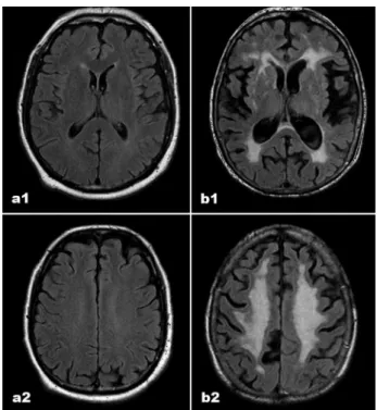
A complete series of MR scans of the brain, with stan-dard and DTI acquisitions, of the two samples was obtained using a 1.5T GE Signa Horizon machine. Axial plane fluid-attenuated inversion recovery (FLAIR) sequence scans were examined to evaluate the extension of the white matter lesions which were classified according to Fazekas’s scoring system37-38. The pathological cases had a Fazekas score=6
and LA≥75% (visual assessment), while the normal subjects yielded lower scores. The scoring was performed by two of the authors in consensus (DMM, EE) (Table 1 and Figure 1). The parameters used for DTI-FA acquisition correspond to those found in international studies on the subject, and are as follows: TR/TE=10000/89.1 msec, matrix=128×128,
FOV=30×24 mm, NEX=1, b=1000 sec/mm2, slice
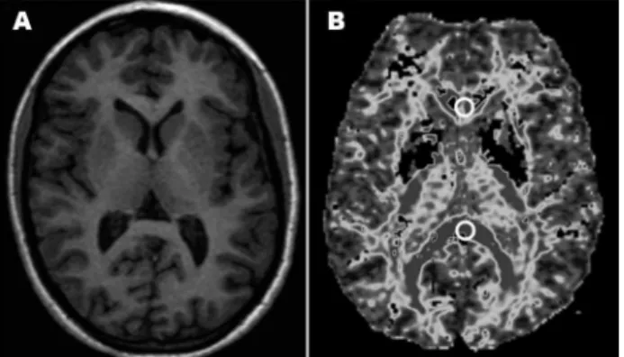
thickness=5 mm, number of slices=30 without gap. Cir-cular ROIs of 60mm2 were localized in the genu and the
splenium of the corpus callosum on one axial plane parallel to the AC-PC line at the basal ganglia level of the DTI-FA maps (total number of ROIs=24 for each group) (Figure 2). The DTI-FA maps were analyzed on an ADW 4.3 work-station using the Functool 4.5.3 (GE Medical Systems). Sta-tistical analysis (basic, ANOVA)39 was performed to
com-pare intra-sample and inter-sample measures of the genu and splenium of the corpus callosum.
Ethics
The present study is part of a larger project on Vascu-lar Cognitive Disorder, approved by the Ethics Committee of IPUB-UFRJ. Informed consent was obtained from the participants or from a family member responsible, before study procedures.
Results
The DTI-FA data of the genu and the splenium showed a significantly lower degree of anisotropy in Binswanger’s disease cases in comparison to normal controls (inter-sample). Considering each sample, there was also signifi-cant lower anisotropy measured between the genu and the splenium (intra-sample).
Table 1. Characteristics of the sample.
NC BD
N 12 12
sex (m/f) 5/7 7/5
age (range) 74.8±5.1 77.6±8.6
education (years: m±sd) 12.4±2.43 9.67±4.56
NINDS-AIREN negative positive
MMSE(a) (score: m±sd) 27.4±2.70 20.2±5.37
CDR(b) (score) 0 1.50±0.64
Hachinski(c) (score) 0.92±0.79 8.75±4.14
Fazekas(d) (score) 2.0±0.85 6.0±0.0
leukoaraiosis (extension %) –– ≥75%
NC, normal controls; BD, Binswanger’s disease; NINDS-AIREN, National Insti-tute of Neurological Disorders and Stroke and Association Internationale pour la Recherché et l’Enseignement en Neurosciences (criteria for clinical diagnosis of vascular dementia); (a)Mini-Mental State Examination (short cognitive screening
tool); (b)CDR, Clinical Dementia Rating scale (global severity stages from 0 to 3); (c)Hachinski, ischemic score (clinical assessment of vascular risk); (d)Fazekas, white
matter lesion scale (severity from 0 to 6).
Figure 1. MR scans in FLAIR acquisition (axial plane) – sections
The obtained data and the significance among the re-gions are depicted in Table 2 and Table 3. Additionally the post-hoc Tukey HSD test was performed for improved rep-resentation of the results (Table 4).
Discussion
The neuropathological characteristics of Binswanger’s disease are extensive subcortical white matter ischemic le-sion, with axonal damage and myelin loss. The corpus cal-losum is also affected, frequently showing atrophy. These changes may be presently revealed by quantitative DTI-FA, an in vivo marker of fiber integrity. These white matter isch-emic changes have been characterized with neuroimaging studies correlated with post mortem brain examination.25,28
The midline corpus callosum, but not its radiations, ap-parently suffers less in view of its rich vascular supply. The development of DTI-FA has provided a qualitative and quantitative evaluation of the white matter, and enabled as-sessment of the integrity of its constitutive fiber tracts.30-31
In spite of the infrequent description of signal changes on conventional neuroimaging,3,17 the DTI-FA shows that
there may be a reduction of the measured values, indicative of fiber loss, related to axonal damage and demyelination, as revealed in neuropathological studies.3,32,40
The present data showed lower DTI-FA values of the two studied segments of the corpus callosum – genu and splenium – in BD in comparison to NC (inter-sample). There was also a differential change between the genu and the splenium in BD patients and in NC (intra-sample). The genu was more significantly affected in comparison to the splenium, suggesting an anterior-to-posterior gradient, as described previously.41
The literature on the issue is very scarce, and the bib-liographical search yielded only a few related international studies. The published papers included data on the cor-pus callosum in Binswanger’s disease, compared to
nor-mal controls and to Alzheimer’s disease patients.42-43 In an
earlier paper, the applied imaging technique was apparent diffusion coefficient (ADC) derived from the diffusion se-quence, that represents the degree of diffusivity, in which ADC values and ratios (for the quantitative assessment of diffusion anisotropy) were calculated. ADCs in the corpus callosum (genu and splenium) were significantly higher in BD patients compared to controls, with disappearance of diffusion anisotropy, in a more significant way in the genu. These results suggest, according to the authors, that the cerebral white matter lesions in BD reflect a decrease in nerve fibers and diffuse myelin loss, and that the loss of nerve fibers in the corpus callosum may play a role in inducing cognitive decline.42
The results are comparable to those of the present study, even allowing for the differences between the tech-niques employed.
A more recent paper43 using the DTI-FA technique,
presented an analysis of the corpus callosum of patients with vascular dementia (VaD) (included with criteria for Binswanger’s disease) in comparison to normal controls and Alzheimer’s disease patients.
Figure 2. MR scans (axial plane) - sections at basal ganglia level: (A)
3DT1 sequence and (B) DTI-FA map. 3DT1 image for topo graphical reference, DTI-FA map is shown to localize ROI placement.
Table 2. Results of quantitative FA in NC vs BD.
Regions ROIs (n)
FA units (mean±sd)
NC BD
Genu 12 0.6041±0.05 0.5555±0.08
Splenium 12 0.7230±0.04 0.6394±0.08
Total 24 0.6635±0.08 0.5975±0.09
Table 3. Statistical significance as shown with ANOVA.
ANOVA summary
Source SS df MS F p
Rows(a) 0.12 1 0.12 27.79 <0.0001
Columns(b) 0.05 1 0.05 11.58 0.0014
Rows × columns 0.01 1 0.01 2.32 0.1349
Error 0.19 44 0
Total 0.37 47
(a)inter-sample (genu and splenium – NC vs BD); (b)intra-sample (genu vs splenium
– NC and BD).
Table 4. Critical values for the Tukey HSD Test.
HSD [0.05] HSD [0.01]
Rows (2) 0.04 0.05
Columns (2) 0.04 0.05
Cells (4) 0.07 0.09
The DTI-FA values of the corpus callosum (genu and splenium) in VaD were significantly lower in comparison to controls, and there were no statistically significant differ-ences between genu and splenium of the corpus callosum in any group.
These results are in part comparable to those of the present study, as the inter-sample differences were statisti-cally significant for both segments of the corpus callosum. In regard to the intra-sample differences between the genu and the splenium however, the present results were signifi-cant, in contrast to the cited study, where this difference may have been due to variation between the samples.
No papers on the subject were found in the national literature by the bibliographical search.
The corpus callosum, as the main neocortical commis-sure, establishes most of the inter-hemispheric connections and information transfer between areas related to cogni-tion, behavior, motor and sensory functions.2,6-9,44 It
partici-pates in the large neural networks that support complex bi-hemispheric functions, and its damage may disrupt these networks and cause inter-hemispheric disconnection.
Disconnection of the anterior brain regions (prefron-tal areas) due to genu damage, and of the posterior brain regions (mainly parieto-occipital areas) due to splenium damage may be related to impairment of high level inter-hemispheric integration,2,6 having a clear significant impact
on the clinical performance of the patients.40 The
interrup-tion of connecinterrup-tions of the genu fibers as well as cortical-prefrontal and subcortical-cortical-prefrontal fibers are of critical importance, where this multiple disconnection of the high-level prefrontal integrative regions may provide a structural basis for the impairment of the complex executive function cognitive domain, and a similar reasoning may be applied in relation to functions of parieto-occipital regions.40-41,45-47
Corpus callosum damage, together with that of the other white matter tracts seen in BD may, contribute to disconnection syndromes, one of the pathophysiological substracts of the VCI-VaD spectrum.48-50
Conclusion
The corpus callosum frequently shows atrophy in Binswanger’s disease as a consequence of extensive cen-trum semiovale white matter ischemic lesion, with axonal damage and myelin loss. The changes of the corpus cal-losum may be currently revealed by quantitative DTI-FA, an in vivo marker of fiber integrity.
The studied regions of the corpus callosum of the brain of Binswanger’s disease patients, namely the genu (prefron-tal interconnections) and splenium (parieto-occipi(prefron-tal inter-connections) showed lower DTI-FA values in comparison to normal controls. The genu is more severely
compro-mised than the splenium. These results are indicative of loss of integrity of fibers that cross the corpus callosum, and suggest their interruption. Such findings represent an inter-hemispheric disconnection process, and compromise of the wide neural networks that are the basis of cognitive, behavioral, motor and sensory integration underlying the diverse clinical manifestations of the Binswanger’s disease subtype of the VCI/VaD continuum.
Acknowledgements – The authors thank Luzinete Alva-renga for her editorial assistance.
References
1. Dejerine J. Anatomie des Centres Nerveux. Paris: J Rueff Ed; 1895:338-344.
2. Doron KW, Gazzaniga MS. Neuroimaging techniques offer new perspectives on callosal transfer and interhemispheric communication. Cortex 2008;44:1023-1029.
3. Tomimoto H, Lin J-X, Matsuo M et al. Different mechanisms of corpus callosum atrophy in Alzheimer’s disease and vascu-lar dementia. J Neurol 2004;251:398-406.
4. Türe U, Yasargil MG, Krisht AF. The arteries of the corpus callosum: a microsurgical anatomic study. Neurosurgery 1996;39:1075-1084.
5. Funnell MG, Corballis PM, Gazzaniga MS. Insights into the functional specificity of the human corpus callosum. Brain 2000;123:920-926.
6. Gazzaniga MS. Cerebral specialization and interhemispheric communication. Does the corpus callosum enable the human condition? Brain 2000;123:1293-1326.
7. Putnam MC, Wig GS, Grafton ST et al. Structural Organiza-tion of the Corpus Callosum Predicts the Extent and Impact of Cortical Activity in the Nondominant Hemisphere. J Neu-rosci 2008;28:2912-2918.
8. Stephan KE, Marshall JC, Penny WD et al. Interhemispheric Integration of Visual Processing during Task-Driven Lateral-ization. J Neurosci 2007; 27:3512-3522.
9. Wahl M, Lauterbach-Soon B, Hattingen E et al. Human Motor Corpus Callosum: Topography, Somatotopy, and Link between Microstructure and Function. J Neurosci 2007;27: 12132-12138. 10. Furuta A, Ishii N, Nishihara Y et al. Medullary Arteries in
Aging and Dementia. Stroke 1991;22:442-446.
11. Kahilogullari G, Comert A, Arslan M et al. Callosal branches of the anterior cerebral artery: an anatomical report. Clin Anat 2008;21:383-388.
12. Moody DM, Bell MA, Challa VR. The corpus callosum, a unique white-matter tract: anatomic features that may ex-plain sparing in Binswanger disease and resistance to flow of fluid masses. Am J Neuroradiol 1988;9:1051-1059.
14. Engelhardt E, Laks J, Cavalcanti JLS et al. Demência vascular. Rev Bras Neurol 2004;40:5-25.
15. Erkinjuntti T. Subcortical Ischemic Vascular Disease and De-mentia. Int Psychogeriat 2003;15(Suppl 1):23-26.
16. Caplan LR and Schoene WC. Clinical features of subcortical arteriosclerotic encephalopathy (Binswanger disease). Neu-rology 1978;28:1206-1215.
17. Babikian V and Ropper AH. Binswanger’s Disease: A Review. Stroke 1987;18:2-12.
18. Jones DK, Lythgoe D, Horsfield MA et al. Characterization of white matter damage in ischemic leukoaraiosis with diffusion tensor MRI. Stroke 1999;30:393-397.
19. Román GC. Senile dementia of the Binswanger type. A vascu-lar form of dementia in the elderly. JAMA 1987;258:1782-1788. 20. Yamanouchi H, Sugiura S, Tomonaga M. Decrease in nerve
fibres in cerebral white matter in progressive subcortical vas-cular encephalopathy of Binswanger type. An electron micro-scopic study. J Neurol 1989;236:382-387.
21. Brown WR, Moody DM, Thore CR et al. Vascular dementia in leukoaraiosis may be a consequence of capillary loss not only in the lesions, but in normal-appearing white matter and cortex as well. J Neurol Sci 2007;257:62-66.
22. Moody DM, Thore CR, Anstrom JA et al. Quantification of Afferent Vessels Shows Reduced Brain Vascular Density in Subjects with Leukoaraiosis. Radiology 2004;233:883-890. 23. Smid J, Nitrini R, Bahia VS, Caramelli P. Clinical
characteriza-tion of vascular dementia: retrospective evaluacharacteriza-tion of an out-patient sample [Caracterização clínica da demência vascular]. Arq Neuropsiquiatr 2001;59:390-393.
24. Young VG, Halliday GM, Kril JJ. Neuropathologic correlates of white matter hyperintensities. Neurology 2008;71:804-811. 25. Awad IA, Johnson PC, Spetzler RF, Hodak JA. Incidental sub-cortical lesions identified on magnetic resonance imaging in the elderly. II. Postmortem pathological correlations. Stroke. 1986;17:1090-1097.
26. Hachinski VC, Potter P, Merskey H. Leuko-araiosis: an an-cient term for a new problem. Can J Neurol Sci 1986;13(Suppl 4):533-534.
27. O’Sullivan M. Leukoaraiosis. Pract Neurol 2008;8:26-38. 28. Rosenberg GA, Kornfeld M, Stovring J and Bicknell JM.
Sub-cortical arteriosclerotic encephalopathy (Binswanger): Com-puterized tomography. Neurology 1979;29:1102-1106. 29. Román GC. Binswanger disease: the history of a silent
epi-demic. Ann N Y Acad Sci 2000;903:19-23.
30. Alexander AL, Lee JE, Lazar M, Field AS. Diffusion tensor imaging of the brain. Neurotherapeutics 2007;4:316-329. 31. Mori S and Zhang J. Principles of diffusion tensor imaging
and its applications to basic neuroscience research. Neuron 2006;51:527-539.
32. O’Sullivan M, Summers PE, Jones DK et al. Normal-appear-ing white matter in ischemic leukoaraiosis: A diffusion tensor MRI study. Neurology 2001;57:2307-2310.
33. Román GC, Tatemichi TK, Erkinjuntti T et al. Vascular demen-tia: diagnostic criteria for research studies: report of the NINDS-AIREN international workshop. Neurology 1993;43: 250-260. 34. Folstein MF, Folstein SE and McHugh PR. “Mini-mental state». A practical method for grading the cognitive state of patients for the clinician. J Psychiatr Res 1975;12:189-198. 35. Hughes CP, Berg L, Danziger WL et al. A new clinical scale for
the staging of dementia. Br J Psychiatry 1982;140:566-572. 36. Hachinski VC, Iliff LD, Zilhka E et al. Cerebral blood flow in
dementia. Arch Neurol 1975;32:632-637.
37. Fazekas F, Chawluk JB, Alavi A et al. MR signal abnormali-ties at 1.5 T in Alzheimer’s dementia and normal aging. Am J Neuroradiol 1987;8:421-426.
38. Pantoni P, Simoni M, Pracucci G et al. Visual Rating Scales for Age-Related White Matter Changes (Leukoaraiosis). Can the Heterogeneity Be Reduced? Stroke. 2002;33:2827-2833. 39. VassarStats: Statistical Computation Web Site.
http://faculty.vas-sar.edu/lowry/VassarStats.html (acessado em junho de 2008). 40. Yamauchi H, Fukuyama H and Shio H. Corpus callosum atrophy in patients with leukoaraiosis may indicate global cognitive impairment. Stroke 2000;31:1515-1520.
41. O’Sullivan M, Morris RG, Huckstep B et al. Diffusion ten-sor MRI correlates with executive dysfunction in patients with ischaemic leukoaraiosis. J Neurol Neurosurg Psychiatry 2004;75:441-447.
42. Hanyu H, Imon Y, Sakurai H et al. Regional differences in diffusion abnormality in cerebral white matter lesions in pa-tients with vascular dementia of the Binswanger type and Alzheimer’s disease. Eur J Neurol1999;6:195-203.
43. Sugihara S, Kinoshita T, Matsusue E, Fujii S, Ogawa T. Useful-ness of Diffusion Tensor Imaging of White Matter in Alzheimer Disease and Vascular Dementia. Acta Radiol 2004;45: 658-663. 44. Ota M, Obata T, Akine Y et al. Age-related degeneration of
corpus callosum measured with diffusion tensor imaging. Neuroimage. 2006;31:1445-1452.
45. Taylor WD, Bae JN, MacFall JR et al. Widespread Effects of Hyperintense Lesions on Cerebral White Matter Structure. Am J Roentgenol 2007;188:1695-1704.
46. Masterman DL, Cummings JL. Frontal-subcortical circuits: the anatomic basis of executive, social and motivated behav-iors. J Psychopharmacol 2007;11:107-114.
47. Schmahmanna JD, Pandya DN. Disconnection syndromes of basal ganglia, thalamus, and cerebrocerebellar systems. Cortex 2008;44:1037-1066.
48. Catani M, Ffytche DH. The rises and falls of disconnection syndromes. Brain 2005;128:2224-2239
49. Catani M, Mesulam M. What is a disconnection syndrome? Cortex 2008; 44:911-913.