Dement Neuropsychol 2013 December;7(4):435-438
History Note
435
Engelhardt E Meynert and the basal nucleus
Meynert and the basal nucleus
Eliasz Engelhardt1
ABSTRACT. Meynert described the “loop of the peduncular foot” (Schlinge des Hirnschenkelfusses), and its ganglion (Ganglion der Hirnschenkelschlinge) and related them to Reil’s Substantia innominata and Gratiolet’s Ansa peduncularis, from which he apparently built up his findings. Koelliker renamed the ganglion with the eponymous designation Meynert’sches Basalganglion
(Meynert’s basal ganglion), a name which endures to the present day, and described its topographical spread in relation to neighboring structures. Meynert and Koelliker also described aspects of cell composition of the ganglion (or nucleus) with a better account of the latter. Both, together with Reil and Gratiolet, were the outstanding personalities of the 19th century who performed the pioneering studies on basal formations of the forebrain. After these works, a considerable body of research appeared in the 20th century, with a focus on Meynert’s basal nucleus and related structures. The development of further knowledge about these structures revealed their great importance in the activity of the brain, as evidenced in both normal and pathological states.
Key words: history, Reil, Gratiolet, Meynert, Koelliker, Substantia innominata, Ansa peduncularis, nucleus basalis, basal nucleus, cholinergic.
MEYNERT E O NÚCLEO BASAL
RESUMO. Meynert descreveu a “alça do pé do pedúnculo” (Schlingedes Hirnschenkelfusses) e seu gânglio (Ganglion der Hirnschenkelschlinge), relacionando-os à Substantia innominata de Reil e à Ansa peduncularis de Gratiolet, a partir dos quais aparentemente desenvolveu seus achados. Koelliker renomeou o gânglio com a designação epônima de Meynert’sches Basalganglion (gânglio basal de Meynert), que perdura até o presente, e descreveu sua extensão topográfica em relação às estruturas vizinhas. Meynert e Koelliker descreveram também aspectos da composição celular do gânglio (ou núcleo), com um relato melhor do segundo. Ambos, juntamente com Reil e Gratiolet, foram as personalidades de destaque do século 19 que realizaram os estudos pioneiros sobre formações basais do prosencéfalo. Após esses, um número considerável de estudos apareceu no século 20, com foco no núcleo basal de Meynert e estruturas relacionadas. O desenvolvimento ulterior do saber sobre as mesmas mostraram sua grande importância na atividade cerebral, como visto em condições normais e patológicas.
Palavras-chave: história, Reil, Gratiolet, Meynert, Koelliker, Substantia innominata, Ansa peduncularis, nucleus basalis,
núcleo basal, colinérgico.
INTRODUCTION
A
s for almost all knowledge in neurosci-ence, a long period elapses, sometimes a century or more, in the transition from anatomical research to the resulting corol-lary application. his is the case for the basal structures of the forebrain, and the included cholinergic nuclei, which involved some of the most outstanding personalities of the 19th and 20th centuries. he highlights of thischronicle will be reviewed and commented in the following paragraphs.
MEYNERT’S CONTRIBUTION
heodor Hermann Meynert (1833-1892) was an outstanding anatomist, neuropathologist, and psychiatrist. He described numerous ner-vous structures, some for the irst time, and also developed theories regarding correla-tions between neuroanatomical and mental processes.1-3
His contribution to the understanding of the basal nucleus is found in his publications where he acknowledges and includes
struc-1Neurologist, Full Professor (retired), Cognitive and Behavioral Neurology Unit –INDC-CDA/IPUB − Federal University of Rio de Janeiro (UFRJ), Rio de Janeiro-RJ,
Brazil.
Eliasz Engelhardt. Av N.S. de Copacabana, 749 / sl 708 – 22050-002 Rio de Janeiro RJ – Brasil. E-mail: [email protected] Disclosure: The authors report no conflicts of interest.
Dement Neuropsychol 2013 December;7(4):435-438
436 Meynert and the basal nucleus Engelhardt E
tures previously observed in the upper mesencephalon by Reil and by Gratiolet4. It is opportune to write a few
words about these two researchers, considering their pioneering descriptions, importance, and Meynert’s quotes (Box 1 and Box 2).
Meynert published his indings, initially in a chap-ter of Stricker’s book, and lachap-ter in his own. In Stricker’s book (1872 - volume 2 - Chapter XXXI - pp 694-808)4
he analyzes the cerebral peduncle and its ganglia (pp 723-734), and describes bundles in the upper mesen-cephalon, underneath the basal ganglia, that constitute a kind of belting, and emphasizes one which he names
Schlinge des Hirnschenkelfusses (loop of the peduncular
foot), with a course transverse and approximately paral-lel to the optic tract (Figs. 245 and 247 Schl). He relates it to the deepest stratum of the Substantia innominata of Reil, or the Ansa peduncularis of Gratiolet (p 734). he description is illustrated (Fig. 245, p 728) (Figure 1), and in the legend he designates the structure, describing his indings (p 734) in a synthetic way, as follows: “he
Substantia innominata of Reil may be divided into 4
lay-ers: (i) the loop of the lenticular nucleus
(Linsenkernsch-linge),…(Schl); (ii) the Ganglion der Hirnschenkelschlinge
(ganglion of the cerebral peduncular loop) (L),…; (iii) the inferior peduncle of the optic thalamus (St), and (iv) the anterior temporal part of the stratum zonale (Z)…”4,8
he description of item (ii) above further details the ganglion: “… a lat extended ganglion (Fig. 245 L), that lays below the cerebral peduncular loop, a mass which represents the 2nd stratum of Reil’s Substantia
innnomi-Box 2. Gratiolet and the Anse pédonculair.
Louis Pierre Gratiolet (1815-1865), a French anatomist, anthropologist and zoologist, in the book published in 1857, regarding the anatomy of the cerebral peduncles and beyond (pp 52-72) describes (abridged) an
arcade fibreuse (fibrous arch), which passes over the peduncular foot, which he named Anse du pédoncule (loop of the peduncle),… adding that it is burrowed along its entire length by a large groove, and “….its bottom is formed by fibres blanches mêlées à beaucoup de substance grise (white fibers mixed with much grey matter), that house the bands of the optic nerves, and it will be for us the goutière de l’anse (groove of the loop)”. He also describes “…beyond the anse, the concavity of the
pavilion pédonculaire*contains a large convex ellipsoid mass which is like the nucleus (le noyeau)**, whose posterior limit meets the anterior border of the Anse pédonculaire to which it is closely united, and where the side of the peduncle touches the midline, an extension of this grey matter joins with a median grey mass that covers the bulge of the intermediary ventricle funnel***– the tuber cinereum.”6
Gratiolet’s mentions the presence of “white fibers mixed with masses of grey matter” related to the bottom of the peduncular loop, behind the optic tract. It is probable that this description was taken by Meynert as a “ganglion” in the course of the anse.
He identifies also, a “large convex ellipsoid mass which is like the nucle-us”, beyond the anse (also corps strié extraventriculaire [deep part of the basal ganglia]) (according to Foville). Thus, Gratiolet situates the anse at the transition of the peduncle and the basal ganglia.
The description employs several unusual terms peculiar to the author, and the accompanying illustrations (Plate XXV – Figs. 1-8, explanatory text pp 38-41) are macroscopic specimens with structures visualized by blunt dissection that are not displayed in a detailed way, hampering any clear understanding.7
*Upper radiating extension of the peduncular foot or couronne de l’éventeil pédonculaire (corona of the peduncular fan) (p 61). **The nucleus (le noyeau) (also corps strié extraventriculaire) (ac-cording to Foville), probably meaning the deep part of the basal ganglia. ***Intermediary ventricle funnel: infundibulum.
Box 1. Reil and Die ungenannte Marksubstanz.
Johann Christian Reil (1859-1813), a German anatomist, physiologist and psychiatrist, in the paper Das Hirnschenkel-System oder die Hirn-schenkel-Organization im Grossen Hirn, published in 1809, describes (pp 147-171) the cerebral peduncle and the surrounding structures and writes in a footnote (p 160 − abridged): “Die ungenannte Marksubstanz
(the unnamed medullary substance) is a medullated formation in con-nection with the anterior rounded extremity of the optic thalami, placed around the cerebral peduncle, from inwards to outwards, above and par-allel the optic nerves, ending in the external wall of the lateral ventricle. This substance becomes evident when the optic nerves are lifted away from the cerebral peduncle until the corpus geniculatum. Its real organi-zation and function are not clear to me, thus I have designated it, for now, unnamed (ungenannte), until I will have it examined in a special way.”5
Apparently he never returned to this subject to analyze its structure and devise an appropriate designation. However, it may be inferred that he meant the presence, in the region, of white matter formations.
Reil’s region was later named Substantia innominata (in Latin).
Two hundred years on, the name is still in use, despite incomplete com-prehension of its complex structure and function.
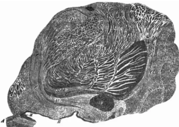
Figure 1. Transverse (coronal) section of the region of the human insula and the basal nuclei (Fig. 245, p 728) (according to Meynert)4 .
Substantia innomminata or Hirnschenkelschlinge and its 4 layers: Schl, L, St, Z (Schl: loop of the
lenticular nucleus [Linsenkernschlinge], L: ganglion of the cerebral peduncular loop
[Hirnschen-kelschlinge], St: inferior peduncle of the optic thalamus, Z: anterior temporal part of the stratum
zonale). Boundary landmark structures. V: grey matter of the 3rd ventricle; L1, L2, L3: lenticular
Dement Neuropsychol 2013 December;7(4):435-438
437
Engelhardt E Meynert and the basal nucleus
nata, or Gratiolet’s Anse pédonculaire…”. here is also
a short description of the cellular component of the ganglion (p 732): “he cells of this ganglion reach the external capsule,…a small number of bundles, isolated or together with others contacting 50 μm long, 15 μm wide fusiform cells…”4.
Twelve years later, in his own book (1884 - pp 87-88)9, he reiterates his former indings and gives some
additional details on anatomical aspects and extent of the ganglion: “…a dense transversely placed particular layer of ganglion cells, parallel to the iber radiation, which extends laterally until the external capsule…it constitutes a lat well bounded special ganglionic forma-tion, which equals in extension the size of the anterior perforated space”…“this formation shows its cells paral-lel to the course of the peduncular loop (Ansa peduncu-laris), being traversed by its bundles, and represents the Ganglion ansae peduncularis (ganglion of the peduncu-lar loop)”. He illustrates the anatomical location of the peduncular loop on a inely dissected brain (Fig. 23 ans, p 49). However, there is no depiction or indication of a ganglionic formation (Fig. 27, p 68), despite a well cut section of the region. He did not ofer further details or elaboration on the anatomic structure of the region, nor on the cellular structure of this ganglion or its connec-tions, adding very little to his initial description.
KOELLIKER’S CONTRIBUTION
Albert von Koelliker (born Rudolf Albert Koelliker) (1817-1905), a Swiss biologist, embryologist, histolo-gist and physiolohistolo-gist, in the 6th edition of his book
published in 1896 (pp 456-458),10, describes in detail
the extension, variation in size and position of the gan-glion in relation to neighboring structures, as follows: “…he Substantia innominata (Reil) Forel is the anterior prolongation of the Zona incerta, where, appearing as a special formation, beside the already mentioned loop of the lenticular nucleus and the inferior thalamic pedun-cle, lays the ganglion of the peduncular loop (Fig. 605, p 457) (Ganglion der Hirnschenkelschlinge [Nap][Nucleus
ansa peduncularis]) (Figure 2), as named by Meynert…” .
After describing several arcuate structures related to the cerebral peduncle, he declares: “his ganglion, which I will name the basal ganglion of Meynert (Meynert’sches
Basalganglion)…”. his designation has remained as an
eponym of this formation to the present day. He fol-lows with a detailed description of the cell clusters of the basal ganglion, analyzing numerous sections, three of them illustrated (igs. 598, 599, and 605), and the changes in size as it spreads out among the bounding structures, the posterior end of the mammillary bodies,
underneath the lenticular nucleus and the radiation of the anterior commissure, above the anterior perforated space and the optic tract, medial to the grey matter of the 3rd ventricle wall and to the septal area, the external
capsule as the lateral limit, and the anterior limit repre-sented by the region of the loor of the inter-hemispher-ic issure, where it gradually terminates.
Regarding the histology of the ganglion, he describes: “About the iner organization of the human Ganglion
ba-sale I cannot report much. Its cells are 20-30 μm large,
multipolar, appearing as two rows, one unstained, and the other strongly pigmented, nearly as those of the
Lo-cus coeruleus. Around the cells everywhere are found well
developed delicately woven ibers, though in the Wei-gert’s preparations I examined there was not any hint about the course of the axons of these cells”.
Reviewing the literature, Koelliker airms that Meynert’s ganglion of the peduncular loop, his “basal ganglion”, as far as he knows, was mentioned only by Forel, Brissaud, and Honegger. However, no relevant in-formation was added to the knowledge on this subject.10
BEYOND THE PIONEERING STUDIES
After Meynert’s and Koelliker’s contributions, the lat-ter responsible for the eponym (nucleus basalis of Meynert – nbM), the region was apparently disregard-ed for a long period. Although additional details of the surroundings of the region were described in the years that followed, several authors made no mention of it at all, possibly due to its unknown relevance3. After some
time, the initially “unnamed medullary substance” was intensely scrutinized, and numerous studies appeared focusing on the histological segmentation of this richly
Figure 2. Frontal (coronal) section of human interbrain (Fig. 605, p 457) (according to Koelliker)10.
Nap: Nucleus ansa peduncularis = ganglion of the peduncular loop (Ganglion der
Hirnschen-kelschlinge). Boundary landmark structures: Ca: anterior commissure; lenticular nucleus (I, II, III:
Globus pallidus; P: putamen); Ce: external capsule; Tr. o: optic tract; Sti: anterior thalamic peduncle (Stilus anterior thalami); Al: ansa lenticularis; Cf: part of Columna fornicis (Säulchen des Gewölbes)
Dement Neuropsychol 2013 December;7(4):435-438
438 Meynert and the basal nucleus Engelhardt E
populated region in several divisions and nuclei, adding other related ones, such as those of the diagonal band and of the septum. he development of speciic histo-logical techniques, allowed the identiication of cells according to their neurotransmitters, and among them the detection of cholinergic neurons in the late 1970s and early 1980s. A new nomenclature was also proposed to identify the several neuronal clusters of the nbM and associated nuclei, and these were also identiied as the major source of long projection neurons for cholinergic innervations11. hese studies were paralleled by
patho-logical demonstration of neuronal loss in the nbM in brains of patients with Alzheimer, and later in other neurodegenerative diseases. During this same period, functional studies about the relationship between cho-linergic deiciency and memory loss were also carried out, which culminated in the proposal of a “cholinergic hypothesis”, and later in the development of speciic therapeutic strategies12.
COMMENTS
he 19th century was the time when four personalities,
engaged in the study of forebrain basal structures, stood out, namely− Reil, Gratiolet, Meynert and Koelliker.
Meynert described the Schlinge des
Hirnschenkel-fusses (the loop of the peduncular foot), and its ganglion
(Ganglion der Hirnschenkelschlinge) as one of the layers
of Reil’s Substantia innominata, or Gratiolet’s Ansa
pe-duncularis. Apparently he drew on Reil’s poorly deined
topography of the area and Gratiolet’s limited report of the region to build up his indings by adding a few structural details and a reduced account on the cellular composition of the ganglion, describing as well as sys-tematizing the components found in this transitional
region between the upper mesencephalon and basal for-mations of the forebrain.
Koelliker renamed the ganglion of the Ansa
peduncu-laris or the Schlinge des Hirnschenkelfusses with the
des-ignation Meynert’sches Basalganglion (Meynert’s basal ganglion), an eponym that endures until the present day, and described the extension, variation in size and position of the basal ganglion in relation to neighboring structures. Most notable in his observation is the revela-tion of the wide extent of the ganglion and its cell clusters, which spreads out in the basal region of the forebrain. Both Meynert and Koelliker described the cell com-position of the ganglion or nucleus. However, it is pos-sible to note some diferences, with a better account by the latter (remembering he was one of the inest histolo-gists of his time), regarding not only the location and ex-tent of the nucleus, but also its cellular component, con-cerning the size and the shape of the neurons that each found. It is possible that the authors described cells from diferent regions, or possibly, from diferent clusters.
he result of these studies was the establishment of the general topography of the region and the description of the extent and some characteristics of the cells found there, mainly of the so-called “nucleus basalis” or “basal
nucleus”. However, the picture given was somewhat
incomplete. Nevertheless, this was a seed thrown in a productive ield, which in the 20th century would reveal
an extraordinary development. A large body of studies appeared, increasing the understanding of the basal nucleus and related formations, beneited by advances in histological techniques and equipment and allied to pharmacological investigation, which revealed the great importance of these structures in the activity of the brain, as evidenced in both normal and pathological states.
REFERENCES
1. Gomes MM, Engelhardt E. Meynert and the biological German psychia-try. Arq Neuropsiquiatr 2012;70:894-896.
2. Seitelberg F. Theodor Meynert (1883-1892). Pioneer and Visionary of Brain Research. J Hist Neurosci 1997;6:264-274.
3. Seitelberger F. Meynert’s basal nucleus. In: Koehler PJ, Bruyn GW, Pearce JMS (editors). Neurological Eponyms. Oxford: Oxford University Press; 2000: 29-36.
4. Meynert T. Vom Gehirn der Säugetiere. In: Handbuch der Lehre von den Geweben des Menschen und der Thiere. Stricker S editor. 2nd
volume. Leipzig: W. Engelmann, 1872 [Retrieved from: http://books. google.com.br/books/about/Handbuch_der_Lehre_von_den_Gewe-ben_des_M.html?id=-9NRAAAAcAAJ&redir_esc=y]
5. Reil JC. Untersuchungen über den Bau des grossen Gehirn im Men-schen. Archive für die Physiologie 1809;9:136-208. [Retrieved from: https://ia700302.us.archive.org/16/items/archivfrdiephys02reilgoog/ archivfrdiephys02reilgoog.pdf]
6. Leuret F, Gratiolet P. Anatomie comparée de systeme nerveux con-sideré dans ses rapports avec l’intelligence. 2nd volume by Gratiolet
P. Paris: JB Baillière et Fils, 1857. [Retrieved from: http://gallica.bnf.fr/ ark:/12148/bpt6k764824]
7. Leuret F, Gratiolet P. Anatomie comparée de systeme nerveux con-sideré dans ses rapports avec l’intelligence. ATLAS. Paris: JBBaillière et Fils, 1857. [Retrieved from: http://jubilotheque.upmc.fr/fonds-atlas/ CH_00000038/document.pdf?name=CH_00000038_pdf.pdf] 8. Pearce JMS. The nucleus of Theodor Meynert (1833–1892). J Neurol
Neurosurg Psychiatry 2003;74:1358.
9. Meynert T. Psychiatrie. Klinik der Erkrankungen des Vorderhirns. Wien: W. Braumüller, 1884. [Retrieved from: http://www.archive.org/details/ psychiatrieklini00meyn]
10. Koelliker A. Handbuch der Gewebelehre des Menschen.Vol 2: Nerven-system des Menschen und der Thiere. 6thed, 1896. [Retrieved from:
http://archive.org/details/handbuchdergeweb02kl]
11. Raghanti MA,Simic G, Watson S, Stimpson CD, Hof PR, Sherwood CC. Comparative analysis of the nucleus basalis of Meynert among pri-mates. Neuroscience 2011;184:1-15.