Charles Bonnet Syndrome
Case series
Sonia Maria Dozzi Brucki
1, Leonel Tadao Takada
2, Ricardo Nitrini
3Abstract – Since its first description in 1760, Charles Bonnet syndrome (CBS) has been reported in many studies. The main characteristics are visual hallucinations, preserved awareness of unreal visions, and absence of psychotic symptoms. CBS can occur with lesions located anywhere along the central visual pathway, from the eye to the calcarine fissure. Objective: To describe patients with CBS and carry out a review of the literature. Methods: Six patients with visual hallucinations were evaluated in an outpatient memory clinic between 2001 and 2008, and their clinical characteristics recorded. Results: Four patients were female, and the mean age was 74.5±16.9 years. Three patients had visual loss secondary to eye disease and three due to cerebral lesions. The visions consisted of animals, persons, moving objects, bizarre creatures or colored forms, and were considered disturbing by five patients. Five patients received treatment, and only three reported partial benefit from the therapy. Complete recovery was not seen in any of the subjects. Conclusions: CBS is relatively rare and its recognition is important to avoid misdiagnoses with psychiatric or dementing illnesses.
Key words: Charles Bonnet syndrome, visual hallucinations, visual loss.
Síndrome de Charles Bonnet: casuística
Resumo – Desde sua descrição em 1760, a Síndrome de Charles Bonnet (SCB) tem sido relatada em vários estudos. Suas características centrais são: alucinações visuais, consciência preservada sobre as alucinações e ausência de sintomas psicóticos. SCB pode ocorrer com lesões desde o olho até o córtex calcarino.Objetivo: Descrever pacientes com SCB e fornecer revisão da literatura. Métodos: Seis pacientes com alucinações visuais foram avaliados em um ambulatório de memória no período de 2001 a 2008 e suas características foram descritas. Resultados: Quatro pacientes eram do sexo feminino, e a idade média foi de 74,5±16,9 anos. Três dos pacientes apresentavam perda visual secundária a lesões oculares e três devido a lesões cerebrais levando a comprometimento visual. O conteúdo das visões consistia de animais, pessoas, objetos em movimento, criaturas bizarras ou formas coloridas. Cinco pacientes receberam tratamento, e apenas três relataram benefício parcial. Melhora completa não foi observada. Conclusões: SCB é relativamente rara e seu reconhecimento é importante para evitar erros diagnósticos com doenças psiquiátricas ou quadros demenciais.
Palavras-chave: síndrome de Charles Bonnet, alucinações visuais, perda visual.
1Department of Psychobiology of Federal University of São Paulo, Brazil. 2Cognitive and Behavioural Neurology Unity - Hospital das Clínicas - University
of São Paulo, SP, Brazil. 3Behavioral and Cognitive Neurology Unit, Department of Neurology, and Cognitive Disorders Reference Center (CEREDIC).
Hospital das Clínicas of the University of São Paulo, School of Medicine, São Paulo, SP, Brazil.
Sônia Maria Dozzi Brucki – Rua Rio Grande 180 / 61 - 04018-000 São Paulo SP - Brazil. E-mail: [email protected] Disclosure: The authors reports no conflicts of interest.
Received January 29, 2009. Accepted in final form February 25, 2009. Charles Bonnet Syndrome (CBS) is characterized by the presence of complex visual hallucinations, frequently associated to visual loss where patients are conscious of the fictitious nature of their hallucinations, and do not pres-ent psychotic symptoms.1 The disorder was termed CBS in
1967 by de Morsier.2
The first report was of Bonnet’s grandfather, who suf-fered from corneal degeneration and complex visual hal-lucinations of humanlike figures, birds, and buildings, yet manifested no cognitive or psychiatric disorders (Figure 1).3
patients with lesions located anywhere from the eye to the calcarine fissure (see below).
The prevalence of CBS varies in the literature, with rates ranging from 0.4 to 15%.5-11 More recently published
studies have reported a lower prevalence of approximately 1% in Asia (0.5% in Japan).12 In contrast, prevalence of
CBS was 17.5% in 200 elderly with visual impairment in Australia,13 and 27.5% in patients with age-related macular
degeneration in the United Kingdom.14 This syndrome has
also been described in a few cases of children with vision loss.15 Its prevalence probably underestimated, due to low
disclosure by the patients, and owing to various medical conditions associated with the syndrome, as well as to the lack of knowledge about this condition among physicians. Our aim is to describe a series of six patients with visual impairment and CBS that were evaluated by our group.
Methods
Patients with visual hallucinations referred for neu-rologic evaluation in an outpatient memory clinic from 2001 to 2008 (Cognitive and Behavioral Neurology Unit from the Hospital das Clínicas – University of São Paulo School of Medicine) or at one of the author’s private
prac-tice (R.N.) and diagnosed with CBS (using the previously reported definition1) had their medical records reviewed.
During the initial evaluation, besides recording of their demographical, clinical and radiological characteristics, patients were assessed to rule out cognitive impairment using scales including the Mini-Mental State Examination (MMSE).16 The patients were subjected to other cognitive
tests if deemed necessary.
Results
The data obtained on the six patients are described in Table 1. Four patients were female, and the mean age (±standard deviation) was 74.5(±16.90) years. Three pa-tients had visual loss due to ocular disease, and three others presented cerebral lesions causing optic chiasmatic com-pression. All patients had significant visual loss, and devel-oped vivid and bizarre hallucinations including animals, persons, objects or horrid and distorted images. Only one patient did not feel disturbed by the visions. Five patients received oral medications, which included acetylcholinest-erase inhibitors, antidepressants and antipsychotics (alone, in combination, or subsequently tried). Although partial benefit was seen in three individuals, complete response was not seen in any of the cases.
In some cases, the hallucinations presented in an un-usual manner. Patient 2 reported seeing a horse wagon moving towards her always while being in the front pas-senger seat of a car. Patient 5 reported visions of land-scapes, with green grass and blue sky, associated with a peaceful sensation. Singularly, he was able to voluntarily change unpleasant visions (tiny creatures crawling on his food, snakes, monsters) with these pleasant hallucinations, providing him some degree of relief.
Discussion
Some risk factors for developing CBS have been de-scribed by many authors: visual impairment, cerebral damage, cognitive deficits, social isolation, sensory depri-vation, and aging.17 Most studies have shown age to be
as-sociated with CBS; where, among 500 low-vision patients the syndrome was significantly associated with an age of over 64 years, occurring in around 3% of patients aged 18 to 49 years and 15% in older elderly (75 to 84 years’ old).7
Teunisse et al. compared elderly subjects with loss of vi-sion and CBS to those without hallucinations, and found that loneliness and low extraversion were predictors for developing CBS.18 In this case series, the mean age was of
Table 1. Patient characteristics with Charles Bonnet syndrome.
Case 1 2* 3 4 5 6
Gender Male Female Female Female Male Female
Age 90 83 68 50 63 93
Cause of visual loss
Ocular disease
Macular degeneration
Optic chiasmatic compression
Optic chiasmatic compression
Optic chiasmatic compression
Macular degeneration Visual acuity N/A 0.1 Light perception Count fingers at
2 meters
Amaurosis Left eye: null; Right eye: 20/400
Frequency Daily Almost daily Daily Daily Daily Daily
Length of symptoms
2 years 1.5 year 3 years 6 years 5 years 1 year
Burden Moderate Not disturbed Mild Moderate Mild Yes
MMSE 26 28 22 23 23 27
Brain imaging
study
N/A N/A Right frontal encephalomalacia, suprasellar tumor with compression of optical chiasma
Bilateral frontal gliosis
Suprasellar tumor with chiasmatic compression,
Right frontal cephalomalacia
Brain CT: normal
Treatment No Donepezil Rivastigmine + Sertraline
Olanzapine Donepezil + sertraline
Galantamine; sertraline; risperidone;
haloperidol Response to
treatment
N/A No benefit Partial benefit Partial benefit Partial benefit No benefit
Type of hallucination
Land division, Asian army, mountains, children
Horse wagon Distorted faces, birds transforming
into hats, pigeons with dog faces, and a persistent fluctuating pilaster
Tiny creatures, black mice, snakes, little elephants bears,
and monsters
Tiny creatures, fantastic beings, some green grass scenes, branches
of herbs
Women dressed in purple clothes;
several identical short men with hat;
yellow and Black dots drawing a net over his home walls
*Patient 2 has been previously reported 41. N/A, Not available; MMSE, Mini-mental state examination.
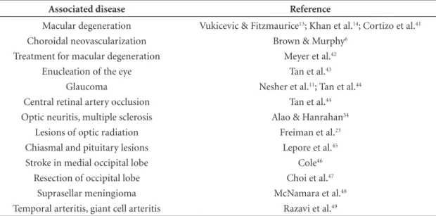
Table 2. Diseases associated to Charles Bonnet syndrome.
Associated disease Reference
Macular degeneration Choroidal neovascularization Treatment for macular degeneration
Enucleation of the eye Glaucoma
Central retinal artery occlusion Optic neuritis, multiple sclerosis
Lesions of optic radiation Chiasmal and pituitary lesions Stroke in medial occipital lobe Resection of occipital lobe
Suprasellar meningioma Temporal arteritis, giant cell arteritis
Vukicevic & Fitzmaurice13; Khan et al.14; Cortizo et al.41
Brown & Murphy6
Meyer et al.42
Tan et al.43
Nesher et al.11; Tan et al.44
Tan et al.44
Alao & Hanrahan34
Freiman et al.23
Lepore et al.45
Cole46
Choi et al.47
McNamara et al.48
74.5±16.9 years, and was in-line with the findings of other reports.5,9,13,19
Complex visual hallucinations have been reported fol-lowing a range of different conditions, and the syndrome stems from a variety of lesions at all levels of the visual sys-tem. The most frequent cause is age-related macular degen-eration and its treatment (Table 2). In our case series, fifty per cent of our patients demonstrated visual impairment secondary to ocular disease and another half to cerebral lesions of visual pathways. Although visual impairment is not mandatory for the diagnosis of CBS, most authors re-port a strong association between the two.17 All cases in the
present series had significant visual disturbance.
Due to the small sample of patients, we cannot draw conclusions regarding gender preponderance in this study. In a review, Menon et al.17 found divergent results
concern-ing gender and CBS among various studies. The frequency of the hallucinations is usually reported as occurring daily or weekly.13,19 Patients may also report continuous
halluci-nations or episodic hallucihalluci-nations with longer intervals.17
In five of our patients, the hallucinations occurred daily. Although previously considered a condition with “pleas-ant or neutral” symptoms,1 later reports have indicated that
CBS can be a cause of emotional burden to patients6,8,10.
Santhouse et al.19 found that in a group of 34 patients, the
hallucinations generated an emotional response in 50%, and in half of these, the experiences were unpleasant. Vukicevic & Fitzmaurice13 reported that the syndrome caused
moder-ate or severe stress in 16 out of 35 patients. In our group, five patients reported a burden associated with the hallucina-tions. Although this burden was referred to as mild or mod-erate in most cases, it highlights the importance of correct diagnosis of CBS (reassurance of the sanity of the patient has positive effects)7 and of the decision on whether to treat
(or at least attempt to treat) the symptoms or otherwise. Ffytche & Howard4 devised hallucination
classifica-tion into eight categories: tessellopsia (overlapping pat-terns, repeated geometry); hyperchromatopsia (vivid and bright colors); prosopometamorphopsia (facial distortions, misshapen and mutilated heads); dendropsia (branching forms); perseveration (persistence of details in another scene); illusory visual spread; polyopia (many equal forms); micro/macropsia. The most common patterns in their pa-tients were tessellopsia in 37% and abnormalities of size in 42% of patients, and of these 58% reported micropsia. Among our patients, we identified descriptions matching some of these categories, such as: tessellopsia (“black dots
drawing a net over his home walls”), hyperchromatopsia (“women dressed in purple clothes”) polyopia (as in “sev-eral identical short men with hats”), micropsia (e.g. “little elephants”) and prosopometamorphopsia (e.g. “distorted faces”). Some patients reported landscapes, animals and/ or objects, which could have been seen sometime in the past by them and thus could represent perseveration or long-term pallinopsia.4
Particular types of hallucinations are related to some areas of the brain, where the experiences of patients with CBS are associated with activity in extra-striate cortex.4,19
Santhouse et al. using fMRI found that, hallucinations in-volving faces were localized in superior temporal sulcus; objects and scenes in the ventral occipito-temporal cortex, for example.19 The notion that these hallucinations could
be a release phenomenon provoked by unusual decreased input was described by Cogan, in 1973.20 By using single
photon emission computed tomography in five patients with CBS secondary to eye disease, Adachi et al. observed that all patients had hyperperfusion in the lateral temporal cortex, striatum and thalamus, and presumed that excessive cortical compensation in these areas could precipitate the syndrome;21 using the same methodology (fMRI), increased
activity in ventral extrastriate visual cortex was observed.19
Burke suggested that hyperactivity in any area will evoke the imagery that is coded by that area.22 The hypothesis
of deafferentation seems a reasonable cause of these hal-lucinations, as they are generated in visual association ar-eas.23 Cone photoreceptor loss, as in macular degeneration,
promotes retino-thalamic-deafferentation, which leads to functional deafferentation of extrastriate cortex.24 Burke
suggested that hallucinations result from deafferentation of visual structures in the brain, or from the effective si-lencing of the principal afferents to these structures.22 The
hypothesis can include two streams of information: one from the periphery to the centre and another in the oppo-site direction. As the flow from the periphery to the centre diminishes, the contrary flow rises.25
Serotonergic activity is related to visual pathways and probably linked to genesis of hallucinations. Serotonin lev-els are lower in the sensory visual deprived cortex.26 Visual
information converging in the geniculate nucleus lateral to the visual cortex is modulated by serotoninergic projections from the brainstem.27 Acetylcholine is another
neurotrans-mitter involved in visual hallucinations, and concentrated in the visual thalamic nuclei and visual cortex.28
halluci-natory episodes last from seconds to days, and duration of CBS may extend to years. In our cases, some therapeutic options were tried, with partial or no success. As positive outcomes are common with the passing of time, partial responses could be due to spontaneous recovery.
Treatments have been used in reports of single cases or case series in the literature. No controlled clinical trials have been published to date. Reports refer to spontaneous regression, and positive results in some cases with phar-macologic treatment. The majority of studies involve the use of antipsychotic or anticonvulsant drugs. Outcomes are variable, probably due to patient heterogeneity. In par-ticular, the therapeutic response may diverge according to anatomical lesion, albeit ocular or in the brain.
• Anticonvulsants-carbamazepine;29 valproate;30
gaba-pentin31
• Haloperidol32
• Atypicalneuroleptics:risperidona,33 olanzapine34
• Selectiveserotoninreuptakeinhibitors:venlafaxine,
citalopram35
• Mirtazapine36
• Anticholinesterasicdrugs:donepezil37
• 5-HT3antagonist–cisapride38
Pilsin et al.39 have raised the question over whether CBS
could be a sign of the initial stages of a dementing illness, having found that patients with CBS performed worse in neuropsychological testing than controls. However, in this study, eight out of fifteen patients had no insight of the illusory nature of their hallucinations, and according to the most frequently accepted definitions,1 patients should
be diagnosed with CBS only if they are conscious of the unrealistic nature of their visions. There are reports of pa-tients initially diagnosed with CBS – with or without mild cognitive impairment – that later developed Alzheimer’s disease.40 The association of CBS and dementia should not
lead to the conclusion that CBS is a risk factor for the de-velopment of dementia, as advanced age is a risk factor for both conditions while this association (CBS and dementia) is only rarely reported. It is indeed necessary that patients manifesting visual hallucinations, even with concomitant visual impairment, should undergo a thorough evaluation to exclude underlying cognitive impairment. Long-term follow-up is also important, so that initial signs of cogni-tive deterioration can be detected, should they appear.
There are a number of limitations of this study. Firstly, information on imaging studies was not available in two cases. It is advisable to rule out the presence of structural
abnormalities that could explain the visual hallucinations; however, in the cases reported, the absence of other focal neurological signs and accompanying symptoms on fol-low-up made the possibility of structural causes less likely. We also possessed no information on the influence of the symptoms of CBS on daily life activities, which could sub-stantiate the impact of the symptoms and thus aid in man-agement decisions (as discussed above). It should be noted however that evaluating the impact on daily life activities in visually impaired patients can be problematic.
Summing up, akin to other reported cases of CBS, our patients’ visions contained vivid and colored pattern, a mixture of images, scenes, abnormal sizes, and the sub-jects showed preserved insight regarding these unrealistic visions. None of the patients responded well to treatment, and among those with some positive response this outcome may not have been due to treatment, but instead to sponta-neous remission or fluctuating course. Despite the paucity of treatment options, awareness and recognition of CBS is of the utmost importance to avoid misdiagnoses with dementing or psychiatric illnesses and to offer patients re-assurance regarding the integrity of their mental status.
References
1. Damas-Mora J, Skelton-Robinson M, Jenner FA. The Charles Bonnet syndrome in perspective. Psychol Med 1982;12: 251-261.
2. DeMorsier G. The Charles Bonnet syndrome: visual hallu-cinations in the aged without mental deficiency. Ann Med Psychol (Paris) 1967;2:678-702.
3. Bonnet C. Essai analytique sur les aculties de l’ame. Copen-hagen: Ferres & Philibert; 1760: 426-429.
4. Ffytche DH, Howard RJ. The perceptual consequences of visual loss: “positive” pathologies of vision. Brain 1999;122:1247-1260. 5. Norton-Willson L, Munir M. Visual perceptual disorders re-sembling the Charles Bonnet syndrome: a study of 434 con-secutive patients referred to a psychogeriatric unit. Fam Pract 1987;4: 27-31.
6. Brown GC, Murphy RP. Visual symptoms associated with choroidal neovascularization: photopsias and the Charles Bonnet syndrome. Arch Ophthalmol 1992;110:1251-1256. 7. Teunisse RJ, Cruysberg JRM, Verbeek A, Zitman FG. The
Charles Bonnet syndrome: a large prospective study in Neth-erlands. Brit J Psychiatry 1995; 166: 254-257.
9. Adachi N. Charles Bonnet syndrome in leprosy: prevalence and clinical characteristics. Acta Psychiatr Scand 1996;93:279-281. 10. O’Reilly R, Chamberlaine C. Charles Bonnet syndrome: inci-dence and demographic and clinical features. Can J Psychiatry 1996;41:259-260.
11. Nesher R, Nesher G, Epstien E, Assia E. Charles Bonnet Syndrome in glaucoma with low vision. J Glaucoma 2001;10:396-400. 12. Shiraishi Y, Terao T, Nakamura J, Tawara A. The rarity of Charles Bonnet Syndrome. J Psychiatr Res 2004;38:207-213. 13. Vukicevic M, Fitzmaurice K. Butterflies and black lacy pat-terns: the prevalence and characteristics of Charles Bonnet hallucinations in an Australian population. Clin Experiment Ophthalmol 2008;36:659-665.
14. Khan JC, Shadid H, Thurlby DA, Yates JR, Moore AT. Charles Bonnet syndrome in age-related macular degeneration: the nature and frequency of images in subjects with end-stage disease. Ophthalmic Epidemiol 2008;15:202-208.
15. Schwartz TL, Vahgei L. Charles Bonnet syndrome in children. J AAPOS 1998;2: 310-313.
16. Brucki SM, Nitrini R, Caramelli P, Bertolucci PH, Okamoto IH. Sugestões para o uso do mini-exame do estado mental no Brasil. Arq Neuropsiquiatr 2003; 61:777-81.
17. Menon GJ, Rahman I, Menon SJ, Dutton GN. Complex visual hallucinations in the visually impaired: the Charles Bonnet syndrome. Surv Ophthalmol 2003;48:58-72.
18. Teunisse RJ, Cruysberg JR, Hoefnagels WH, Kuin Y, Verbeek AL, Zitman FG. Social and psychological characteristics of elderly visually handicapped patients with the Charles Bonnet Syndrome. Compr Psychiatry 1999;40:315-319.
19. Santhouse AM, Howard RJ, ffytche DH. Visual hallucina-tory syndromes and the anatomy of the visual brain. Brain 2000;123:2055-2064.
20. Cogan DG. Visual hallucinations as release phenomena. Arch Klin Exp Ophthalmol 1973;188:139-150.
21. Adachi N, Watanabe T, Matsuda H, Onuma T. Hyperperfusion in the lateral temporal cortex, the striatum and the thalamus during complex visual hallucinations: single photon emission computed tomography findings in patients with Charles Bon-net syndrome. Psychiatry Clin Neurosci 2000;54:157-162. 22. Burke W. The neural basis of Charles Bonnet hallucinations: a
hypothesis. J Neurol Neurosurg Psychiatry 2002;73:535-541. 23. Freiman TM, Surges R, Vougioukas VI et al. Complex visual hallucinations (Charles Bonnet syndrome) in visual field de-fects following cerebral surgery. J Neurosurg 2004;101:846-853. 24. Plummer C, Kleinitz A, Vroomen P, Watts R. Of Roman chari-ots and goats in overcoats: the Syndrome of Charles Bonnet. J Clin Neurosc 2007;14:709-714.
25. Villa FJ. The Charles Bonnet Syndrome. An R Acad Nac Med (Madr) 2008;125: 387-395.
26. Qu Y, Eysel UT, Vandesande F, Arckens L. Effect of partial sensory deprivation on monoaminergic neuromodulators in striate cortex of adult cat. Neuroscience 2000;101:863-868. 27. Seeburg DP, Liu X, Chen C. Frequency-dependent
modula-tion of retinogeniculate transmission by serotonin. J Neurosci 2004;24:10950-10962.
28. Manford M, Andermann F. Complex visual hallucinations. Clin-ical and neurobiologClin-ical insights. Brain 1998;121:1819-1840. 29. Görgens K, Liedtke M. Charles Bonnet syndrome. Psychiatr
Prax 1998; 25: 85-86.
30. Hori H, Terao T, Shiraishi Y. Treatment of Charles Bonnet syndrome with valproate. Int Clin Psychopharmacol 2000; 15:117-119.
31. Paulig M, Mentrup H. Charles Bonnet syndrome: complete remission of complex visual hallucinations treated by gaba-pentin. J Neurol Neurosurg Psychiatry 2001;70: 813-814. 32. Valencia C, Franco JG. Charles Bonnet syndrome:
re-port of one case managed with haloperidol. Rev Med Chil 2008;136:347-350.
33. Maeda K, Shirayama Y, Nukina S, Yoshioka S, Kawahara R. Charles Bonnet syndrome with visual hallucinations of child-hood experience: successful treatment of 1 patient with ris-peridone. J Clin Psychiatry 2003;64:1131-1132.
34. Alao AO; Hanrahan B. Charles Bonnet syndrome: visual hallucination and multiple sclerosis. Int J Psychiatry Med 2003;33:195-199.
35. Lang UE, Stogowski D, Schulze D et al. Charles Bonnet Syn-drome: successful treatment of visual hallucinations due to vision loss with selective serotonin reuptake inhibitors. J Psy-chopharmacol 2007;21:553-555.
36. Siddiqui E, Ramaswamay S, Petty F. Mirtazapine for Charles Bonnet syndrome. Can J Psychiatry 2004;49:787-788. 37. Ukai S, Yamamoto M, Tanaka M, Takeda M. Treatment of
typical Charles Bonnet syndrome with donepezil. Int Clin Psychopharmacol 2004;19:355-357.
38. Ranen NG, Pasternak RE, Rovner BW. Cisapride in the treatment of visual hallucinations caused by vision loss: the Charles Bonnet syndrome. Am J Geriatr Psychiatry 1999;7: 264-265.
39. Pliskin NH, Kiolbasa TA, Towle VL et al. Charles Bonnet Syndrome: an early marker for dementia ? J Am Geriatr Soc 1996;44:1055-1061.
41. Cortizo V, Rosa AAM, Soriano DS, Takada LT, Nitrini R. Charles Bonnet syndrome: visual hallucinations in patients with ocu-lar diseases- case report. Arq Bras Oftalmol 2005;68: 129-132. 42. Meyer CH, Mennel S, Hörle S, Schmidt JC. Visual halluci-nations after intravitreal injection of bevacizumab in vas-cular age-related mavas-cular degeneration. Am J Ophthalmol 2007;143:169-170.
43. Tan CS, Sabel BA, Eong KG. Charles Bonnet syndrome (vi-sual hallucinations) following enucleation. Eye 2006;20: 1394-1395.
44. Tan CS, Sabel BA, Goh KY. Visual hallucinations during visual recovery after central retinal artery occlusion. Arch Neurol 2006;63:598-600.
45. Lepore FE. Spontaneous visual phenomena with visual loss:
104 patients with lesions of retinal and neural afferent path-ways. Neurology 1990;40:444-447.
46. Cole M. When the left brain is not right the right brain may be left: report of personal experience of occipital hemianopia. J Neurol Neurosurg Psychiatry 1999; 67:169-173.
47. Choi EJ, Lee JK, Kang JK, Lee SA. Complex visual hallucina-tions after occipital cortical resection in a patient with epilep-sy due to cortical dysplasia. Arch Neurol 2005;62:481-484. 48. McNamara ME, Heros RC, Boller F. Visual hallucinations
in blindness: the Charles Bonnet syndrome. Int J neurosci 1982;17:13-15.