312
C
ASER
EPORTAdie-Holmes Syndrome
Síndrome de Adie-Holmes
Renato Antunes Schiave Germano
1, Barbara Leite de Souza
2, Flavio Augusto Schiave Germano
3, Rogerio Masahiro
Kawai
4, Caroline Schiave Germano
5, Milena Naomi Chibana
1, Jorge Estefano Germano
4Received for publication 19/06/2014 - Accepted for publication 04/08/2014 The authors declare no conflict of interest
1 Ophthalmology Department, Hospital das Clínicas, Faculdade de Medicina, Universidade de São Paulo, SP, Brazil. 2 Ophthalmology Residency Program, Centro de Excelência em Oftalmologia, Bauru (SP), Brazil.
3 Academic Medical Course, Universidade Nove de Julho, São Paulo, SP, Brazil. 4 Physician, Centro de Excelência em Oftalmologia, Bauru (SP), Brazil
5 Academic Medical Course, Faculdade de Ciências Médicas, Santa Casa de Misericórdia, São Paulo, SP, Brazil.
R
ESUMOA Síndrome de Holmes-Adie É caracterizada pela presença de pupila tônica associada à diminuição ou ausência dos reflexos tendíneos profundos. Em alguns casos pode haver disfunção nervosa autônoma. O mecanismo que causa a desordem não é totalmen-te conhecido, mas acredita-se que seja causada pela desnervação do suprimento pós-ganglionar para o esfínctotalmen-ter da pupila e para o músculo ciliar, que pode ocorrer após doença viral. Tipicamente afeta adultos jovens e é unilateral em 80% dos casos, embora possa se desenvolver no olho contralateral em meses ou anos. Nós relatamos o caso de uma mulher apresentando sinais típicos desta síndrome, em que o teste farmacológico foi fundamental para o diagnóstico.
Descritores: Síndrome de Adie/diagnóstico; Pupila tônica; Exame Físico; Relatos de casos
A
BSTRACTThe Holmes-Adie syndrome is characterized by the presence of tonic pupil associated with absence or diminution of deep tendon reflexes. In some cases there may be autonomous nerve dysfunction. The mechanism that causes the disorder is not fully known, but is believed to be caused by denervation of the postganglionic supply to the sphincter of the pupil and the ciliarymuscle which can occur following viral disease. Typically it affects young adults and is unilateral in 80% of cases, although it may develop in the contralateral eye in months or years. We report a case of a woman presenting typical signs of this syndrome, in which pharmacological test was essential for diagnosis.
Keywords: Adie syndrome/diagnosis; Pupil disordes/diagnosis; Physical examination; Case reports
Rev Bras Oftalmol. 2015; 74 (5): 312-4
313
Rev Bras Oftalmol. 2015; 74 (5): 312-4
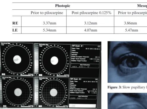
Table 1
Pupillary diameter measurement in both photopic and mesopic conditions
I
NTRODUCTIONT
he Holmes-Adie syndrome was described simultaneously in 1931 by Gordon Morgan Holmes and William John Adie. It is characterized by the presence of tonic pupil associated with absence or diminution of deep tendon reflexes. In some cases there may be autonomous nerve dysfunction.1The mechanism that causes the disorder is not fully known, but is believed to be caused by denervation of the postganglionic supply to the sphincter of the pupil and the ciliarymuscle which can occur following viral disease. Typically it affects young adults and is unilateral in 80% of cases, although it may develop in the contralateral eye in months or years.2, 3
C
ASER
EPORTFemale, 43 years old, white, searched an ophthalmologist complaining her left eye pupil was “bigger” than the right eye one for over a month. No other ophthalmological complaints. She reported associated vertigo. She searched for neurological aid and a MRI of
Adie-Holmes Syndrome
the brain was performed without changes. She went to the otorhinolaryngologist, where she was diagnosed with Meniere’s Syndrome, and she began treatment with Labirin® (betahistine di
hydrochloride). She abandoned treatment after developing headache as a side effect of the drug. Personal background reports thalassemia minor, flu shot one year ago and infection treatment of H. pylori for 6 months. She is a folic acid user.
Ophthalmologic exam showed visual acuity of 20/15 in the right eye (RE) and 20/20 in the left eye (LE) without correction. Biomicroscopy shows significant anisocoria, left greater than right. Pupillary diameter measured by iTrace Ray Tracing Corneal Topography and WavefrontAberrometer (Tracey Technologies) is shown in Table 1 and Figure 1.
Fundus biomicroscopy was unchanged. Direct pupillary reflex present in the right eye and slow in the left eye. Slow consensual reflex in both eyes, with no relative afferent defect in either eye.(Figures 2 and 3).
The pupillary near reflex was slow in the left eye, with slow dilation. (Figure 4)
The patient also had absent tendinous reflex in the right and normal in the left.
Figure 1: iTrace in physiological mesopic conditions
Figure 2: Normal pupillary light reflex of the right eye
Figure 3: Slow pupillary light reflex of the left eye
D
ISCUSSIONThe tonic pupil is usually noticed by others and not by the patient himself. The difference in pupil size (anisocoria) can be found in a variety of ophthalmic disorders, which may
Figure 4: Pupillary near reflex
Photopic Mesopic
Prior to pilocarpine Post pilocarpine 0.125% Prior to pilocarpine Post pilocarpine 0.125%
RE 3.37mm 3.12mm 3.86mm 3.30mm
314
Corresponding author:
Renato Antunes Schiave Germano
Rua Galeno de Almeida, 107 - apto.: 182 A , São Paulo - SP.
Rev Bras Oftalmol. 2015; 74 (5): 312-4
Germano RAS, de SouzaBL, GermanoFAS, KawaiRM, Germano CS, ChibanaMN, Germano JE
be benign or not, such as physiological anisocoria, Horner’s syndrome, Adie’s pupil, pharmacological anisocoria, pupillary supranuclear disorders, problems of the iris, systemic diseases or paralysis of the third cranial nerve.4
In our case, after clinical and complementary orbit USG and cranial MRI, which was normal, was done testing the instillation of pilocarpine 0.125%5, which was positive for the left eye. We
believe that drug tests, such as that performed above, assist in laboratory diagnosis of tonic pupil syndrome.
R
EFERENCES1. Martinelli P. Holmes–Adie syndrome. Lancet. 2000; 356(9243):1760–1. 2. Ulrich J. Morphological basis of Adie’s syndrome. Eur Neurol. 1980;
19(6):390–5.
3. Mak W, Cheung RT.The Holmes-Adie plus syndrome. J ClinNeurosci. 2000; 7(5):452
4. Wilhelm H. Neuro-ophthalmology of pupillary function—practical guidelines. J Neurol. 1998;245(9):573-83.
