THE CLINICAL PROFILE OF CHILDHOOD
OPTIC NEURITIS
Marco Aurélio Lana-Peixoto
1, Gustavo Cardoso de Andrade
2For the Brazilian Committee for Treatment and Research in Multiple Sclerosis (BCTRIMS)
ABSTRACT - Purpose: To report the clinical features and outcome of a series of children with optic neuritis. Methods: We reviewed the medical records of patients up to 16 years old with optic neuritis. Group 1 comprised children seen up to two weeks after the onset of visual loss; Group 2 comprised patients already harboring optic atrophy. Results: There were 15 boys and 12 girls. The mean age was 10.9 years. Bilateral optic neuritis occurred in 10. Optic disc pallor was found in 35%, edema in 46%, and 19% had normal fundus. During follow-up visual acuity improved in all but one eye in Group 1, and in six of seven eyes in children in Group 2. Just one child converted to multiple sclerosis. Conclusions: This study shows that the clinical features of childhood optic neuritis differ from those observed in adults. In children it has a better visual outcome and a lower conversion rate to multiple sclerosis than in adults.
KEY WORDS: childhood optic neuritis, visual outcome, multiple sclerosis.
O quadro clínico da neurite óptica na infância
RESUMO - Objetivos: Descrever as características clínicas e evolutivas da neurite óptica na infância. Métodos: Foram revistos os prontuários de crianças até 16 anos de idade neurite óptica. Os pacientes foram divididos em dois grupos. Grupo 1 referia a crinças examinadas até duas semanas após o início da perda visual; Grupo 2 foi formado por crianças já apresentando atrofia óptica ao primeiro exame. Resultados: Foram encontrados 15 meninos e 12 meninas, com idade média de 10,9 anos. Neurite óptica bilateral foi observada em 10 crianças. O disco óptico estava pálido em 35% dos casos, edemaciado em 46% e normal em 19%. Exame durante follow-up demonstrou melhora da acuidade visual em todos, exceto em um caso no Grupo 1 e em 6 de 7 olhos no Grupo 2. Apenas uma criança desenvolveu esclerose múltipla. Conclusões: Este estudo demonstrou que as características clínicas da neurite óptica na infância diferem das observadas em adultos. Em crianças, a recuperação visual é melhor, e a taxa de conversão para a esclerose múltipla é mais baixa.
PALAVRAS-CHAVE:neurite óptica na infância, recuperação visual, esclerose múltipla.
1Associate Professor, Department of Neuro-Ophthalmology, Hospital São Geraldo, Federal University of Minas Gerais Medical School,
Belo Horizonte MG, Brazil. 2Fellow, Department of Neuro-Ophthalmology, Hospital São Geraldo, Federal University of Minas Gerais
Medical School, Belo Horizonte, Brazil.
Received 13 December 2000, received in final form 9 March 2001. Accepted 20 March 2001.
Dr. Marco Aurélio Lana-Peixoto - Rua Padre Rolim 769 / 130 andar - 30130-090 Belo Horizonte MG – Brasil. Tel/Fax: 55 31 3222 3935.
E-mail: lanapma@zaz.com.br
Childhood optic neuritis is a rare condition that differs from adult-onset optic neuritis in some clini-cal and evolutive aspects. It is widely accepted that in children, attacks of optic neuritis usually occur following a febrile illness, tend to affect both eyes, are frequently associated with swollen discs, improve rapidly, and have a low conversion rate to multiple sclerosis (MS)1-7. On the other hand, optic neuritis in adults is usually unilateral, predominantly affects the retrobulbar portion of the optic nerve, and presents a high conversion rate to MS8, 9.
Although these facts have been accepted on a worldwide basis few series of children with optic neuritis have been studied. In addition to that it is well known that genetic and environmental factors
play an important role in the prevalence and clinical expression of demyelinating diseases. Our previous studies have demonstrated that in Brazil MS has some clinical features different from those most com-monly seen in other Western countries10, 11. Further-more, isolated demyelinating optic neuritis in Brazil converts to MS in a much lower rate as compared to those reported in North America and Europe11. It is, therefore possible that in our country idiopathic optic neuritis in children may harbor different character-istics from those reported in other geographical ar-eas or population groups.
METHOD
The medical records of all patients with ages up to 16 years and the diagnosis of optic neuritis who were exam-ined by one of us in a 16-year period were reviewed. The inclusion criteria for the diagnosis of idiopathic optic neu-ritis were: (1) history of acute loss of central vision; (2) no evidence of neurologic, metabolic, toxic, vascular, heredi-tary, or compressive etiology; (3) no evidence of retinal exudates or other retinal lesions; (4) at least two of the following: a relative afferent pupillary defect, a color vi-sion deficit, a nerve fiber bundle defect on field examina-tion, and a swollen or pale optic disc. Cases of neuromy-elitis optica or MS in which optic neuritis was not the ini-tial symptom were also discarded.
The selected cases fulfilling the above criteria for the diagnosis of optic neuritis were then divided into two groups according to the interval between the onset of the visual loss and the neuro-ophthalmic examination at our Department. Group 1 comprised children seen up to two weeks after the onset of visual loss; Group 2 comprised patients first examined by us two weeks or later following visual loss and already harboring optic disc pallor.
Patients were further classified as having unilateral or bilateral optic neuritis. Bilateral simultaneous optic neuri-tis was defined as involvement of both eyes either at the same time or within a period of two weeks, after which it was considered as sequential bilateral optic neuritis. Con-version to MS was defined as the occurrence of other signs of central nervous deficit according to Poser’s Committee criteria12.
The charts were then analyzed in relation to children’s age at onset of the visual loss, sex, history of a prodrome, occurrence of eye pain, laterality, best corrected visual acuity (VA) at the first examination, color vision assessed by Ishihara’s pseudoisochromatic plates, fundoscopic ex-amination and visual field exex-amination by Goldmann’s or automated perimetry.
Long-term follow-up noted recurrence of visual loss, final visual acuity, color plates, ophthalmoscopic appear-ance of the optic nerves, interval medical history and neu-rological examination to detect any symptom or sign of central nervous system involvement which could herald the development of MS. Cerebrospinal fluid (CSF) exami-nation, whenever available was analyzed in relation to number and types of cells, glucose and protein concen-tration, and protein electrophoresis. As the presence of oligoclonal bands in the CSF was not searched by isoelec-tric focusing electrophoresis it was not considered in the present study. Most patients did not have a brain MRI as this test was not available to them at the time of onset of the optic neuritis. When it was performed the presence of T2-weighted hyperintense areas in the optic nerves or brain white matter was noted.
RESULTS
Twenty-seven patients (Table 1) fulfilled the set of criteria as described above for the diagnosis of
childhood optic neuritis. There were 15 boys and 12 girls with ages ranging from 3 to 16 years (mean 10.9 years). Three patients were under six; eight were between six and 10, and 16 were 11 to 16.
There were 37 involved eyes as 10 children (37%) had bilateral optic neuritis (eight bilateral simulta-neous, and two bilateral sequential) and 17 (63%) had unilateral attacks (8 in the right eye and 9 in the left eye). Recurrent optic neuritis occurred in one child.
In 10 patients (37%) an infection had occurred within two weeks before the onset of optic neuritis. In nine of these patients there was a febrile condi-tion secondary to an upper respiratory infeccondi-tion, whereas in one patient the onset of optic neuritis was associated with immunization as part of treat-ment for recurring furunculosis due to Staphylococ-cus aureus.
A positive history of eye pain could be collected from the medical records of 10 children (37%), pre-ceding by a few days or in association with the on-set of visual loss.
Neuro-ophthalmic examination was performed within a two-week period following the onset of the visual loss in 19 patients, whereas in the remaining eight children it was done after an interval up to nine years after visual failure had been noticed. There were 24 affected eyes in Group 1 and 13 in Group 2. In Group 1 VA was normal in one eye; 20/50 or bet-ter in six (25%); 20/60 to 20/200 in seven (29%); and 20/400 or worse in 11 eyes (46%). In Group 2 VA was 20/50 or better in two eyes (15%); 20/60 to 20/200 in seven (54%); and 20/400 or worse in four (31%).
All children with ages or VA suitable for color vi-sion assessment showed some degree of color crimination deficit. Fundoscopic examination dis-closed optic disc edema in 17 eyes (46%) and pallor in 13 (35%). One child had a pseudo Foster-Kennedy syndrome. In six patients - seven eyes (19%) - the fundus appearance was normal suggesting retrob-ulbar optic neuritis. Two of these patients had bilat-eral simultaneous optic neuritis; one with a bilatbilat-eral simultaneous retrobulbar optic neuritis, and the other with optic disc edema in one eye and a ret-robulbar involvement in the fellow eye.
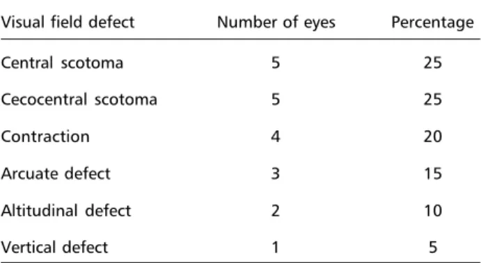
Visual fields by perimetry were recorded in 20 affected eyes. In all of them a defect could be found (Table 2). In 50% of these eyes a central or cecocentral scotoma was detected.
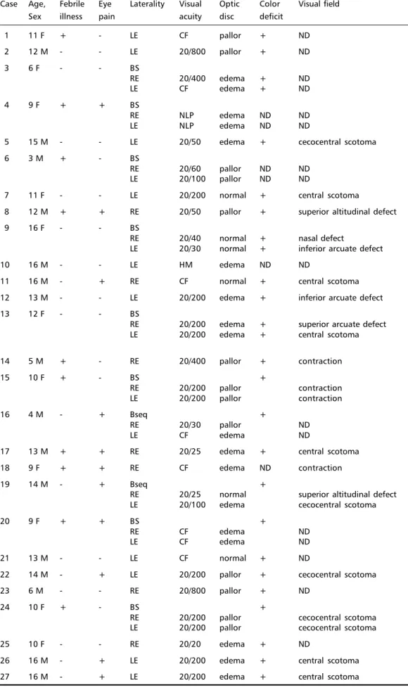
Table 1. Children with optic neuritis.
Case Age, Febrile Eye Laterality Visual Optic Color Visual field Sex illness pain acuity disc deficit
1 11 F + - LE CF pallor + ND
2 12 M - - LE 20/800 pallor + ND
3 6 F - - BS
RE 20/400 edema + ND
LE CF edema + ND
4 9 F + + BS
RE NLP edema ND ND
LE NLP edema ND ND
5 15 M - - LE 20/50 edema + cecocentral scotoma
6 3 M + - BS
RE 20/60 pallor ND ND
LE 20/100 pallor ND ND
7 11 F - - LE 20/200 normal + central scotoma
8 12 M + + RE 20/50 pallor + superior altitudinal defect
9 16 F - - BS
RE 20/40 normal + nasal defect
LE 20/30 normal + inferior arcuate defect
10 16 M - - LE HM edema ND ND
11 16 M - + RE CF normal + central scotoma
12 13 M - - LE 20/200 edema + inferior arcuate defect
13 12 F - - BS
RE 20/200 edema + superior arcuate defect LE 20/200 edema + central scotoma
14 5 M + - RE 20/400 pallor + contraction
15 10 F + - BS +
RE 20/200 pallor contraction
LE 20/200 pallor contraction
16 4 M - + Bseq +
RE 20/30 pallor ND
LE CF edema ND
17 13 M + + RE 20/25 edema + central scotoma
18 9 F + + RE CF edema ND contraction
19 14 M - + Bseq +
RE 20/25 normal superior altitudinal defect LE 20/100 edema cecocentral scotoma
20 9 F + + BS +
RE CF edema ND
LE CF edema ND
21 13 M - - LE CF normal + ND
22 14 M - + LE 20/200 pallor + cecocentral scotoma
23 6 M - - RE 20/800 pallor + ND
24 10 F + - BS +
RE 20/200 pallor cecocentral scotoma LE 20/200 pallor cecocentral scotoma
25 10 F - - RE 20/20 edema + ND
26 16 M - + LE 20/200 edema + central scotoma
27 16 M - + LE 20/200 edema + central scotoma
In five children protein electrophoresis disclosed in-creased content of γ-globulin.
Brain MRI was performed in six cases, showing normal results in three, and signs of sinusitis in one. In one child there was a T2-weighted hyperintense lesion in the involved optic nerve, whereas in an-other there were a few T2-weighted hyperintense lesions scattered in the brain white matter.
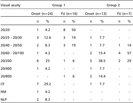
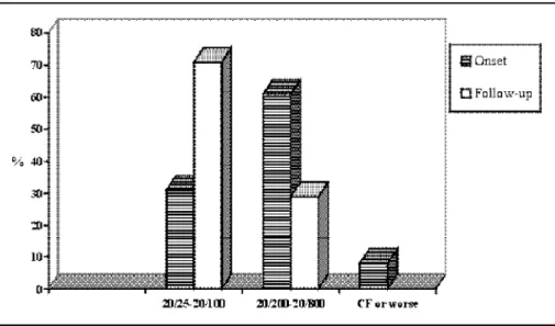
Seventeen children - 12 in Group 1 and five in Group 2 - could be evaluated in follow-up of vari-able extension. The follow-up period ranged from 1 to 41 months (median 13 months). Visual acuity improved in all but one eye in Group 1, and in six out of seven eyes in Group 2 examined at follow-up (Table 3). In Group 1 50% of patients recovered nor-mal VA whereas 88% had VA 20/50 or better during follow-up. The VA was 20/200 or worse in 6% at follow-up as compared to 25% at the initial exami-nation, and there was no patient with VA worse than 20/800 at follow-up as compared to 42% at the on-set. The only patient in this Group who did not get better had retrobulbar neuritis and VA of 20/200 and was followed for 30 months. Another patient with acute anterior optic neuritis and sinusitis had VA of CF (counting fingers) at the onset which turned out to 20/800 after six months. All other Group 1 pa-tients got improvement greater than two lines at the Snellen chart. Four eyes in Group 1 patients got one line improvement in their visual acuity; two had two lines improvement and one eye had no VA change during follow-up. In Group 2 no change in the percentage of patients with VA 20/50 or better was observed, whereas 85% of patients examined at follow-up had VA between 20/60 to 20/200, as compared to 54% at first examination. No patient in this Group was found with VA worse than 20/200 at follow-up as compared to 29% with this VA found at the initial examination. Figures 1 and 2 show the VA at the initial evaluation and follow-up in Group 1 and Group 2 patients.
Just one child in this series converted to MS. He was a 16-year-old boy who developed ataxia and decreased sensation in the right upper limb 18 months after the onset of acute retrobulbar optic neuritis. CSF examination had been normal but MRI had shown an abnormal higher signal intensity area on the involved optic nerve. Repeated MRI then sho-wed a number of abnormal T2-weighted hyperin-tense areas in the brain white matter.
DISCUSSION
To the best of our knowledge this study is the first series of childhood optic neuritis published in the Brazilian literature. We have used a restrictive set of criteria for the diagnosis of optic neuritis in this series. We did not include, therefore, patients in whom papillitis was associated with macular edema and retinal exudates as this condition, commonly de-nominated neuroretinitis, has a variety of etiologies, a different pathogenesis other than demyelination, and is not associated with the development of MS. We have also discarded cases with any clinical or laboratory evidence of any neurologic infectious dis-ease or autoimmune condition which could be di-rectly related to the development of the optic neuri-tis. Finally, in order to analyze the outcome of isolated optic neuritis, patients with previous diagnosis of MS or with neuromyelitis optica were also discarded.
This series confirms that isolated optic neuritis in children has some peculiar findings which may dis-tinguish it from the more commonly seen adult-on-set condition, although our data differ in some as-pects from those reported in most series in the lit-erature1-5. To start with optic neuritis was slightly mo-re common in boys than in girls in the pmo-resent series, whereas in all other studies there was predominance of females1-6. This difference remains to be explained.
The mean age at onset of the disease was 10.9 years in our study, 9.3 years in the series of Leern-snyder et al.’s4 , 8.6 years in the Kriss et al. series5 and 11 years in the series reported by Lucchinetti et al.6 .
Attacks of optic neuritis in children are common after a febrile illness. A positive history of a febrile condition, usually an upper respiratory infection can be elicited in more than one third of the patients (37% in our series; 46% in Kriss et al.’s series5 and 36% in the cohort reported by Lucchinetti et al.6). The lower association with a febrile illness in our series and in that reported by Lucchinetti et al. may be the result of the more restrictive diagnostic crite-ria used in our study but it is still higher than the
Table 2. Visual field examination in childhood optic neuritis.
Visual field defect Number of eyes Percentage
Central scotoma 5 25
Cecocentral scotoma 5 25
Contraction 4 20
Arcuate defect 3 15
Altitudinal defect 2 10
frequency of a preceding viral syndrome found in adult optic neuritis13.
A history of pain associated with the visual loss was found in only 10 of our patients (37%). Lucchi-netti et al.6 observed the same frequency in their cohort and it dramatically contrasts with the 92% frequency of pain found in adult optic neuritis13. This striking difference may be due to the difficulty in getting information from children. This issue had not been addressed by other authors.
The high frequency of bilateral simultaneous in-volvement is another differential characteristics of
childhood optic neuritis. Bilateral optic neuritis in adults is very rare14. Ten children (37%) in the present study had bilateral optic neuritis (eight bilateral si-multaneous and two bilateral sequential). The fre-quency of bilateral involvement in our cohort is lower than those reported by other authors. Bilateral si-multaneous optic neuritis was reported by Leern-snyder et al.4 in 71%, by Kriss et al5 in 64%, and by Lucchinetti et al.6 in 42% of their cases. The higher frequency of bilateral simultaneous optic neuritis in some studies may suggest a stronger implication of an infection agent in those series.
Table 3. Evolution of visual acuity in childhood optic neuritis.
Visual acuity Group 1 Group 2
Onset (n=24) FU (n=16) Onset (n=13) FU (n=7)
n % n % n % n %
20/20 1 4.2 8 50 - - -
-20/25 - 20/30 3 12.6 3 19 1 7.7 -
-20/40 - 20/50 2 8.3 3 19 1 7.7 1 14
20/60 - 20/100 1 4.2 - - 2 15.4 4 57
20/200 6 25 1 6 5 38.5 2 29
20/400 1 4.2 - - 1 7.7 -
-20/800 - - 1 6 2 14.4 -
-CF 7 29.2 - - 1 7.7 -
-HM 1 4.2 - - -
-NLP 2 8.3 - - -
-n: number; FU: follow-up
In order to assess the visual outcome of optic neu-ritis in children we divided our cases into two groups according to the interval between the onset of the visual loss and the time they were first examined by us. In children seen in the acute phase of their visual loss (Group 1), VA was more severely affected than in those evaluated after development of optic atro-phy (Group 2), suggesting that there had been some degree of recovery in the latter group. A striking re-covery of the VA was observed in group 1 patients. Marked recovery of vision following childhood op-tic neuritis had already been observed by other au-thors 1-6, suggesting that children’s optic nerve has a high capacity for remyelination. Still more impor-tant is our observation that there was visual recov-ery in almost all patients whose optic discs were al-ready pale at the time we first examined them. In some of these patients optic neuritis might have occurred months or even years earlier. The same find-ing was reported by Lucchinetti et al.6. These find-ings favor the hypothesis that children retain a ca-pability for remyelination of their optic nerves for a long time.
Optic disc edema is found more commonly in children than in adults with optic neuritis. We found swollen discs in 46% of our cohort, whereas Kennedy and Carrol reported 70%, Leersnyder et al.4 85%, Kriss et al.5 74% and Lucchinetti et al.6 47%. The lower percentage of children with optic disc edema in ours and in the cases reported by Lucchinetti et al.6, as compared with frequencies observed by other in-vestigators, may be due to inclusion in both of our series of patients with optic disc atrophy. If these cases had been excluded in our series the frequency of optic disc edema would rise to 71%, a figure
simi-lar to those found by most authors. By contrast, optic disc edema has been observed in only 35% of cases of adult-onset optic neuritis13.
Visual field testing in our series demonstrated a dense central or cecocentral scotoma in 50%, whe-reas there was some impairment of color vision in all children. Kriss et al5 found central scotoma and color vision deficits in 98% of their cases.
CSF examination in childhood optic neuritis may show slight pleocytosis and elevated protein con-tent, but more frequently is normal2,4,5. In our pa-tients CSF examination was unrevealing except for increased content of γ-globulin, suggesting an in-flammatory reaction in the CNS. As there is no marker for the diagnosis of demyelinating optic neuritis a major importance of CSF examination is to rule out other neurological disorders which may cause visual disturbances and optic disc changes simulating id-iopathic optic neuritis.
Cranial MRI using advanced techniques demon-strates signal abnormalities in 84% of the symptom-atic and 20% of the asymptomsymptom-atic optic nerve with optic neuritis15. Abnormalities in the cerebral white matter in these patients can be detected in 56 to 72% of cases16.
Conversion to MS is a dreadful perspective in pati-ents with isolated demyelinating optic neuritis. In adult-onset optic neuritis the rate of conversion var-ies in different geographical regions reaching rates in New England area as high as 74% of women and 34% of men within 10 years after the initial attack9. In Brazil we found a 10.7% rate after a mean of 4.8 years of follow-up, a figure similar to those described in Japan and other Eastern countries,
ing that environmental and genetic aspects play a role not only in the clinical expression of the disea-se, but also in the rate of its development from iso-lated idiopathic optic neuritis10. In the present series just one child (4%) developed MS during follow-up, a rate lower than one-half the one we found in our previous adult series10. The conversion rate we ob-served in children is close to that reported by Parkin et al7 who found that only one of his 19 patients developed further evidence of MS after a mean fol-low- up of 26 years. Kriss et al5 found a conversion rate of 15% (6/33) in their series, whereas Kennedy and Carter1 reported an overall frequency of MS of 16.5% (3/18).
Lucchinetti et al.6 studied the risk factors for de-veloping MS in a cohort of 79 children with optic neuritis during a mean follow-up of 19.4 years. Life table analysis showed that 10% of the children pro-gressed to MS by 10 years of follow-up, 19% by 20 years and 26% by 40 years. Bilateral sequential or recurrent optic neuritis increased the risk of devel-oping MS; gender, age, fundoscopic findings, visual acuity or family history did not predict the develop-ment of multiple sclerosis, whereas presence of in-fection within two weeks before the onset of optic neuritis decreased the risk. Our findings are in agree-ment with these observations confirming that optic neuritis in children is a more benign condition and has a lower rate conversion to MS.
Our study also demonstrates a number of clini-cal peculiarities of optic neuritis in children distin-guishing it from that seen in adults. Aside from the predominance of boys and a still lower conversion rate to multiple sclerosis in our cohort, we were un-able to find other particular distinguishing features of optic neuritis in Brazilian children as compared with other series in the literature.
Although the clinical picture of childhood optic neuritis can be well delineated the nature of the in-flammatory process in children’s optic nerve, as well as the reasons for its better visual and neurological outcome remain unknown. It is possible that idio-pathic optic neuritis in children is a different pro-cess than that occurring in adults. Further studies are necessary to clarify these issues.
REFERENCES
1. Kennedy C, Carter S. Relation of optic neuritis to multiple sclerosis in children. Pediatrics 1961;28:377-387.
2. Taylor D, Cuendet F. Optic neuritis in childhood. In Hess RF, Plant GT (eds).. Optic Neuritis. Cambridge: Cambridge Press, 1986;73-85. 3. Meadows SP. The Downe Memorial Lecture: Retrobulbar and optic
neuritis in childhood and adolescence. Trans Ophthalmol Soc UK 1969;89:603-638.
4. Leersnyder H, Bursztyn J, Ponsot G et al. Névrites optiques de l’énfant. Aspects cliniques et évolutifs. A propos de 14 observations. Arch Fr Pediatr 1981;38:563-568.
5. Kriss A, Francis DA, Cuendet F et al. Recovery after optic neuritis in chidhood. J Neurol Neurosurg Psychiatry 1988;51:1253-1258. 6. Lucchinetti CF, Kiers L, O’Duffy A, et al. Risk factors for developing
mul-tiple sclerosis after childhood optic neuritis. Neurology 1997;49:413-418. 7. Parkin PJ, Hierons R, McDonald WI. Bilateral optic neuritis. A long
term follow-up. Brain 1985;197:951-964.
8. McDonald WI. Doyne Lecture: The significance of optic neuritis. Trans Ophthalmol Soc UK 1983;103:230-246.
9. Rizzo JF, Lessell S. Risk of developing multiple sclerosis after uncom-plicated optic neuritis. Neurology 1988; 38:185-190.
10. Lana-Peixoto MA, Lana-Peixoto MI. The risk of multiple sclerosis de-veloping in patients with isolated idiopathic optic neuritis in Brazil. Arq Neuropsiquiatr 1991;49:377-383.
11. Lana-Peixoto MA, Lana-Peixoto MI. Is multiple sclerosis in Brazil and Asia alike? Arq Neuropsiquiatr 1992;50:419-425.
12. Poser CM, Paty DW, Scheinberg L et al. New diagnostic criteria for multiple sclerosis: guidelines for research protocols. Ann Neurol 1983;13:227-231.
13. Optic Neuritis Study Group. The clinical profile of optic neuritis: optic neuritis treatment trial. Arch Ophthalmol 1991;109:1673-1678. 14. Perkin GD, Rose CF. Optic Neuritis and its differential diagnosis.
Ox-ford: Oxford University Press, 1979; 217-220.
15. Miller DH, Newton MR, van der Poel JC et al. Magnetic resonance imaging of the optic nerve in optic neuritis. Neurology 1988, 38:175-179.