LETTER TO THE EDITOR
IDepartment of Internal Medicine, Radiology Division, School of Medicine of
Ribeirão Preto, University of Sao Paulo - Ribeirão Preto/SP, Brazil.
IIDepartment of Gynecology and Obstetrics, School of Medicine of São José
do Rio Preto (FAMERP) - São José do Rio Preto/SP, Brazil.
IIIVictorio Valeri Institute of Medical Diagnosis - Ribeirao Preto/SP, Brazil. IVDepartment of Surgery and Anatomy, School of Medicine of Ribeirão Preto,
University of Sao Paulo - Ribeirão Preto/SP, Brazil. Tel.: 55 16 3602.2643
Email: [email protected]
MR IMAGING FEATURES OF PERITONEAL
ADENOMATOID MESOTHELIOMA: A CASE REPORT
doi: 10.1590/S1807-59322009000300020
Cynthia Maria Coelho Lins,I Jorge Elias Jr,I Adilson Ferreira Cunha,II Valdair Francisco Muglia,I Carlos Ribeiro Monteiro,I
Fábio V. Valeri,III Omar FeresIV
Adenomatoid mesothelioma of the peritoneum (AMP) is a rare benign tumor originating from mesothelial cells.1 Most
frequently, AMP occurs between 26 and 55 years of age, at a mean age of 41 years.1 In contrast to diffuse malignant
mesothelioma, which has been linked to asbestos exposure, the etiology of AMP has not been established.2 Only a
minority of patients have symptoms related to the tumor. AMP may present local recurrence, but it has no potential for malignant transformation.3 Although there are many case
reports of abdominal mesotheliomas, to date, there have been no reports of MR imaging features of AMP. In this article, we present the MR imaging features of a case of AMP with histopathological correlation.
CASE REPORT
A 25-year-old woman presented with pelvic pain. The patient had had a cesarean section 3 years before her visit and an appendectomy about 6 months earlier than her onset. Routine ultrasound exam showed an expansive pelvic lesion suggesting an adnexal origin, most likely an ovarian neoplasm, and this finding was confirmed by two other ultrasound exams, each one performed at a different facility. A pelvic-abdominal MR exam was requested for lesion characterization. Laboratory tests were within normal values, except for CA125 of 37.9 µ/ml (normal range 0.0 to 35.0 µ/ml).
Imaging Findings
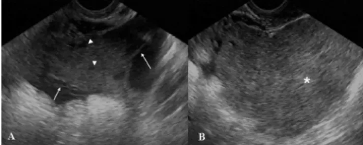
Transvaginal pelvic sonography showed a retrouterine
adnexal mass extending towards the left parauterine region with a complex echotexture containing a homogeneous solid component about 5.0 cm in size and small cystic areas intermingled with linear septa. The mass measured 10.8 x 6.1 x 10.5 cm (volume of 359.7 cm3). Color Doppler imaging
demonstrated vascularization of the solid area and of some septa in the cystic region, with a resistive index ranging from 0.60 to 0.70. Ovaries were identified in neither ultrasound exam. There was no sign of ascites (Figure 1).
intravenous injection of contrast agent was performed to search for calcification and showed a low homogeneous coefficient of attenuation for the lesion, ranging from – 2 to +10 U.H, with no evidence of calcification (Figure 3).
Patient outcome
The patient was submitted to open abdominal surgical resection of the lesion, which had adherences to the rectal
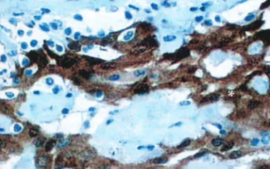
anterior wall but no invasion of the uterus or adnexa. Macroscopic anatomopathological examination revealed various irregular fragments of brownish, friable tissue that measured 15.0 x 12.0 x 4.0 cm and weighed 170.0 g. Microscopic analysis revealed a well-differentiated mesothelial neoplasia, which is detailed in Table 1. Immunohistochemistry evaluation was carried out in the histological sections with an immuno-peroxidase reaction via the avidin-biotin peroxidase method, with the primary antibodies calretinin + CEA- and BerEp4- (Figure 4).
A follow-up MR exam revealed an expansive, predominantly cystic lesion with high protein content located in the posterior cul-de-sac; this lesion had shown a progressive increase in volume over 3 years and was characterized as a recurrent tumoral lesion (Figure 5).
Figure 2 - Pelvic MR exam. Axial GRE T1-weighted (A), axial TSE 512 T2-weighted (B), sagittal post-contrast GRE T1-weighted image and (D) sagittal TSE T2-weighted images. There is a large, expansive, well-delimited lesion with lobulated contours; the T1-weighted sequence shows homoge-neous signal intensity predominately with a low signal, and T2-weighted sequences are heterogeneous with small high-intensity foci (arrow). The le-sion dislocated the ovaries (arrowheads, B) anterolaterally and the uterus (*, A,C and D) anteriorly. After intravenous injection of paramagnetic contrast agent, there was a heterogeneous enhancement of the lesion that was more evident peripherally (arrowheads, C)
Figure 3 - Computed tomography with no intravenous contrast agent revealed that there was no calcification within the mass (arrows)
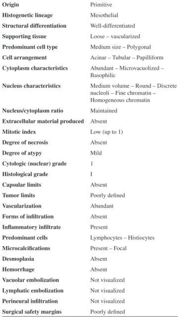
Table 1 - Histopathological findings of the reported case
Origin Primitive
Histogenetic lineage Mesothelial
Structural differentiation Well-differentiated
Supporting tissue Loose – vascularized
Predominant cell type Medium size – Polygonal
Cell arrangement Acinar – Tubular – Papilliform
Cytoplasm characteristics Abundant – Microvacuolized – Basophilic
Nucleus characteristics Medium volume – Round – Discrete nucleoli – Fine chromatin – Homogeneous chromatin
Nucleus/cytoplasm ratio Maintained
Extracellular material produced Absent
Mitotic index Low (up to 1)
Degree of necrosis Absent
Degree of atypy Mild
Cytologic (nuclear) grade 1
Histological grade I
Capsular limits Absent
Tumor limits Poorly defined
Vascularization Abundant
Forms of infiltration Absent
Inflammatory infiltrate Present
Predominant cells Lymphocytes – Histiocytes
Microcalcifications Present – Focal
Desmoplasia Absent
Hemorrhage Absent
Vacuolar embolization Not visualized
Lymphatic embolization Not visualized
Perineural infiltration Not visualized
The lesion was resected surgically, and there was no sign of recurrent disease on subsequent follow-up exams. The patient is currently asymptomatic.
DISCUSSION
Mesotheliomas are rare tumors originating from mesothelial cells of serosal membranes such as the pleura, peritoneum,4 pericardium, and tunica vaginalis.1
Simultaneous pleural and peritoneal involvement occurs in 30-45% of cases, whereas disease limited to the peritoneum occurs in 10 to 20% of the patients.5 Peritoneal
mesotheliomas can be classified as benign (adenomatoid, fibrous),3,6 borderline (multicystic, well-differentiated
papilliferous),1,4, 7,8 and malignant (epithelioid, sarcomatoid
or biphasic/mixed),8 and their characteristics are described
in Table 2.
Peritoneal adenomatoid mesothelioma (AMP) is a benign neoplasia9,10 of unknown etiology that primarily involves
the genital tract of both sexes,9,10 occurring more frequently
among males.1,3,8,11-13
We report here a case of a 25-year-old woman diagnosed with AMP that was confined to the peritoneum. Among women, adenomatoid tumors are more commonly encountered in the myometrium (posterior wall of the uterus), in the fallopian tubes, in paraovarian connective
tissue,1,10-12 and rarely in the ovaries.1,9 In our patient, the
uterus and ovaries were free of disease, and the lesion was confined to the peritoneum.
Among males, in most cases the tumor is detected in the inferior pole of the epididymis,9,10 but it can also involve
the ejaculatory duct, sperm cord, tunica albuginea, tunica vaginalis, testicular parenchyma, prostate, and rarely the spermatic funiculus.1,9-13
Adenomatoid tumors have also been detected in the omentum, mesentery, pancreas, liver, bladder, mediastinal lymph nodes, pleura, heart, and adrenal glands.3,9,11,13
The cause of the apparent predominance in the genital tract when compared to other mesothelial locations has not been explained.3 Historically, adenomatoid
tumors have always attracted interest regarding their histological origin, and several hypotheses have been proposed. Immunohistochemical studies favor mesothelial histogenesis.1,10 The “adenomatoid” designation was
introduced by Golden and Ash in 19451,9,10 because of the
arrangement of the cells in a cohesive manner, forming tubules and canaliculi. Four histological patterns of adenomatoid tumors have been identified and classified as adenoid, angiomatoid, solid or cystic.1,3 The histological
pattern of the present case was classified as solid. The peak incidence of this tumor is between the 3rd and 5th decades of life, between 26 and 55 years (mean: 41 years), with an extremely rare occurrence in children.1,10
AMP is an uncommon tumor,3 usually asymptomatic,9
and is incidentally discovered during radiologic exams, surgeries or autopsies.3,9 It is typically a single polypoid or
nodular small lesion (2.0 cm or less)1,14 that can measure
up to 13 cm in diameter in a few cases.1 Adenomatoid
tumors are usually solid, not encapsulated and often contain small cystic lesions (0.4 to 1.5 cm).12 When present, signs
and symptoms are abdominal pain, loss of weight, loss of appetite, nausea, fluid accumulation in the peritoneal space (ascites), and a pelvic mass.8
In the present case, the tumor was 15 cm at its widest diameter on pathological examination, exceeding the size of previously reported masses. This might explain why the patient was symptomatic; additionally, after surgical resection, symptoms disappeared for a period of approximately one year. After that, the lesion recurred, and the patient was submitted to a new surgical intervention. The patient has now been free of the disease for 7 years.
Usually, surgical resection is the treatment of choice for AMP. Accurate diagnosis and staging are important because of the obvious therapeutic implications.15 Although benign,
AMP is a source of great concern due to the differential diagnosis of malignant entities.10
In the present case, in view of the location of the tumor Figure 4 - The histopathologic section shows calretinin staining for
well-differentiated mesothelial cells (*), which confirms the mesothelial origin of the tumor
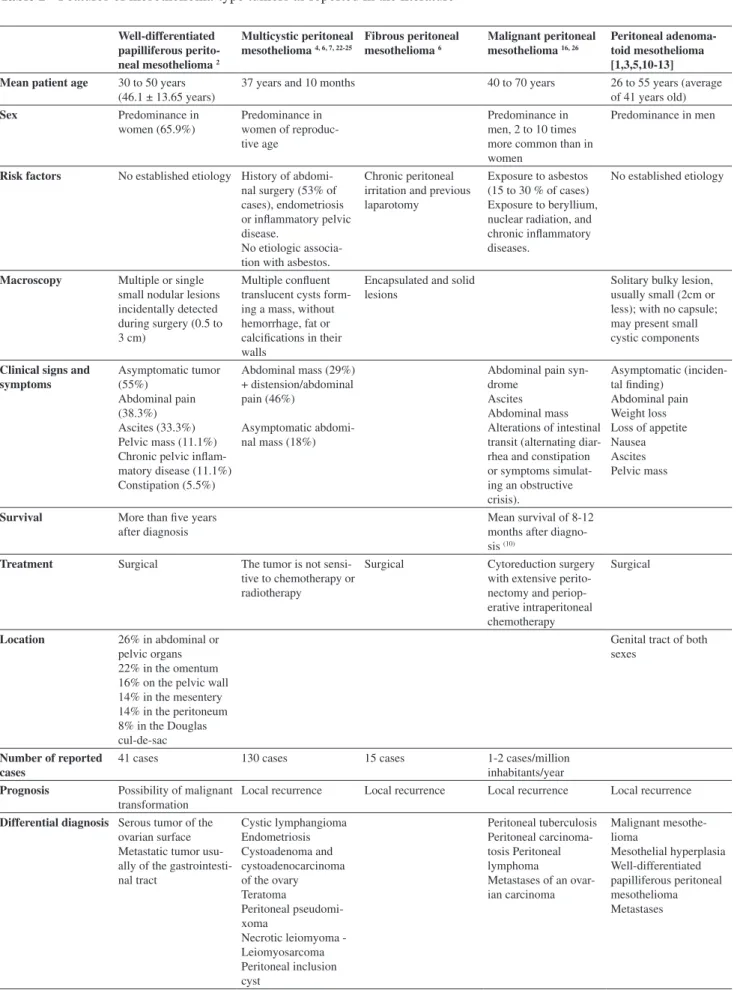
Table 2 - Features of mesothelioma-type tumors as reported in the literature
Well-differentiated papilliferous perito-neal mesothelioma 2
Multicystic peritoneal mesothelioma 4, 6, 7, 22-25
Fibrous peritoneal mesothelioma 6
Malignant peritoneal mesothelioma 16, 26
Peritoneal adenoma-toid mesothelioma [1,3,5,10-13] Mean patient age 30 to 50 years
(46.1 ± 13.65 years)
37 years and 10 months 40 to 70 years 26 to 55 years (average
of 41 years old)
Sex Predominance in
women (65.9%)
Predominance in women of reproduc-tive age
Predominance in men, 2 to 10 times more common than in women
Predominance in men
Risk factors No established etiology History of abdomi-nal surgery (53% of cases), endometriosis or inflammatory pelvic disease.
No etiologic associa-tion with asbestos.
Chronic peritoneal irritation and previous laparotomy
Exposure to asbestos (15 to 30 % of cases) Exposure to beryllium, nuclear radiation, and chronic inflammatory diseases.
No established etiology
Macroscopy Multiple or single small nodular lesions incidentally detected during surgery (0.5 to 3 cm)
Multiple confluent translucent cysts form-ing a mass, without hemorrhage, fat or calcifications in their walls
Encapsulated and solid lesions
Solitary bulky lesion, usually small (2cm or less); with no capsule; may present small cystic components
Clinical signs and symptoms Asymptomatic tumor (55%) Abdominal pain (38.3%) Ascites (33.3%) Pelvic mass (11.1%) Chronic pelvic inflam-matory disease (11.1%) Constipation (5.5%)
Abdominal mass (29%) + distension/abdominal pain (46%)
Asymptomatic abdomi-nal mass (18%)
Abdominal pain syn-drome
Ascites Abdominal mass Alterations of intestinal transit (alternating diar-rhea and constipation or symptoms simulat-ing an obstructive crisis).
Asymptomatic (inciden-tal finding)
Abdominal pain Weight loss Loss of appetite Nausea Ascites Pelvic mass
Survival More than five years after diagnosis
Mean survival of 8-12 months after diagno-sis (10)
Treatment Surgical The tumor is not sensi-tive to chemotherapy or radiotherapy
Surgical Cytoreduction surgery with extensive perito-nectomy and periop-erative intraperitoneal chemotherapy
Surgical
Location 26% in abdominal or pelvic organs 22% in the omentum 16% on the pelvic wall 14% in the mesentery 14% in the peritoneum 8% in the Douglas cul-de-sac
Genital tract of both sexes
Number of reported cases
41 cases 130 cases 15 cases 1-2 cases/million
inhabitants/year
Prognosis Possibility of malignant transformation
Local recurrence Local recurrence Local recurrence Local recurrence
Differential diagnosis Serous tumor of the ovarian surface Metastatic tumor usu-ally of the gastrointesti-nal tract
Cystic lymphangioma Endometriosis Cystoadenoma and cystoadenocarcinoma of the ovary Teratoma
Peritoneal pseudomi-xoma
Necrotic leiomyoma - Leiomyosarcoma Peritoneal inclusion cyst Peritoneal tuberculosis Peritoneal carcinoma-tosis Peritoneal lymphoma
Table 3 - Positivity of immunohistochemical markers in adenocarcinomas and mesotheliomas11, 25
Adenocarcinomas (%)
Mesotheliomas (%)
CEA 90-100 0-10
B72.3 81 0-5
BEREP4 90-100 0-11
CD15(LEU-M1) 58-100 0-10
Calretinin 6-9 42-100
in the cul-de-sac and its histopathological characteristics (cells clustered in a papillary formation), it was necessary to establish a differential diagnosis with adenocarcinoma.1
Histology revealed medium-sized polygonal cells in an acinar, tubular and papilliform cell arrangement, a low mitotic index (up to 1), a mild grade of atypia, abundant vascularization, and absence of necrosis, suggesting an adenomatoid tumor.14
Immunohistochemistry revealed positivity for calretinin, which labels mesothelial cells in 60 to 100% of cases1,9,13
and rarely labels adenocarcinomas (0 to 28%). In addition, the cells of the neoplasia reported here were negative for BerEp4, which labels epithelial cells that are not present in mesotheliomas.2,5 The present case was negative for CEA
immunoreagent, which frequently labels pulmonary and gastrointestinal carcinomas and is detected in only 0 to 35% of serous ovarian carcinomas.2,16 Thus, negativity of this
marker is of no help for differentiation between adenomatoid tumors and adenocarcinomas. The possibility of the latter was ruled out due to immunohistochemistry compatible with an adenomatoid tumor, and by MRI and laparotomy findings that revealed disease-free ovaries. Another possible differential diagnosis for this case, arising from its location in a cul-de-sac, would be a metastatic tumor. However, CEA negativity and calretinin positivity do not favor this possibility, as demonstrated in Table 3. Other differential diagnoses are cysts of peritoneal inclusion, hemangiomas, lymphangiomas,12 mesothelial hyperplasia, malignant
mesotheliomas,9 and well-differentiated papilliferous
mesotheliomas.
Mesothelial hyperplasia has been associated with peritoneal insults such as hernia, ectopic tubal pregnancy, and abdominal cirrhosis and tuberculosis3 and is accompanied
by adherences and chronic inflammation.14 This entity
rarely produces tumoral masses and does not have the
tubulopapilliferous complex or the labyrinth architecture of mesotheliomas.14 The differential diagnosis with
malignant mesothelioma and well-differentiated papilliferous mesothelioma is made on the basis of the distinct histological characteristics of these tumors when compared to AMP.17
MR has become a valuable noninvasive technique for evaluation of the female pelvis,18-20 with advantages over
computed tomography and ultrasound for diagnosis and for staging various pathological conditions of the pelvis (leiomyoma, adenomyosis, carcinoma of the endometrium and of the uterine cervix, carcinoma of the vagina, ovarian cysts, endometriosis, teratomas, polycystic ovaries, and other ovarian masses).18,20
MR has proven to be a highly sensitive modality for characterization of pelvic masses, allowing physicians to determine whether the pelvic mass is uterine or of adnexal origin and also to characterize most adnexal masses.20
MR can also provide multiplanar information, revealing additional information when compared to CT or US. This is especially true along the pelvic walls and the presacral space.18,20 MR is also especially useful for surgical planning15
and patient follow-up. Low et al21 studied 24 patients with
suspected peritoneal tumors and found that MR had higher sensitivity, specificity and accuracy than CT in the detection of tumors (84%, 87% and 86%, compared to 54%, 91% and 74%, respectively, for CT) and was superior for detection of carcinomatosis and of tumors measuring less than 1 cm in diameter (75% to 80% for MR and 22% to 33% for CT). Post-contrast T1-weighted images with fat suppression were proven to be the most sensitive MR technique for detecting peritoneal disease. MR and CT showed identical performance for detection of tumors measuring more than 2 cm and 1 to 2 cm in diameter.21
REFERENCES
1. Hanada S, Okumura Y, Kaida K. Multicentric adenomatoid tumors involving uterus, ovary, and appendix. J Obstet Gynaecol Res. 2003;29:234-8.
2. Hoekman K, Tognon G, Risse EK, Bloemsma CA, Vermorken JB. Well-differentiated papillary mesothelioma of the peritoneum: a separate entity. Eur J Cancer. 1996; 32A:255-8.
3. Hayes SJ, Clark P, Mathias R, Formela L, Vickers J, Armstrong GR. Multiple adenomatoid tumours in the liver and peritoneum. J Clin Pathol. 2007;60:722-4.
4. Wong WL, Johns TA, Herlihy WG, Martin HL. Best cases from the AFIP: multicystic mesothelioma. Radiographics. 2004;24:247-50. 5. Davidson B. Biological characteristics of cancers involving the serosal
cavities. Crit Rev Oncog. 2007;13:189-227.
6. Adachi T, Sugiyama Y, Saji S. Solitary fibrous benign mesothelioma of the peritoneum: report of a case. Surg Today. 1999;29:915-8. 7. Sawh RN, Malpica A, Deavers MT, Liu J, Silva EG. Benign cystic
mesothelioma of the peritoneum: a clinicopathologic study of 17 cases and immunohistochemical analysis of estrogen and progesterone receptor status. Hum Pathol. 2003;34:369-74.
8. Daya D, McCaughey W.T. Pathology of the peritoneum: a review of selected topics. Semin Diagn Pathol. 1991;8:277-89.
9. Isotalo PA, Nascimento AG, Trastek VF, Wold LE, Cheville JC. Extragenital adenomatoid tumor of a mediastinal lymph node. Mayo Clin Proc. 2003;78:350-4.
10. Gokce G, Kilicarslan H, Ayan S, Yildiz E, Kaya K, Gultekin EY. Adenomatoid tumors of testis and epididymis: a report of two cases. Int Urol Nephrol. 2001;32:677-80.
11. Cajaiba MM, Senise SM, Osório CABdT, Pinto CAL. Adenomatoid Tumor of Myometrium: Report of Three Cases. Applied Cancer Research. 2005;25:3.
12. Ghossain MA, Chucrallah A, Kanso H, Aoun NJ, Abboud J. Multilocular adenomatoid tumor of the ovary: ultrasonographic findings. J Clin Ultrasound. 2005;33:233-6.
13. Hamamatsu A, Arai T, Iwamoto M, Kato T, Sawabe M. Adenomatoid tumor of the adrenal gland: case report with immunohistochemical study. Pathol Int. 2005;55:665-9.
14. Goldblum J., Hart W.R. Localized and diffuse mesotheliomas of the genital tract and peritoneum in women. A clinicopathologic study of nineteen true mesothelial neoplasms, other than adenomatoid tumors, multicystic mesotheliomas, and localized fibrous tumors. Am J Surg Pathol. 1995;19:1124-37.
15. Devine C, Szklaruk J, Tamm EP. Magnetic resonance imaging in the characterization of pelvic masses. Semin Ultrasound CT MR. 2005;26:172-204.
16. Trupiano JK, Geisinger KR, Willingham MC, Manders P, Zbieranski N, Case D;et al. Diffuse malignant mesothelioma of the peritoneum and pleura, analysis of markers. Mod Pathol. 2004;17:476-81. 17. Bhandarkar DS, Smith VJ, Evans DA, Taylor TV. Benign cystic
peritoneal mesothelioma. J Clin Pathol. 1993;46:867-8.
18. van Ruth S., Bronkhorst MW, van Coevorden F, Zoetmulder FA. Peritoneal benign cystic mesothelioma: a case report and review of the literature. Eur J Surg Oncol. 2002;28: 192-5.
19. Szklaruk J, Tamm EP, Choi H, Varavithya V. MR imaging of common and uncommon large pelvic masses. Radiographics. 2003;23:403-24. 20. Olson MC, Posniak HV, Tempany CM, Dudiak CM. MR imaging of
the female pelvic region. Radiographics. 1992;12:445-65.
21. Low RN, Barone RM, Lacey C, Sigeti JS, Alzate GD, Sebrechts C.P. Peritoneal tumor: MR imaging with dilute oral barium and intravenous gadolinium-containing contrast agents compared with unenhanced MR imaging and CT. Radiology 1997;204:513-20.
22. Bui-Mansfield LT, Kim-Ahn G, O’Bryant LK. Multicystic mesothelioma of the peritoneum. AJR Am J Roentgenol. 2002;178:402.
23. Ozgen A, Akata D, Akhan O, Tez M, Gedikoglu G, Ozmen MN. Giant benign cystic peritoneal mesothelioma: US, CT, and MRI findings. Abdom Imaging. 1998;23:502-4.
24. Romero JA, Kim EE, Kudelka AP, Edwards CL, Kavanagh JJ. MRI of recurrent cystic mesothelioma: differential diagnosis of cystic pelvic masses. Gynecol Oncol. 1994;54:377-80.
25. Soreide JA, Soreide K, Korner H, Soiland H, Greve OJ, Gudlaugsson E. Benign peritoneal cystic mesothelioma. World J Surg. 2006;30:560-6. 26. Puvaneswary M, Chen S, Proietto T. Peritoneal mesothelioma: CT and