Jo
rn
a
l B
ra
s
ile
ir
o
d
e
P
a
to
lo
g
ia
e
M
e
d
ic
in
a
L
a
b
o
ra
to
ria
l
365
Jo
rn
a
l B
ra
s
ile
ir
o
d
e
P
a
to
lo
g
ia
e
M
e
d
ic
in
a
L
a
b
o
ra
to
ria
l
Recebido em 19/ 03/ 03 Aceito para publicação em 07/ 05/ 03
Mucous gland adenoma of the bronchus in a 5-year-old child:
case report and review of the literature
Adenoma de células mucosas do brônquio em criança de 5 anos: relat o de caso e revisão da lit erat ura
Luiz Cesar Peres1 Eumenia Costa da Cunha Castro2
y
key words
unitermos
abstract
Mucous gland adenoma is a rare benign tumor arising from the mucous secreting glands of the larger airway mucosa. The majority of the cases are seen in the bronchus, but it has also been described in the trachea or peripheral airways. The tumor is composed of mucus-secreting cells, usually forming glands that grow into the lumen as to form an obstructive mass. There is no predilection for sex or age. We report here a case seen in a 5-year-old girl, to our knowledge the youngest in the consulted literature, who presented to Hospital das Clínicas of Faculdade de Medicina de Ribeirão Preto for investigation and treatment of recurrent pneumonia and lung atelectasis. Bronchoscopy showed a mass obstructing the lower left bronchus and a biopsy of the lesion revealed a benign, although uncharacteristic lesion. Surgical removal of the affected segment, the recommended treatment for this benign lesion, evidenced a mucous gland adenoma. The child recovered well and was completely free of disease when she was lost for follow-up when she was 6 years old.
Adenoma
Adenoma of the bronchus
Mucous gland adenoma
Child
resumo
O adenoma de células mucosas é uma neoplasia benigna rara que se origina nas glândulas da mucosa das vias aéreas maiores. A maioria dos casos é vista nos brônquios, mas já foram descritos casos na traquéia e nas vias respiratórias distais. O tumor é constituído por células mucossecretoras, que geralmen-te formam glândulas que crescem para dentro da luz, formando massa obstrutiva. Não há predileção por sexo ou idade. Nós relatamos aqui um caso observado em uma menina de 5 anos, o mais jovem relato na literatura consultada, que se apresentou no Hospital das Clínicas da Faculdade de Medicina de Ribeirão Preto para investigação e tratamento de pneumonia recorrente e atelectasia do pulmão. O exame broncoscópico mostrou massa obstruindo o brônquio inferior esquerdo e a biópsia revelou lesão benigna incaracterística. A remoção cirúrgica do segmento afetado, que é o tratamento recomendado para esta lesão benigna, evidenciou um adenoma de células mucosas. A criança recuperou-se bem e estava completamente livre de doença quando deixou o seguimento com 6 anos de idade.
Adenoma
Adenoma brônquico
Adenoma de células
mucosas
Criança
1. Associate professor of the Pathology Departament of Faculdade de Medicina de Ribeirão Preto/Universidade de São Paulo. 2. Reader professor of General Pathology of Faculdade de Medicina do Triângulo Mineiro. This work was conducted at the Departamento de Patologia, Faculdade de Medicina de Ribeirão Preto/Universidade de São Paulo, Ribeirão Preto, São Paulo, Brazil.
R
io
d
e
J
a
n
e
iro
, v
. 3
9
, n
. 4
, p
. 3
6
5
-3
6
9
, 2
0
0
366
R
io
d
e
J
a
n
e
ir
o
,
v.
3
9
,
n
.
4
,
2
0
0
3
Jo
rn
a
l
B
ra
s
il
e
ir
o
d
e
P
a
to
lo
g
ia
e
M
e
d
ic
in
a
L
a
b
o
ra
to
ri
a
l
The surgical specimen consisted of a segment of bronchus measuring 1.5cm in length with a mean diameter of 0.8cm. The wall measured 0.2cm and showed no abnormality. A nodular mass of 0.7cm in diameter exhibiting a smooth surface emerged from the mucosa and filled the lumen. The cut surface of the lesion showed a glistening homogeneous tissue.
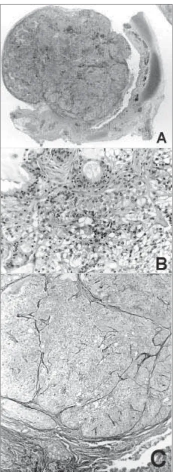
Histology of hematoxylin and eosin (H&E) stained sections of this polypoid neoplasm (Figure 1A) revealed that it was composed of large vacuolated cells with oval nuclei displaced to the periphery (Figure 1B). The cells were immersed in a basophilic matrix. Delicate fibrous septa with blood vessels separated the cells in ill-defined lobules evidenced by the reticulin staining (Figure 1C). Well-formed mucus-secreting glands were only occasionally identified. The mucus was PAS positive-diastase resistant and Alcian-blue pH 2.5- but not pH 1.0-positive, the same as the remaining mucous glands of the submucosa. The neoplasia was restricted to the submucosa and close to normal mucous glands. No mitosis, hemorrhage or necrosis was identified. There was erosion and squamous metaplasia of the surface of the epithelium. The immunohistochemical study, presented in Table 1, was performed with the Streptavidin-Biotin method using the following antibodies: AE1AE3 (Boehringer®), CEA (Dako®), EMA (Dako®), NSE (Dako®), 34bE12 (Dako®) and 35bH11 (Dako®). Final diagnosis was mucous gland adenoma of the bronchus.
The child recovered well and was thriving after 16 months, when she was lost for follow up, with a normally expanding left lung and a normal looking bronchial lumen by bronchoscopy.
Discussion
Mucous gland adenoma has been identified from childhood to adult life and the previous youngest patient in the consulted literature was aged 7 (Table 2). This is a benign lesion and should be clearly separated from the other low malignant tumors that may arise in this region (3, 12, 14, 15) – carcinoid, cylindroma and mucoepidermoid tumour – incorrectly referred to as bronchial adenoma (5).
It is usually seen in the main bronchi although there are reports of tumors located in the peripheral airways (15) or even in the trachea (6). In the present case, the lesion was located in the left inferior lobe bronchus bulging to obstruct the main left bronchus. Because of its intraluminal growth, these lesion may induce a valvular
Introduction
Mucous gland adenoma is a rare benign tumor originated from the mucous secreting glands of the larger airway mucosa (7). This tumor, first reported in 1882 by Muller as a pathologic entity separate from carcinoma of the lung (7), was first named bronchial adenoma arising from mucous gland. Different nomenclature and origin of the tumor over the years have brought confusion to the pathologists’ interpretation of the lesion (7, 10-12, 14, 15). This tumor has also been previously named adenomatous polyp (12), adenoma of mucous gland type (15), bronchial cystadenoma (3) or papillary cystic adenoma (13), but now the name mucous gland adenoma of the bronchus is preferred (1, 4, 6). This tumor has incorrectly been described under the term bronchial adenoma with three other low malignant neoplasms, namely: cylindromas, carcinoid and mucoepidermoid carcinoma (5, 7), and many authors have already stressed the inadequacy of such designation (1, 5, 6). It is seen in all age groups with no sex predilection (13). We report here a case of mucous gland adenoma of the bronchus in a 5-year-old patient, the youngest in English literature, as far as we know.
Case report
367
R
io
d
e
J
a
n
e
iro
,
v.
3
9
,
n
.
4
,
2
0
0
3
Jo
rn
a
l B
ra
s
ile
ir
o
d
e
P
a
to
lo
g
ia
e
M
e
d
ic
in
a
L
a
b
o
ra
to
ria
l
mechanism of two different types: ball valve, in which the tumor mass blocks the airway in inspiration and opens in expiration, and check valve, in the opposite way (12). In the present case, the first mechanism was the most likely since the patient developed atelectasis of the left lung. Pedunculated lesions may also change their position with body posture modifying the patient’s symptomatology.
Clinically, patients may exhibit signs of a low growing intrabronchial tumor reflected by cough, wheezing, hemoptisis, recurrent pneumonia, emphysema (12), asthma (13) and atelectasis (12) until a definite diagnosis is made by bronchoscopy and biopsy. In our case, a small superficial biopsy showed only squamous metaplasia overlying a lesion made up of large vacuolated cells. A provisional diagnosis of a benign hamartomatous lesion was offered.
Histologically, mucous gland adenoma of the bronchus may be of three different types: orderly tubular glands, papillary and cystic adenomas (10) or, as in our case, small cystic mucinous lesion (13). The mucus is histochemically similar to the normal epithelial mucin (9). Our results show that the mucus was PAS positive-diastase resistant and Alcian blue pH 2.5- but not pH 1.0-positive as the mucous gland of the remaining normal mucosa. Squamous metaplasia or erosion of the respiratory epithelium is frequently seen (14-15), as in our case. The erosion explains the common hemorrhage and hemoptisis seen in these patients.
The immunohistochemical study was consistent with the literature (9) (Table 2). Tumor cells were positive for AE1AE3, CEA and EMA in the same way as the normal mucous gland cells but presented a weaker positivity for 34bE12 and 35bH11. On the other hand, CEA was positive in the vacuolated tumor cells as well as in the mucus.
The histochemical and immunohistochemical patterns exhibited by this tumor may help in characterizing a biopsy specimen taken from a similar lesion as derived from the normal bronchial mucous glands (Table 2).
The tumor does not extend beyond the bronchial cartilage, although it may distort it. There are no mitosis, hemorrhage, necrosis, and infiltration of normal tissues or metastasis, characterizing this lesion as a completely benign tumor. The treatment of choice is total local excision (3-4) with the most conservative technique as in this case. Our patient was 6 years old when lost for follow-up completely free of disease after 16 months of the surgical treatment.
Figura – Photomicrography of the mucous gland adenoma of the bronchus. Note the
368
R
io
d
e
J
a
n
e
ir
o
,
v.
3
9
,
n
.
4
,
2
0
0
3
Jo
rn
a
l
B
ra
s
il
e
ir
o
d
e
P
a
to
lo
g
ia
e
M
e
d
ic
in
a
L
a
b
o
ra
to
ri
a
l
Case Reference Age Macroscopy Microscopy Histochemestry IH
1, 2, 3 Kroe & Pitcock, 11, 14, No description No description No description NO
1967 (9) 81/ 2
4 Emory e t al., 14 Oval, irregularly Irregularly shaped Mucicarmine NO 1973 (4) shaped,pedunculated aggregate of cells positive
polypoid lesion forming mucus filled,
(1.1 x 1cm) intact, tubular or glandular-like spaces
5, 6 Heard e t al., 7, 12 Single mass Acini and ducts Mucus positive for NO 1985 (8) (2 to 6.5cm), firm, cut resembling those of mucicarmine and PAS;
surface pale grey and normal bronchial gland focally alcianophilic slightly translucent Main tumor cell: with Alcian
blue-Compression but no mucous epithelial diastase-PAS
infiltration of the lung cell of the gland sequence and PAS
elsewhere
7 Dickstein e t al., 12 Polypoid lesion Tumor composed of NO NO 1993 (2) (1.5 x 0.6cm) with base well differentiated
attached to the wall of monomorphic columnar
the bronchus. Bronchial cells, forming microcystic wall dilatation distal to structures and acini in
the tumor forming a a fibrotic stroma. No
cyst-like structure filled evidence of mitotic
with mucinous material figures or invasion
8 Peres e t al. 5 Smooth surfaced nodular Large vacuolated Mucus PAS Tumor cells (present case) mass (0.7cm) attached to tumor cells in a positive-diastase positive for
the bronchial mucosa basophilic matrix, resistant and Alcian- AE1AE3, CEA
Glistening homogeneous separated ill blue pH 2.5- but not and EMA like cut surface defined lobules by pH 1.0-positive, the the normal
delicate fibrous septa. same as the remaining mucous gland
Tumor restricted to the mucous glands of the cells. Weak
submucosa and close to submucosa positivity for normal mucous glands. 34βE12 and
No mitosis or invasion 35βH11. Mucus
and vacuolated
tumor cells positive for
CEA
IH = immunohistochemestry.
Review of the English literature of mucous gland adenoma in children Table 2
Tumor glands Vacuolated cells Normal epithelium Mucus
AE1 AE3 ++ ++ ++
-CEA ++ + ++ ++
EMA ++ ++ ++
-NSE - - -
-34βE12 + - ++
-35βH11 + - ++
-++: diffuse; +: focal; -: no reaction; AE1AE3: cytokeratin pool; CEA: carcinoembryonic antigen; EMA: epithelial membrane antigen; NSE: neuron specific enolase; 34βE12: light weight cytokeratin; 35βH11: heavy weight cytokeratin.
369
R
io
d
e
J
a
n
e
iro
,
v.
3
9
,
n
.
4
,
2
0
0
3
Jo
rn
a
l B
ra
s
ile
ir
o
d
e
P
a
to
lo
g
ia
e
M
e
d
ic
in
a
L
a
b
o
ra
to
ria
l
References
1. Allen Jr, M .S. et al. M ucous gland adenoma of the bronchus.
J. Thor. Cardiovasc. Surg., 6 7(6): 966-8, 1974.
2. D ickstein, P.J. et al. Bronchial mucous gland adenoma presenting as bronchogenic cyst. Ped. Pulmonol. 1 6: 370-4, 1993. 3 Edwards, C .W . & M athhews, H .R. M ucous gland adenoma of
the bronchus. Thorax, 3 6: 147-8, 1981.
4. Emor y, W .B. et al. M ucous gland adenoma of the bronchus.
Am. Rev. Resp. D is., 1 0 8: 1407-10, 1973.
5. England, D.M. & H ochholzer, L. Truly benign “bronchial adenoma”. Repor t of 10 cases of mucous gland adenoma with immuno hist o chemical and ult r ast r uct ur al findings.
Am. J. Surg. Pathol., 1 9(8): 887-99, 1995.
6. Fergusson, C .J. & Cleeland, J.A. M ucous gland adenoma of the trachea: case repor t and literature review. J. Thor. Cardiovasc. Surg., 9 5: 347-50, 1988.
7. Gilman, G.A. et al. M ucous gland adenoma of the bronchus. Repor t of a case with study of secretion. Am. J. Clin. Path., 2 6: 151-4, 1956.
Correspondence to Luiz Cesar Peres Departamento de Patologia
Faculdade de Medicina de Ribeirão Preto/Universidade de São Paulo
Av. Bandeirantes 3900
CEP 14049-900 – Ribeirão Preto-SP, Brazil Tel.: (16) 602-3123
Fax: (16) 633-1068 e-mail: lcperes@fmrp.com.br
8. H eard, B.E. et al. Pathology of seven mucous gland adenomas of t he bro nchial glands w it h par t icular reference t o ultrastructure. H istopathol., 9: 687-01, 1985.
9. Kroe, D.J. & Pitcock, J.A. Benign mucous gland adenoma of the bronchus. Arch. Pathol., 8 4: 539-42, 1967.
10. Markel, S.F. et al. N eoplasms of bronchus commonly designated as adenomas. Cancer, 1 7(5): 590-608, 1964.
11. Ramsey, J.H . & Reimann, D.L. Bronchial adenomas arising in mucous glands. Illustrative case. Am. J. Pathol., 2 9: 339-51, 1953.
12. Rosenblum, P. & Klein, R.I. Adenomatous polyp of the right main bronchus producing atelectasis. J. Ped., 7: 791-6,1935. 13. Smith, A.J. Benign epithelial tumours of the bronchus. South.
M ed. J., 5 8: 1535-9, 1965.
14. Sniffen, R.C . et al. Mucoepidermoid tumors of the bronchus arising from surface epithelium. Am. J. Pathol., 3 4: 671-83, 1958. 15. W einberger, M.A. et al. Peripheral bronchial adenoma of mucous