ABSTRACT
http://dx.doi.org/10.1590/1678-775720130570
to prevent root dentin demineralization
Vinícius Rangel GERALDO-MARTINS1, Cesar Penazzo LEPRI1, Juliana Jendiroba FARAONI-ROMANO2, Regina Guenka PALMA-DIBB2
1- Department of Biomaterials, School of Dentistry, Uberaba University, Uberaba, MG, Brazil.
2- Department of Restorative Dentistry, Ribeirão Preto School of Dentistry, University of São Paulo, Ribeirão Preto, SP, Brazil.
Corresponding address: Regina Guenka Palma-Dibb - Departamento de Odontologia Restauradora - Faculdade de Odontologia de Ribeirão Preto - Universidade de São Paulo - Av. do Café S/N - Monte Alegre - 14040-904 - Ribeirão Preto - SP - Brazil - Phone/Fax +55 (16) 3602-4016 - e-mail: rgpalma@ forp.usp.br
!
The use of erbium lasers to prevent caries in enamel has shown positive results. However, it is not known if Er,Cr:YSGG laser can also be used to increase acid resistance of root dentine, which is another dental tissue susceptible to the action of cariogenic bacteria. !"#$%&'()%*+!-/%0"12-3!& Material and Methods: One hundred human root dentin samples were divided into 10 groups (G) and treated as follows: G1: no treatment; G2: NaF; G3: laser (4.64 J/cm2)
with water cooling (WC=5.4 mL/min); G4: laser (4.64 J/cm2) without WC; G5: laser (8.92
J/cm2) with WC; G6: laser (8.92 J/cm2) without WC; G7: laser (4.64 J/cm2) with WC and
NaF; G8: laser (4.64 J/cm2) without WC and NaF; G9: laser (8.92 J/cm2) with WC and
NaF; G10: laser (8.92 J/cm2) without WC and NaF. The NaF gel was applied alone or after
4 min of irradiation. After 14 days of acid challenge, the samples were sectioned and the Knoop microhardness (KHN) test was done at different depths (30, 60, 90 and 120 μm) from the outer dentin surface. Data were analyzed by one-way ANOVA and Fisher’s test (α=5%). Results: The results showed that G8 and G10 presented higher KHN than the G1
for the depths of 30 and 60 μm, indicating an increase of the acid resistance of the dentin in up to 35% (p<0.05). Conclusions: The use of Er,Cr:YSGG laser irradiation at 4.64 J/ cm2 and 8.92 J/cm2 without water cooling and associated with 2% NaF can increase the
acid resistance of human root dentin.
Keywords: Lasers. Fluorides. Hardness. Dentin.
INTRODUCTION
The increase in lifespan associated with the development and application of preventive dentistry concepts have contributed to the maintenance of the teeth in the oral cavity of elderly population. This dental longevity is accompanied by an increasing frequency of exposed root surfaces as a result of periodontal diseases, mechanical injury and/or surgical treatments. When dentin exposition is combined with some situations common to senility, such as reduced salivary flow due to diseases or drugs and the inability to have proper Z/ [ on tooth surfaces, increase the risk of root caries
occurrence4. Epidemiological studies have shown
that the incidence and prevalence of root caries in elders are high. Recent studies pointed out that almost half of the community-dwelling elders had root caries, which indicates the need of preventing this disease in this population7.
Since root caries is caused, among other reasons, by a bacterial biofilm that adhere to exposed root surfaces, carious lesions can be prevented or even inactivated through the adoption of preventive actions such as the reduction of \ [Z
316.
in public water supply represented important advances in caries prevention21. Although the use
3 3 factor to reduce the prevalence of dental caries in both rich and poor countries, it has not led to the elimination of dental caries, which remains an widespread disease affecting all age groups and still represents a risk factor for the occurrence of ]Z tooth development20. Thus, new techniques and
3Z/ ZZ laser irradiation, for example, have been studied in order to prevent the beginning or progression of carious lesions.
The possibility of increasing the acid resistance of dental hard tissues after laser irradiation was
first demonstrated in the 1970s, using a CO2
laser24. Since then, studies to verify the effects of
the interaction between different wavelengths and enamel and dentin were conducted.
The Er,Cr:YSGG (erbium, chromium: yttrium-scandium-gallium-garnet) laser works at a wavelength of 2.78 μm and it has been investigated for preventive purposes, even though they are usually applied to cavity preparation due to the mechanism of ablation. However, for caries prevention, it is important that the laser does not ablate the treated surface, but change the tissue morphologically or chemically. Therefore, to achieve the preventive effect, studies have been performed with low energy densities (sub-ablative parameters) and without water cooling1,2,9,10,14. It
was demonstrated that the irradiation of the enamel surface with an Er,Cr:YSGG laser device, using an
energy density of 8 J/cm2 3 [
temperature increase to change the chemical structure of enamel, turning it into a less soluble
structure9,10&+Z3
ablation and also induce a greater mineral loss during an acid challenge17. Recent studies combined
33 irradiation, and they found that this association decreased the enamel demineralization more alone9,22.
In this way, it is plausible that the association / effective in increasing the acid resistance of enamel9,10. Thus, it is interesting to know whether
these effects also occur in root dentin. A past study performed by Hossain, et al.17 (2001) showed that
the Er,Cr:YSGG increases the acid resistance of dentin.However, the authors concluded that when they were doing cavity preparations on dentin, they used energy densities higher than those used to obtain the preventive effect on the dental tissues17.
Since the purpose of irradiation for caries prevention
is to promote an increase in the tissue surface temperature, only minor morphological or chemical changes must occur. Gao, et al.11 (2006) irradiated
root dentin samples with a CO2 laser (1.14 J/cm2
per3-
/%0"12-to verify if this treatment was capable /%0"12-to increase the acid resistance of that tissue. The authors /Z[Z effect of CO2
on the inhibition of root demineralization, possibly 3\ dentin11. In this manner, there is evidence that
lasers can be used to protect root dentin against demineralization.
The aim of this paper was to analyze the effects of Er,Cr:YSGG laser irradiation associated /%0"12-33 on root caries prevention. The null hypothesis is that Er,Cr:YSGG laser irradiation combined /33 _ surface microhardness of root dentin after an acid challenge.
MATERIAL AND METHODS
Experimental design
The focus of this study was the treatment of root dentin surface using 10 groups of treatment: "-` %0 Z (NaF); Er,Cr:YSGG laser (4.64 J/cm2) with water
cooling (WC=5.4mL/min); Er,Cr:YSGG laser (4.64 J/cm2) without water cooling; Er,Cr:YSGG laser
(8.92 J/cm2) with water cooling; Er,Cr:YSGG laser
(8.92 J/cm2) without water cooling; Er,Cr:YSGG
laser (4.64 J/cm2) with water cooling and NaF;
Er,Cr:YSGG laser (4.64 J/cm2) without water cooling
and NaF; Er,Cr:YSGG laser (8.92 J/cm2) with water
cooling and NaF; Er,Cr:YSGG laser (8.92 J/cm2)
without water cooling and NaF. The experimental units consisted of 100 root dentin blocks from the buccal and lingual surface of human molars. The specimens were randomly assigned to 10 groups (n=10) according to the surface treatment. The quantitative response variable was the dentin longitudinal Knoop microhardness test.
Preparation of the samples
Inc., Westlake, OH, USA) with a diamond disk (Isomet; 10.2x0.03 cm, arbor size 1.27 cm, series 15HC diamond; Buehler Ltd., Lake Bluff, IL, USA) in low speed. Then, the roots were sectioned to obtain 100 fragments of 5x5x3 mm and a 9 mm2 area in
the buccal surface in each one of the 100 root dentin samples was delimited. Around this area, two layers of varnish sealer (Colorama Maybelline Ltda, São Paulo, SP, Brazil) were applied.
Experimental groups
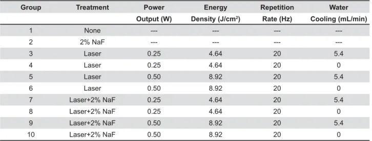
One hundred root dentin samples were randomly divided in 10 groups (n=10). In each sample, the delimitated area was treated according to Table 1. Group 1 received no treatment (control). The samples of group 2 were treated with 2% NaF during 4 min and then stored in a humid environment at 37°C until the next step of the experiment.
The samples of the groups 3-10 were irradiated with an Er,Cr:YSGG laser device (WaterLase Millennium™, Biolase Technologies Inc., San Clemente, CA, USA). This equipment emits photons /Z%&'(~&3/ []%*+!&3/*)& beam diameter at the focal area for the handpiece "%-/**~"3-& consistent spot size with the hand irradiation, [ / [] 3 handpiece, and kept a distance of 1 mm from the Z& for laser irradiation in each group is shown in Table 1. The handpiece was positioned perpendicularly to the root dentin surface, and the samples were irradiated once in each direction, slowly moving the handpiece horizontally and vertically, in order to promote homogeneous irradiation and to cover the entire sample area. The irradiation was performed manually, scanning the dentin surface with a uniform motion during 30 seconds. The output power was measured with a power meter (TM-
744D, Tenmars Electronics Co. Ltd., Taipei, Taiwan). Z3' /// 5 mL/min, which is the minimum amount that the equipment permits. In groups 7-10, the 2% NaF was applied after irradiation during 4 min. At the end of these treatments, all samples were kept in a humid environment at 37°C. Following, the samples of all groups were submitted to an acid challenge.
Acid challenge
For the acid challenge, samples were submitted 3+_Z3[3 described protocols13,14.The demineralization
solution (pH=4.3) consisted of 2 mmol/L of Ca and 2 mmol/L of phosphate in buffer solution of acetate 0.075 mol/L, and the remineralization solution (pH=7.0) consisted of 1.5 mmol/L of Ca, 0.9 mmol/L of phosphate and 150 mmol/L of potassium chloride. First of all, each specimen was immersed in 3 ml of demineralization solution for 6 hours at 37°C. Then, the specimens were washed with distilled water for 1 minute and immersed in the remineralizing solution for 18 hours at 37°C. This cycle was carried out for 14 days. At the end of each 5 consecutive days of cycling, the samples were immersed in remineralizing solution for 2 days.
Microhardness test
At the end of the pH cycling, the samples were sectioned longitudinally through the exposed area. The samples were embedded in epoxy resin, with the cut face exposed. The exposed surfaces were polished with 600 and 1,200 grit silicon carbide paper (Saint-Gobain Abrasivos Ltda, São Paulo, SP, Brazil) in a polishing machine (MetaServ 250, Buehler Ltd., Lake Bluff, IL, USA) under water cooling followed by 0.3 μm alumina paste applied over a felt disc using the same polishing machine. Next, the specimens were ultrasonically cleaned. Subsequently, each sample was assessed with the
Group Treatment Power Energy Repetition Water
Output (W) Density (J/cm2) Rate (Hz) Cooling (mL/min)
1 None --- --- ---
---2 2% NaF --- --- ---
---3 Laser 0.25 4.64 20 5.4
4 Laser 0.25 4.64 20 0
5 Laser 0.50 8.92 20 5.4
6 Laser 0.50 8.92 20 0
7 Laser+2% NaF 0.25 4.64 20 5.4
8 Laser+2% NaF 0.25 4.64 20 0
9 Laser+2% NaF 0.50 8.92 20 5.4
10 Laser+2% NaF 0.50 8.92 20 0
Knoop microhardness examination of the dentin, Z * ~ / * ~ / * ~ %*~& three measurements of Knoop microhardness were done, and the distance between measurements was **~3\33Z each other. A static load of 10 g/15 s was applied.
Statistical analysis
First, the assumptions of equality of variances (modified Levene’s test) and the normality of the error distributions (Shapiro-Wilk test) were checked for the response variables tested. Since 3/[_/1 "$0- / 33 Z Z ( * software (Origin Lab Corporation, Northampton, MA, USA). Fisher’s test was used at the 5% Z[Z means.
RESULTS
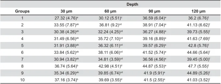
Table 2 shows the KNH for all groups. Statistical analysis compared treatments at each depth, and no comparisons were made among depths. At 30 μm, groups 8 (0.25 W + 2% NaF without water cooling) and 10 (0.5 W + 2% NaF without water cooling) showed a surface more resistant to acids than group 1 (control) (p<0.05). The data found in the other experimental groups were not different from the KHN found for group 1. At 60 μm, groups 8 and 10 also showed higher KHN than group 1 (p<0.05). The results found in control group were similar to those found in the other experimental groups. At 90 and 120 μm, no statistically differences were found among all groups.
DISCUSSION
W h i l e t h e c o m b i n a t i o n o f f l u o r i d e a n d Er,Cr:YSGG laser has been studied to inhibit enamel demineralization, there is a lack of investigation on the use of that method on root dentin to increase acid resistance. The present study assessed the use of laser and 2% NaF, combined or individually, to increase the acid resistance of root dentin surface. The null hypothesis of the present study was rejected, since the results showed that the samples irradiated with 0.25 W and 0.50 W without water cooling, followed by 2% NaF application, presented surfaces more resistant to acids when compared to untreated samples. This situation occurred for the depths of 30 and 60 μm.
The data found for the depths of 90 and 120 μm were not statistically different. This probably occurred because of the short period used for the acid challenge, which did not allowed the penetration of the demineralizing solution through the deeper layers of root dentin (90 and 120 μm). For this reason, the dentin layers deeper than 120 μm were not analyzed. Thus, further studies should be conducted using a longer period of acid challenge to know if these deeper layers of dentin would be affected by the acid challenge.
3 had already been demonstrated. Previous studies suggested that the treatment and prevention of root ]Z
Z23,25,26.
Additionally, a previous in vitro study showed that 33 (APF) application and 1100-ppm-F dentifrice used 3 times a day provided a synergistic effect, Z [ and reducing root dentin demineralization25.
Those studies suggested that only the use of
Depth
Groups 30 μm 60 μm 90 μm 120 μm
1 27.32 (4.76)a 30.12 (5.51)c 36.59 (6.04)e 36.2 (6.76)f
2 33.55 (7.87)ab 36.81 (9.2)cd 38.91 (7.04)e 41.13 (6.62)f
3 30.38 (4.26)ab 32.24 (4.25)cd 36.27 (4.88)e 39.73 (5.55)f
4 31.49 (6.56)ab 35.72 (7.10)cd 39.16 (8.89)e 41.63 (7.69)f
5 31.91 (3.88)ab 36.32 (6.11)cd 39.57 (6.29)e 42.8 (5.76)f
6 33.84 (5.62)ab 38.11 (6.06)cd 41.52 (5.74)e 44.66 (5.64)f
7 30.94 (3.82)ab 34.81 (3.59)cd 36.56 (4.56)e 39.45 (5.00)f
8 36.74 (5.64)b 42.98 (4.51)d 44.87 (5.53)e 47.7 (5.55)f
9 35.34 (6.29)ab 39.85 (6.74)cd 41.9 (5.91)e 44.89 (5.26)f
Z 3 against the attack of acidogenic bacteria. In fact, the results of the present study suggested that the association between the Er,Cr:YSGG laser irradiation 3 resistant to acids than the simple application of NaF. 233 individually resulted in comparable and low levels of root caries inhibition. When Er,Cr:YSGG laser / // about 35% of root dentin demineralization reduction when comparing to the control group.
The mechanism of the synergistic effect of / application is still unknown. It was reported that laser treatment significantly increase fluoride 3\ **0 '0 [ bound forms, respectively11. During a cariogenic
challenge, the loosely bound calcium fluoride
(CaF2-
demineralization and enhance remineralization. The [ Z structure may increase crystal stability and acid resistance. A study has revealed a contraction in the a-axis dimension and an improvement in the enamel crystallinity induced by laser irradiation, with a further reduction of the axis of hydroxyapatite _ treatment6. Probably some of those effects also
occur in dentin. In addition, the firmly bound / greater substantivity than that of the loosely bound
11. In this way, these factors may be the
main reasons for the increase of the acid resistance of root dentin.
It is important to note that, in the present study, 2% NaF was applied after irradiation to observe whether the irradiated dentin surface enhances the &Z effect could be observed on samples irradiated without water cooling.
Regarding the energy densities used, although / Z[ among the treated groups, the higher parameters seemed to promote better results than the lower ones when only the laser was used. There were no previous research that used the Er,Cr:YSGG laser for root caries prevention, so the parameters used in this study were based on those used for the enamel irradiation with the same wavelength. Previous studies showed that enamel irradiation with an energy density of 8 J/cm2 was necessary
33[Z to modify the chemical structure of the enamel, turning it into a less soluble structure8,10,22. Lower
energy densities should not be able to increase the temperature of the surface up to 300-450°C, and, consequently, less soluble compounds would not
be formed8.
Another hypothesis suggested the “organic blocking” theory, when the partial denaturation of the organic matrix caused by laser irradiation may block the diffusion pathway in enamel, resulting in a retardation of enamel demineralization19. Blocking
the diffusion pathway may affect the porosity of the enamel and the microsurface area. The organic ZZ[ in the pore volume and surface area in enamel after laser irradiation, may be one of the key players in the laser induced blocking of diffusion pathway and subsequent prevention of enamel demineralization11,27.
As shown previously3, the laser Er,Cr:YSGG also
produces the sealing of dentinal tubules through Z [ / reduces the permeability of that tissue and thus hinders the penetration of the acid into the deeper layers of dentin.
These theories support the results obtained in the present study. The absence of water cooling may lead to an increase in the temperature of the surface, promoting the same effects that occur in enamel, as described before, and this also can be observed here when groups that used the same energy density are individually compared. The possibility of using minimum levels of water (5.0 mL/min) during irradiation was thought to avoid thermal damages in the tissue. The researchers are aware that the presence of large amounts of water hinders the preventive effect promoted by Er,Cr:YSGG. Somehow, the presence of water was more disadvantageous when used with lower parameters, as evidenced by the results obtained in groups 3 and 7.
A previous study showed that the dentin surface,
/&2 and with the
same Er,Cr:YSGG device under water cooling (15.0 mL/min), presented opened dentinal tubules12.
Although the amount of water used by these authors was almost three times higher than that [ZZZ3 of water cooling does not made easier for the laser to promote the preventive effect in the root dentin, since opened dentinal tubules facilitate the acid penetration, causing the carious lesions extend more into the dentin5. Conversely, when water
cooling is not used, the dentin undergoes a melting process that involves, among other things, the obliteration of the dentinal tubules3. This hinders
the penetration of acids into the dentin, making this tissue more resistant to demineralization. This hypothesis explains why the results obtained in the absence of cooling were a little bit better even for the same energy density.
energy density of 8.92 J/cm2 was used without
/ Z& [Z ZZ areas of root dentin overheated. That factor is of extreme importance because it indicates that Z/
Z[Z15,18.
2Z ablation and also induce a greater mineral loss during an acid challenge17.
Therefore, analyzing the results obtained in the present study, it was possible to verify, after the Knoop microhardness test, that the use of Er,Cr:YSGG laser associated with 2% NaF application promoted an increase in the acid resistance of the human root dentin in up to 35%. However, laser irradiation without water cooling promoted thermal damage on dentin. On the other hand, the presence of water, even in small amounts, did not favor the preventive effect of the Er,Cr:YSGG laser irradiation. Further studies using lower energy densities and lasers with different wavelengths are needed, not [Z / / happens in the irradiated tissue that makes it more resistant to acids.
CONCLUSIONS
Considering the limitations of the present study, it can be concluded that the association between of human root dentin if that irradiation is provided without water cooling.
REFERENCES
1- Apel C, Birker L, Meister J, Weiss C, Gutknecht N. The caries-preventive potential of subablative Er:YAG and Er:YSGG laser radiation in an intraoral model: a pilot study. Photomed Laser Surg. 2004;22:312-7.
%_3!++\1& changes in human dental enamel after subablative erbium laser irradiation and its potential use for caries prevention. Caries Res. 2005;39:65-70.
3- Aranha AC, Eduardo CP. In vitro effects of Er,Cr:YSGG laser on dentine hypersensitivity. Dentine permeability and scanning electron microscopy analysis. Lasers Med Sci. 2012;27:827-34. 4- Bahrami G, Vaeth M, Kirkevang LL, Wenzel A, Isidor F. Risk factors for tooth loss in an adult population: a radiographic study. J Clin Periodontol. 2008;35:1059-65.
5- Cummins D. Dentin hypersensitivity: from diagnosis to a breakthrough therapy for everyday sensitivity relief. J Clin Dent. 2009;20:1-9.
_Z+& crystalline structure of human enamel: a pilot study. Proc SPIE. 2005;5687:42-9.
7- Du M, Jiang H, Tai B, Zhou Y, Wu B, Bian Z. Root caries patterns and risk factors of middle-aged and elderly people in China. Community Dent Oral Epidemiol. 2009;37:260-6.
8- Fowler BO, Kuroda S. Changes in heated and in laser-irradiated human tooth enamel and their probable effects on solubility. Calcif Tissue Int. 1986;38:197-208.
9- Freitas PM, Rapozo-Hilo M, Eduardo CP, Featherstone JD. In vitro evaluation of erbium,chromium:yttrium-scandium-gallium-garnet laser-treated enamel demineralization. Lasers Med Sci. 2010;25:165-70.
10- Fried D, Featherstone JDB, Visuri SR, Seka W, Walsh JT. The caries inhibition potential of Er:YAG and Er:YSGG laser radiation. Proc SPIE. 1996;2672:73-8.
11- Gao XL, Pan JS, Hsu CY. Laser-fluoride effect on root demineralization. J Dent Res. 2006;85:919-23.
%___Z& of etching with Er,Cr:YSGG laser on the microtensile bond strength of adhesives to dentin. J Oral Lasers Appl. 2010;10:79-86. 13- Geraldo-Martins VR, Lepri CP, Palma-Dibb RG. Effect of different root caries treatments on the sealing ability of conventional glass ionomer cement restorations. Lasers Med Sci. 2012;27:39-45. _ _ 3 _ & Er,Cr:YSGG laser irradiation on enamel caries prevention. Lasers Med Sci. 2013;28(1):33-9.
15- Geraldo-Martins VR, Tanji EY, Wetter NU, Nogueira RD, Eduardo CP. Intrapulpal temperature during preparation with the Er:YAG laser: an in vitro study. Photomed Laser Surg. 2005;23:182-6. _[Z[+& 3Z&&%**'`(*_& 17- Hossain M, Kimura Y, Nakamura Y, Yamada Y, Kinoshita JI, Matsumoto K. A study on acquired acid resistance of enamel and dentin irradiated by Er,Cr:YSGG laser. J Clin Laser Med Surg. 2001;19:159-63.
18- Hossain M, Nakamura Y, Yamada Y, Kimura Y, Matsumoto N, Matsumoto K. Effects of Er,Cr:YSGG laser irradiation in human enamel and dentin: ablation and morphological studies. J Clin Laser Med Surg. 1999;17:155-9.
19- Hsu CY, Jordan TH, Dederich DN, Wefel JS. Effects of low-energy CO2 laser irradiation and the organic matrix on inhibition of enamel demineralization. J Dent Res. 2000;79:1725-30. 20- Ismail AI, Hasson H. Fluoride supplements, dental caries and /&&%**(` '_ 68.
21- Meenakshi, Maheshwari RC. Fluoride in drinking water and its removal. J Hazard Mater. 2006;137:456-63.
22- Moslemi M, Fekrazad R, Tadayon N, Ghorbani M, Torabzadeh H, Shadkar MM. Effects of Er,Cr:YSGG laser irradiation and && 2009;31:409-13.
23- Paraskevas S, Danser MM, Timmerman MF, van der Velden & incidence of root caries in periodontal maintenance patients. A 2 year evaluation. J Clin Periodontol. 2004;31:965-71.
24- Stern RH, Vahl J, Sognnaes RF. Laser enamel: ultrastructural observations of pulsed carbon dioxide laser effects. J Dent Res. 1972;51:455-60.