DOI 10.5897/AJPP2013.3598
ISSN 1996-0816 © 2013 Academic Journals http://www.academicjournals.org/AJPP
African Journal of Pharmacy and
Pharmacology
Full Length Research Paper
Biological screening of extracts of Brazilian Asteraceae
plants
Cintia Cristina de Carvalho
1,
Kamilla Nunes Machado
1,
Paulo Michel Pinheiro Ferreira
2,3,
Cláudia Pessoa
4,
Thaisa Helena Silva Fonseca
5,
Maria Aparecida Gomes
5and Andréa Mendes
do Nascimento
1*
1
Departamento de Química, Instituto de Ciências Exatas e Biológicas, Universidade Federal de Ouro Preto, Campus Universitário Morro do Cruzeiro, Bauxita, CEP 35400-000, Ouro Preto, MG, Brazil.
2
Departamento de Ciências Biológicas, Campus Senador Helvídio Nunes de Barros, Universidade Federal do Piauí, Rua Cícero Duarte 905, Bairro Junco, CEP 64607-670, Picos, PI, Brazil.
3
Programa de Pós-Graduação em Ciências Farmacêuticas, Núcleo de Tecnologia Farmacêutica, Universidade Federal do Piauí, Ininga, CEP 64.049-550, Teresina, PI, Brazil.
4
Departamento de Fisiologia e Farmacologia, Faculdade de Medicina, Universidade Federal do Ceará, CEP 60430-270, Fortaleza, CE, Brazil.
5
Departamento de Parasitologia, Universidade Federal de Minas Gerais, CEP 31270-901, Belo Horizonte, MG, Brazil.
Accepted 28 June, 2013
Natural products are a very productive source of leads for the development of medicines. Seven Brazilian Asteraceae adult plants were randomly chosen. The current study was designed to evaluate the antiprotozoal and cytotoxic activities in vitro of 21 extracts obtained. Phytochemical properties of the most active extracts also were checked. Cytotoxic activity was evaluated by 3-(4,5-dimethylthiazol-2-yl)-2,5-diphenyltetrazolium bromide (MTT) method against human tumor cell lines (HCT-116, OVCAR 8 and SF-295). The antiprotozoal activity was evaluated against Trichomonas vaginalis, Entamoeba histolytica and Giardia lamblia. None of the extracts showed antiprotozoal activity. However, 76% of the extracts displayed moderate to high in vitro cytotoxic activities against human cancer cell which suggests that they are a promising source of anticancer compound, since none of the extracts showed hemolytic activity. Terpenoids, flavonoids, saponins, tannins, besides other compound classes, were identified and may be responsible for their antitumor activity. Cytotoxic assays indicate the anticancer potential of Asteraceae species from Brazil.
Key words: Drug prospecting, Antiprotozoal agents, Cytotoxicity, Drug screening assays, Brazilian plant.
INTRODUCTION
Currently, there still persists many difficulties and challenges in cancer therapy such as drug resistance, toxicity and low specificity of drugs (Mesquita et al., 2009). Plants have a long history of use in the cancer treatment. Active constituents of Catharanthus roseus (L.) G.Don (Apocynaceae), Angelica gigas Nakai (Apiaceae), Podophyllum peltatum L. (Berberidaceae), Taxus brevifolia Nutt. (Taxaceae), Ochrosia elliptica
Labill. (Apocynaceae), and Camptotheca acuminata Decne. (Cornaceae) have been used in the treatment of advanced stages of various malignancies (Patel et al., 2009). Over 50% of drugs used in clinical trials for anticancer activity were isolated from natural sources or are related to them (Majumdar, 2012). The three anaerobic protozoa, Entamoeba histolytica, Giardia lamblia and Trichomonas vaginalis are highly prevalent
human-infective parasites with a worldwide prevalence (Cantillo-Ciau et al., 2010). The most effective and commonly used drug in the treatment of these three protozoans is metronidazole. However, this substance has unpleasant side effects such as a metallic taste, headache, dry mouth, urticaria, pruritus, and dark-colored urine (Pérez et al., 2012). Due to these undesired side effects and taking into account the possibility of the development of resistant strains of the T. vaginalis, E. histolytica, and G. lamblia against metronidazole, there is a clear need for new, effective, and safer antiprotozoal agents.
Natural products, especially of plant origin, represent an excellent starting point for research. In traditional medicine there are also several plants used to treat vaginitis (Girón et al., 1988) and amoebic dysentery (Bautista et al., 2011). Amaral et al. (2006) described 153 plant species from 69 families that were evaluated for their giardicidal activity. It was found that the majority of extracts and fractions obtained from plant species employed in popular medicine for the treatment of diarrhea and dysentery exhibited in vitro giardicidal activity, and these were mainly from species belonging to the Asteraceae family.
Asteraceae is the largest family of angiosperms and it comprises 1535 genera and about 23 thousand species distributed in 3 subfamilies and 17 tribes (Bremer, 1994). The plants of the Asteraceae family are very common in the open formations of Brazil, mainly in the cerrado, where the family is well represented by approximately 250 genera and 2000 species (Guimarães et al., 2012). Asteraceae species have been used in the Brazilian folk medicine for several therapeutic purposes. For example, species of the genus Lychnophora, popularly known as "arnica", are widely used in Brazilian folk medicine as anti-inflammatory, to treat bruise, pain, rheumatism and for insect bites (Ferrari et al., 2012). Species of the genus Mikania, known as "guaco", they are widely used in Brazil in the formulation of syrups for the treatment of the respiratory system (Guimarães et al., 2012). Among the native plants of Brazil, species of genus Baccharis, popularly known as "carqueja", has been used as diure-tic, tonic, digestive, protective and stimulate of the liver, antianemic, anti-rheumatic, obesity control, diabetes, hepatitis and gastroenteritis (Morais and Castanha, 2011).
Aiming to explore the rich Brazilian biodiversity, we initiated a bioprospection of plants from the Asteraceae family occurring in the state of Minas Gerais, Brazil, by screening plant extracts for cytotoxic and antiprotozoal activities.
MATERIALS AND METHODS
Plant material
Seven plants belonging to the Asteraceae family were collected in Ouro Preto-MG, Brazil (April 2010 to April 2012), and were identi-
Carvalho et al. 2001
fied by comparison with voucher specimens present in the
herbarium, previously identified. Voucher specimens for each plant
collected were deposited at the Herbarium José Badini, Universidade Federal de Ouro Preto-UFOP (Table 1).
Extract preparation
Approximately 4 g of the powdered aerial plant material of each specimen was extracted at room temperature by maceration with hexane (100 ml, 3 consecutive extractions over 24 h) followed by extraction using ethyl acetate (100 ml, 3 consecutive extractions over 24 h) and ethanol (100 ml, 3 consecutive extractions over 24 h). The colored solution from each of the plant material was filtered and finally concentrated by vacuum evaporation. The concentrated extract obtained was preserved for further use.
Antiprotozoal activity
E. histolytica, strain HM1:IMSS (ATCC 30459), T. vaginalis, strain JT, were maintained in YI-S medium (Diamond et al., 1995) and G. lamblia, strain Portland (ATCC 30888), was grown in Diamond’s
modified TYI-S-33 medium (Diamond et al., 1978). All protozoans were placed in individual vials containing axenic trophozoites cultures (2.4 × 10⁵E. histolytica, 6 × 10⁴T. vaginalis and 1.2 × 10⁵
G. lamblia inoculums) in log growth phase.
The extracts were dissolved in 1 ml of dimethylsulfoxide (DMSO). Aliquots of 300 µl of each solution were diluted in 5 ml of culture medium and added in the glass tubes (13 × 100 mm) containing
trophozoites, reaching a final concentration test of 17 μg/ml in a final volume of 6 ml.
The vials were incubated for 48 h at 37°C. All assays were performed in triplicate and repeated twice. Three vials were used as negative control (inoculum + medium) and three as positive control (Metronidazole, Sigma-Aldrich®). Protozoans viability was qualitatively measured using an inverted microscope (Nikon TMS), to observe trophozoites motility and adherence by comparing with the positive and negative controls.
Cytotoxic assay
The antiproliferative potential of the seed extracts was evaluated by the MTT assay (Mosmann 1983) against 3 human tumor cell lines: HCT-116 (colorectal carcinoma), OVCAR 8 (ovarian) and SF-295 (glioblastoma), all obtained from the National Cancer Institute (Bethesda, MD, USA). All cell lines were maintained in RPMI 1640 medium supplemented with 10% fetal bovine serum, 2 mM glutamine, 100 U/ml penincillin and 100 μg/ml streptomycin, at 37°C with 5% CO2. Tumor cell growth was quantified by the ability of
living cells to reduce the yellow dye 3-(4,5-dimethyl-2-thiazolyl)-2,5-diphenyl-2H-tetrazoliumbromide (MTT) to a purple formazan product. Briefly, cells were plated in 96-well plates (0.7 × 10⁵ cells/ml) and extracts (50 μg/ml) were added to each well. After 72 h of incubation, the supernatant was replaced by fresh medium containing MTT (0.5 mg/mL), the formazan product was dissolved in 150 μl DMSO and absorbance was measured at 595 nm (DTX
880 Multimode Detector, Beckman Coulter). Doxorubicin (0.3 μg/ml, Sigma Aldrich) was used as positive control. The results are summarized in Table 1.
Hemolytic test
Table 1. Tumor cell proliferation inhibition (%) of crude extracts of seven plant species belonging to Brazilian Asteraceae family determined by MTT assay after 72 h of incubation at the concentration of 50 μg/ml.
Species (Voucher no.) Extract Cell proliferation inhibition (%)* HCT-116 OVCAR 8 SF-295
Stevia urticifolia Thunb. (OUPR 24049)
H 63.0 ± 0.5 61.2 ± 0.3 55.7 ± 1.1
EtAc 97.8 ± 7.4 97.9 ± 0.1 69.6 ± 4.0
Et 5.2 ± 0.2 4.9 ± 1.4 41.8 ± 1.1
Vernonia polyanthes Less. (OUPR 26355)
H 17.6 ± 0.9 10.9 ± 1.9 53.5 ± 2.6
EtAc 99.4 ± 1.2 92.5 ± 2.7 82.8 ± 0.8
Et 15.9 ± 2.1 –2.1 ± 2.2 45.6 ± 1.2
Vernonia crotonoides (DC.) Sch.Bip. (OUPR 25896)
H 55.8 ± 5.6 32.6 ± 13.0 36.9 ± 5.2
EtAc 100.7 ± 0.8 98.6 ± 0.7 89.6 ± 2.8
Et 7.1 ± 1.1 4.5 ± 0.8 53.1 ± 0.9
Moquinia racemosa DC. (OUPR 26602)
H 58.1 ± 0.5 51.9 ± 2.1 97.8 55.7 ± 1.2
EtAc 98.3 ± 1.7 0.6 96.9 ± 0.4
Et 23.3 ± 0.0 5.8 ± 0.3 52.3 ± 0.4
Mutisia campanulata Less (OUPR 26754)
H 47.4 ± 0.5 49.1 ± 0.6 58.3 ± 0.1
EtAc 3.3 ± 0.9 6.4 ± 0.3 45.3 ± 0.2
Et 24.2 ± 4.2 5.4 ± 7.1 49.6 ± 0.8
Acanthospermum australe (Loefl.) Kuntze (OUPR 25895)
H 48.7 ± 0.1 75.3 ± 0.7 54.5 ± 6.5
EtAc 96.5 ± 5.6 96.4 ± 0.0 85.4 ± 0.4
Et 12.0 ± 2.8 2.2 ± 4.0 42.5 ± 3.0
Calea fruticosa (Gardner) Urbatsch, Zlotsky & Pruski (OUPR 26290)
H 72.7 ± 3.2 95.3 ± 0.5 45.2 ± 0.3
EtAc 99.4 ± 0.2 96.7 ± 1.0 96.2 ± 2.4
Et 95.7 ± 19.5 95.6 ± 19.4 61.2 ± 10.9
H, hexane; EtAc, ethyl acetate; Et, ethanol. *Results are expressed as mean ± standard error mean (S.E.M.) from two independent experiments for colorectal carcinoma (HCT-116), ovarian (OVCAR 8), and glioblastoma (SF-295) human cancer cells. All cell lines were plated with RPMI 1640 medium supplemented with 10% fetal bovine serum, 2 mM glutamine, 100 U/ml penincillin and 100 μg/ml streptomycin, at 37°C with 5% CO2. Doxorubicin (0.3 μg/ml) was used as
positive control. High activity: > 75%; moderated activity: 50 to 75%; low activity: < 50 %.
was the negative control that contained only the vehicle (1% DMSO), and in the second well 50 ml of test substance that was diluted in half was added. The extracts were tested at concentrations ranging from 1.56 a 200 µM. The last well received 50 ml of 0.2% triton X-100 (in 0.85% saline) to obtain 100% hemolysis. Then, each well received 50 ml of a 2% suspension of mouse or human erythrocytes in 0.85% saline containing 10 mM CaCl2. After incubation at room temperature for 1 h, and
centrifugation, the supernatant was removed and the liberated hemoglobin was measured spectroscopically as absorbance at 540 nm.
Phytochemical screening
Chemical tests were carried on the most active extracts (that exhibited high in vitro cytotoxic activities, cell growth inhibition between 75 to 100%) to identify the phytoconstituents, that is, alkaloids, flavonoids, saponins, tannins, and terpenoids, as per the standard procedure (Edeoga et al., 2005; Egwaikhide and Gimba,
2007; Abalaka et al., 2011). The results are summarized in Table 2.
Statistical analysis
The analysis of cell proliferation (in vitro cytotoxic assays) and hemolytic potentialwere determined by non-linear regression using the Graphpad program (Intuitive Software for Science, San Diego, CA).
RESULTS
Carvalho et al. 2003
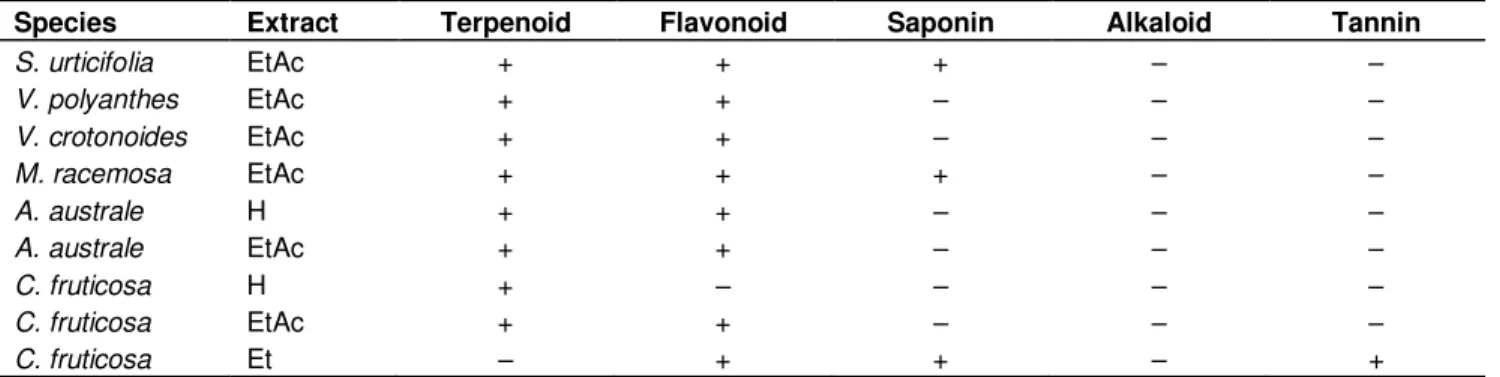
Table 2. Phytochemical analysis of the most active extracts.
Species Extract Terpenoid Flavonoid Saponin Alkaloid Tannin
S. urticifolia EtAc + + + – –
V. polyanthes EtAc + + – – –
V. crotonoides EtAc + + – – –
M. racemosa EtAc + + + – –
A. australe H + + – – –
A. australe EtAc + + – – –
C. fruticosa H + – – – –
C. fruticosa EtAc + + – – –
C. fruticosa Et – + + – +
H, Hexane; EtAc, ethyl acetate; Et, ethanol. Key: + = present ; – = absent
human tumor cell lines when compared to those from hexane or ethanol. Calea fruticosa (Gardner) Urbatsch, Zlotsky and Pruski was the only plant species that revealed promising in vitro cytotoxic action with their hexane, ethyl acetate and ethanol extracts. All extracts were found to be ineffective against all the tested protozoa. The results of phytochemical screening of the most active extracts are shown in Table 2. Alkaloids were not present in any extract. The presence of tannins was observed only in C. fruticosa (ethanolic extract). The presence of terpenoids has been detected in all extracts except for the ethanolic extract of C. fruticosa. Flavonoids were also observed in all extracts except for the hexanic extract of C. fruticosa. Only Stevia urticifolia Thunb. (ethyl acetate extract), Moquinia racemosa DC. (ethyl acetate extract) and C. fruticosa (ethanolic extract) showed the presence of saponins.
DISCUSSION
The 16 extracts that showed moderate to high in vitro cytotoxic activities against human cancer cells were considered promising anticancer compound sources. Researches for antineoplasic compounds have demon-strated the great pharmacological relevance of the plant extracts (Ferreira et al., 2011). According to the American National Cancer Institute, the limit to be considered a promising crude extract for further purification is a value
lower than 50 μg/ml and cell proliferation inhibition is
higher than 90% (Suffness and Pezzuto, 1990; Ferreira et al., 2011). In relation to the cytotoxic or antitumor activity of these plant species, rare findings are available. For example, Acanthospermum australe extracts were capable to increase the survival of Ehrlich ascites tumor-bearing mice and stimulated myelepoiesis, which can influence on antitumor immune responses (Mirandola et al., 2002).
Studies indicate that some plant substances like polyphenols, epicatechins, steryl glycosides and triterpe-noid saponins cause damage to red cell membranes and
produce hemolysis (Costa-Lotufo et al., 2002). The mechanical stability of erythrocyte membrane a good indicator of insults by vegetal substances (Sharma and Sharma, 2001; Santos et al., 2010). Then, hemolysis detection is an useful and cheap technique which can displays the effect of increasing concentrations and can to be sigmoidally related to the logarithm contact time, emphasizing the membrane stability as a biological complex to maintain its structure under stress conditions, such as oxidation, hipotonicity, pH changes, heat and in presence of osmotic active solutes (Van Ginkel and Sevanian, 1994; Sharma and Sharma, 2001; Freitas et al., 2008). Herein, none of the extracts tested caused
hemolysis even at the highest concentration (200 μg/ml),
suggesting that the mechanism of cytotoxicity is probably related to a more specific pathway. Guo and Gao (2013) described the antiproliferative effects of SPV (total saponin extract from Patrinia villosa) and FPV (total flavonoid extract from P. villosa) on four cancer cell lines and concluded that the mechanisms involved in cancer chemoprevention by FPV and SPV extracts were cell cycle arrest and induction of apoptosis. Targeting cell cycle and apoptotic pathways has emerged as an attractive approach for treatment of cancer (Aslantürk and Çelik, 2013).
Vernonia polyanthes Less., known as assa-peixe, have been studied and their pharmacological properties which include antinociceptive and anti-inflammatory (Temponi et al., 2012), antibacterial (Silva et al., 2012), antifungal and leishmanicidal (Braga et al., 2007), have been established. Fixed acids, alkaloids, aminoacids, couma-rins, steroids, triterpenes, anthraquinones, flavonoids, saponins and tannins were detected in infusions of V. polyanthes and may be responsible for their pharmacolo-gical effects (Temponi et al., 2012). Acanthospermum australe (Loefl.) Kuntze is an annual shrub widely distributed in South America. In Brazil, where it is
popularly known as “carrapichinho” or “carrapicho
-de-carneiro”. Its aerial parts are used in folk medicine as a
(Lorenzi and Matos, 2002). Previous phytochemical investigations of A. australe have led to the isolation of germacranolides, melampolides, diterpene lactones and 6-methoxyflavonoids (Bohlmann et al., 1979, 1981; Matsunaga et al., 1996). Antiviral (Rocha Martins et al., 2011) and antitumor properties (Mirandola et al., 2002) were already described for this species of plant. Vernonia crotonoides (DC.) Sch.Bip. is a synonym of Eremanthus crotonoides (DC.) Sch.Bip. (Robinson, 1999). Bohlmann et al. (1982) revealed that germacrene D,
bicycloger-macrene, α-humulene, caryophyllene, lupeol and its
acetate derivative, taraxasterol and its acetate, stigmasterol, and sesquiterpene lactones are present in aerial parts of E. crotonoides. Lobo et al. (2012) evaluated the antiproliferative effects of extracts and sesquiterpene lactone from E. crotonoides against two brain tumor cell lines. Dichloromethane fraction was cytotoxic to both glioblastoma multiforme cell lines. Centratherin alone was also evaluated against both U251 and U87-MG cells, which showed IC50 values comparable with those obtained for the commercial anticancer drug doxorubicin.
To our knowledge, no further research was carried out with the species S. urticifolia, M. racemosa, Mutisia campanulata Less, and C. fruticosa.
In this study, the evaluation of the extracts (hexane, ethyl acetate and ethanol) against E. histolytica, T. vaginalis and G. lamblia, was carried out. However, none of the extracts showed antiprotozoal activity.
The Asteraceae plant species tested showed important activity against human tumor cell lines examined. These findings are the base for further studies to isolate (guided by biological assays) and elucidate, the structure of the bioactive compounds assessed from these plants.
Our results are a contribution to a better understanding of the Brazilian biodiversity, which indicate that these natural sources may become an important source for therapeutic agents.
ACKNOWLEDGEMENT
We are grateful to Universidade Federal de Ouro Preto (UFOP) and to Brazilian agencies Conselho Nacional de Desenvolvimento Científico e Tecnológico (CNPq), Fundação de Amparo à Pesquisa de Minas Gerais (FAPEMIG), Fundação de Amparo à Pesquisa do Estado do Piauí (FAPEPI) and Fundação Cearense de Amparo à Pesquisa (FUNCAP) for financial support.
REFERENCES
Abalaka ME, Adeyemo SO, Daniyan SY (2011). Evaluation of the antimicrobial potentials of leaf extracts of Khaya senegalensis. J. Pharm. Res. Opinion,1(2):48-51.
Amaral FMM, Ribeiro MNS, Barbosa-Filho JM, Reis AS, Nascimento FRF, Macedo RO (2006). Plants and chemical constituents with giardicidal activity. Rev. Bras. Farmacogn, 16(Supl.):696-720. Aslantürk OS, Çelik TA (2013). Investigation of antioxidant, cytotoxic
and apoptotic activities of the extracts from tubers of Asphodelus aestivus Brot. Afr. J. Pharm. Pharmacol., 7(11):610-621.
Bautista E, Calzada F, Ortega A, Yépez-Mulia L (2011). Antiprotozoal activity of flavonoids isolated from Mimosa tenuiflora (Fabaceae-Mimosoideae). J. Mex. Chem. Soc., 55(4):251-253.
Berlinck RGS, Ogawa CA, Almeida AMP, Andrade MAS, Malpezzi ELA, Costa LV, Hajdu EM, Freitas JC (1996). Chemical and pharmaco-logical characterization of halitoxin from Amphimedon viridis
(PORIFERA) from the southeastern brazilian coast. Comp. Biochem. Physiol.,115C(2):155-163.
Bohlmann F, Jakupovic J, Dhar AK, King RM, Robinson H (1981). Two sesquiterpene and three diterpene lactones from Acanthospermum australe. Phytochemistry,20(5):1081-1083.
Bohlmann F, Jakupovic J, Zdero C, King RM, Robinson H (1979). Neue melampolide und cis,cis-germacranolide aus vertretern der subtribus melampodiinae. Phytochemistry,18(4):625-630.
Bohlmann F, Singh P, Zdero C, Ruhe A, King RM, Robinson H (1982). Furanoheliangolides from two Eremanthus species and from Chresta sphaerocephala. Phytochemistry,21(7):1669-1673.
Braga FG, Bouzada MLM, Fabri RL, Matos MO, Moreira FO, Scio E, Coimbra ES (2007). Antileishmanial and antifungal activity of plants used in tradicional medicine in Brazil. J. Ethnopharmacol. 111(2):396–402.
Bremer K (1994). Asteraceae: Cladistics and Classification. Timber Press, Inc., Portland.
Cantillo-Ciau Z, Moo-Puc R, Quijano L, Freile-Pelegrín Y (2010). The tropical brown alga Lobophora variegata: a source of antiprotozoal compounds. Mar Drugs 8(4):1292-1304.
Costa-Lotufo LV, Cunha GMA, Farias PAM, Viana GSB, Cunha KMA, Pessoa C, Moraes MO, Silveira ER, Gramosa NV, Rao VSN (2002). The cytotoxic and embryotoxic effects of kaurenoic acid, a diterpene isolated from Copaifera langsdorffii oleo-resin. Toxicon, 40(8):1231-1234.
Diamond LS, Clark CG, Cunnick CC (1995). YI-S, a casein-free medium for axenic cultivation of Entamoeba histolytica, related Entamoeba, Giardia intestinalis and Trichomonas vaginalis. J. Eukaryot. Microbiol. 42(3):277-278.
Diamond LS, Harlow DR, Cunnick CC (1978). A new medium for the axenic cultivation of Entamoeba histolytica and other Entamoeba. T. R. Soc. Trop. Med. H. 72(4):431-432.
Edeoga HO, Okwu DE, Mbaebie BO (2005). Phytochemical constituents of some Nigerian medicinal plants. Afr. J. Biotechnol. 4(7):685-688.
Egwaikhide PA, Gimba CE (2007). Analysis of the phytochemical content and anti-microbial activity of Plectranthus glandulosis whole plant. Middle-East J. Sci. Res., 2(3-4):135-138.
Ferrari FC, Grabe-Guimarães A, Carneiro CM, Souza MR, Ferreira LC, Oliveira TT, Saúde-Guimarães DA (2012). Toxicological evaluation of ethanolic extract of Lychnophora trichocarpha, Brazilian arnica. Rev. Bras. Farmacogn. 22(5):1104-1110.
Ferreira PMP, Farias DF, Vianna MP, Souza TM, Vasconcelos IM, Soares BM, Pessoa C, Costa-Lotufo LV, Moraes MO, Carvalho AFU (2011). Study of the antiproliferative potential of seed extracts from Northeastern Brazilian plants. An. Acad. Bras. Cienc. 83(3):1045-1058.
Freitas MV, Netto RCM, Huss JCC, Souza TM, Costa JO, Firmino CB, Penha-Silva N (2008). Influence of aqueous crude extracts of medicinal plants on the osmotic stability of human erythrocytes. Toxicol. Vitro 22(1):219-224.
Girón LM, Aguilar GA, Cáceres A, Arroyo GL (1988). Anticandidal activity of plants used for the treatment of vaginitis in Guatemala and clinical trail of Solanum nigrescens preparations.J. Ethnopharmacol. 22(3):307-313.
Guimarães LGL, Cardoso MG, Silva LF, Gomes MS, Andrade MA, Souza JA, Miranda CASF, Andrade J, Machado SMF, Figueiredo AC, Barroso JG, Mansanares ME, Nelson, DL (2012). Chemical analyses of the essential oils from leaves of Mikania glauca Mart. e x Baker. J. Essent. Oil Res. 24(6):599-604.
Guo L, Gao X (2013). Antitumor effects and mechanisms of total saponin and total flavonoid extracts from Patrinia villosa (Thunb.) Juss. Afr. J. Pharm. Pharmacol. 7(5):165-171.
Amorim LMF, Burth P, Rocha L, Santos MG, Harmerski L, Lopes NP, Pinto AC (2012). Antiproliferative activity of Eremanthus crotonoides
extracts and centratherin demonstrated in brain tumor cell lines. Rev. Bras. Farmacogn. 22(6):1295-1300.
Lorenzi H, Matos FJA (2002). Medicinal plants in Brazil: native and exotic. Pantarum Institute, São Paulo.
Majumdar SH (2012). Antitumor potential of Semecarpus anacardium
against Ehrlich ascites carcinoma in nude mice. Int. J. Pharm. Biol. Sci. 3(4):820-829.
Matsunaga K, Saitoh M, Ohizumi Y (1996). Acanthostral, a novel antineoplastic cis-cis-cis-germacranolide from Acanthospermum australe. Tetrahedron Lett. 37(9):1455-1456.
Mesquita ML, Paulab JL, Pessoa C, Moraes MO, Costa-Lotufo LV, Grougnetd R, Michel S, Tillequind F, Espindolaa LS (2009). Cytotoxic activity of Brazilian Cerrado plants used in traditional medicine against cancer cell lines. J. Ethnopharmacol. 123(3):439-445. Mirandola L, Justo GZ, Queiroz ML (2002). Modulation by
Acanthospermum australe extracts of the tumor induced
hematopoietic changes in mice. Immunopharmacol. Immunotoxicol. 24 (2): 275-288.
Morais LAS, Castanha RF (2011). Composição química do óleo essencial de duas amostras de carqueja (Baccharis sp.) coletadas em Paty do Alferes – Rio de Janeiro. Rev. Bras. Pl. Med. Sepc. Issue13:628-632.
Mosmann T (1983). Rapid colorimetric assay for cellular growth and survival: application to proliferation and cytotoxicity assays. J. Immunol. Methods, 65(1-2):55-63.
Patel S, Gheewala N, Suthar A, Shah A (2009). In-vitro cytotoxicity activity of Solanum nigrum extract against hela cell and vero cell line. Int. J. Pharm. Pharm. Sci. Suppl 1.1:38-45.
Pérez GS, Ramos-López MA, Sánchez-Miranda E, Fresán-Orozco MC, Pérez-Ramos J (2012). Antiprotozoa activity of some essential oils. J. Med. Plants Res.6 (15):2901-2908.
Robinson H (1999). Generic and Subtribal Classification of American Vernonieae. Smithson Contrib. Bot. 89:1-116.
Carvalho et al. 2005
Rocha Martins LR, Brenzan MA, Nakamura CV, Dias Filho BP, Nakamura TU, Cortez LER, Cortez DAG (2011). In vitro antiviral activity from Acanthospermum australe on herpesvirus and poliovirus. Pharm. Biol. 49(1):26-31.
Santos AG, Ferreira PMP, Vieira-Júnior GM, Perez CC, Tininis AG, Silva GH, Bolzani VS, Costa-Lotufo LV, Pessoa C, Cavalheiro AJ (2010). Casearin X, its degradation product and other clerodane diterpenes from leaves of Casearia sylvestris: evaluation of cytotoxicity against normal and tumour human cells. Chem. Biod. 7(1): 205-215.
Sharma P, Sharma JD (2001). In vitro hemolysis of human erythrocytes by plant extracts with antiplasmodial activity. J. Ethnopharmacol. 74(3):239-243.
Silva NCC, Barbosa L, Seito LN, Fernandes Junior A (2012). Antimicrobial activity and phytochemical analysis of crude extracts and essential oils from medicinal plants. Nat. Prod. Res. 26(16):1510–1514.
Suffness M, Pezzuto JM (1990). Assays related to cancer drug discovery. In: Hostettamann K (Ed), Methods in plant biochemistry: assays for bioactivity. Acad. Press, London, pp.71-133.
Temponi VS, Da Silva JB, Alves MS, Ribeiro A, De Pinho JJRG, Yamamoto CH, Pinto MAO, Del-Vechio-Vieira G, De Sousa OV (2012). Antinociceptive and anti-inflammatory effects of ethanol extract from Vernonia polyanthes leaves in rodents. Int. J. Mol. Sci. 13(3):3887-3899.