Joana Margarida de Andrade Poejo
Degree in Biochemistry
Evaluation of
Opuntia spp.
bioactive products
as promising natural chemotherapeutical
agents- an
in vitro
approach
Dissertation to obtain a Master Degree in Biotechnology
Supervisor: Ana Teresa Serra, Ph.D, IBET/ITQB-UNL
Co-Supervisor: Catarina Duarte, Ph.D, IBET/ITQB-UNL
Júri
Presidente: Prof. Doutora Cecília Roque
Arguente: Prof. Doutora Maria Eduardo
Vogal: Doutora Ana Teresa Serra
Evaluation of Opuntia spp. bioactive products as promising natural chemotherapeutical agents- an in vitro approach
Copyright Joana Poejo, FCT/UNL, UNL
Acknowledgments
Gostaria de agradecer a todas as pessoas que directa ou indirectamente contribuíram para me ajudar na realização deste trabalho.
Em primeiro lugar, gostaria de agradecer à Dra. Ana Teresa Serra por me ter acompanhado incansavelmente desde o primeiro ao último dia. Agradeço a constante orientação prestada, os conselhos e ideias novas, a paciência inesgotável, o apoio e o encorajamento demonstrado nas fases mais difíceis. Obrigada por me teres ajudado a crescer tanto profissionalmente como pessoalmente.
À Dra. Catarina Duarte por me ter dado oportunidade para desenvolver o trabalho no seu laboratório. Obrigada pela sua confiança, pelo incentivo e apoio demonstrados durante este ultimo ano.
Agradeço a toda a equipa do laboratório de Nutracêuticos e Libertação Controlada. À Dra. Ana Matias pela sua ajuda e partilha de excelentes conhecimentos e ideias. À Sara Nunes, minha companheira “Opuntiana”, agradeço os momentos divertidos e de cumplicidade que facilitaram de algum modo a execução do trabalho. Obrigada também pela tua ajuda e conselhos. À Ana Nunes agradeço a excelente companhia, o apoio, o incentivo e as palavras amigas nos momentos mais solitários do Verão. Agradeço à Patrícia Almeida pela sua preciosa ajuda e por estar sempre disponível. Ao Agostinho Alexandre agradeço pela sua valiosa e indispensável ajuda no projecto
“Opuntia”. À Dora Pereira agradeço os ensinamentos iniciais, a simpatia e a boa disposição sempre demonstrada. Agradeço à Dra. Maria do Rosário Bronze e à Catarina Semedo pela ajuda prestada na parte analítica do projecto.
Agradeço a todos os meus amigos que de alguma forma me ajudaram a superar os momentos mais difíceis, que sempre me apoiaram e ajudaram quando mais precisei, agradeço especialmente à Sofia Fortalezas.
Ao Filipe pelo seu apoio incondicional e compreensão. Por me ter ajudado a ultrapassar muitos obstáculos, por me ter ouvido quando mais precisei.
À minha família: pais e irmãos especia lment e. Agradeço aos meus irmãos pelo apoio, incentivo e motivação. Aos meus pais por sempre me terem apoiado ao longo de todo este percurso, por me incentivarem e acreditarem em mim do princípio ao fim. Obrigada por me terem ajudado a ultrapassar mais uma etapa importante da minha vida.
Obrigada por tudo!
Abstract
Recently, prevention of cancer through dietary intervention has received an increasing interest. In particular, dietary polyphenols mainly found in fruits and vegetables have become very attractive as chemopreventive and therapeutic agents.
The main aim of this work was to investigate the potential activity of Portuguese Opuntia spp. on colon cancer therapy, as this fruits have been reported to be rich sources of bioactive compounds.
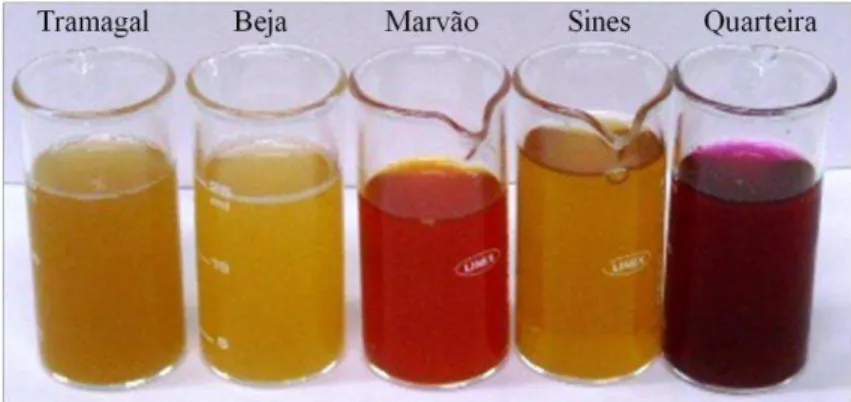
Opuntia spp. fruits were collected in different sites of Portugal, namely Tramagal, Beja, Marvão Sines and Quarteira and processed in order to obtain fruit juices, polyphenol-rich concentrates and residues extracts from fruit juices production. All Opuntia products were investigated for antiproliferative activity of human colon cancer cells (HT29). Opuntia juices from Tramagal, Sines and Quarteira were the most effective on cancer cell growth inhibition due to the highest content in total polyphenol content and betalains.
The most promising extracts were derived from Quarteira, Sines and Beja fruit juices residues using conventional solvent extraction, adsorption technology and pressurized liquid extraction, respectively. These extracts showed high antiproliferative effect inducing cell cycle arrest in different checkpoints. Additionally, the same extracts showed to induce ROS production in cancer cells, playing an important role in activation of pro-apoptic cell death mechanisms.
The capacity of natural extracts in reverting chemoresistance of tumor cells was evaluated on a drug-resistant HT29 cell culture. The product derived from Beja residue using pressurized liquid extraction with 60%CO2 and 40%EtOH showed to increase cells sensitivity to the drug.
Opuntia spp. fruits reveled to be promising natural sources for the production of functional products as well as for the development of novel chemotherapeutic agents.
Keywords: Colon cancer; polyphenols; Opuntia spp.; antiproliferative effect; chemoresistance.
Resumo
Recentemente tem surgido um grande interesse na prevenção do cancro através da dieta. Em particular, os polifenóis presentes nas frutas e nos vegetais são reconhecidos como sendo compostos muito atractivos na área da quimioprevenção e terapia do cancro.
O principal objectivo deste trabalho consistiu na avaliação da potencial actividade de frutos de
Opuntia spp. Portugueses para terapia do cancro do cólon, uma vez que este frutos são reconhecidos como sendo fontes ricas em compostos bioactivos.
Os frutos de Opuntia spp. foram recolhidos em diferentes regiões de Portugal, nomeadamente Tramagal, Beja, Marvão, Sines e Quarteira, e foram processados para obter sumos de fruta, extractos concentrados em polifenóis e extractos de resíduo obtidos a partir do processamento dos sumos de fruta. Todos os produtos foram analisados em termos de actividade antiproliferativa numa linha celular humana de cancro do cólon (HT29). Os sumos de fruta provenientes de Tramagal, Sines e Quarteira foram os mais efectivos na inibição do crescimento das células cancerígenas devido ao elevado conteúdo em polifenóis e à presença de betalainas.
Os extractos mais promissores foram obtidos a partir dos resíduos dos sumos de Quarteira, Sines e Beja, usando tecnologias limpas como a extracção convencional, tecnologia de adsorção e a extracção com solventes pressurizados. Os extractos obtidos demonstraram um efeito antiproliferativo elevado e induziram a paragem do ciclo celular em fases distintas. Para além disso induziram a produção de espécies reactivas de oxigénio (ROS) a nível celular, desempenhando assim um papel importante na activação de mecanismos pro-apoptóticos de morte celular.
Neste trabalho foi também avaliada a capacidade dos extractos naturais em reverter a quimioresistência em células resistentes à doxorubicina, uma droga anticancerígena. O extracto obtido por extracção com solventes pressurizados, nomeadamente 60%CO2 e 40% EtOH, a partir do resíduo
de Beja, demonstrou aumentar a sensibilidade à droga nas células resistentes.
Os resultados obtidos nesta tese demonstraram que os frutos de Opuntia spp. são fontes promissoras para a produção de produtos funcionais e para o desenvolvimento de novos agentes naturais para aplicação em quimioterapia.
Palavras-chave: cancro do cólon; polifenóis; Opuntia spp.; actividade antiproliferativa; quimioresistência.
Index
1. Introduction ………..………. 1
1.1 Cancer worldwide ……….... 1
1.1.1Colorectal Cancer ………... 1
1.1.1.1 Epidemiology, etiology and causes ………... 1
1.1.1.2 Chemoteraphy and chemoprevention: a perfect combination ………. 2
1.1.1.3 Food and nutrition on cancer chemoprevention ………..….... 4
1.2 Polyphenols and health-promoting effects ………...………...… 7
1.3 Opuntia spp. ……….….. 11
1.4 Nutraceuticals and functional foods ………...………..…. 12
1.4.1 Extraction methods ……….………..…. 13
1.5 Aim and rational of the thesis ……….…... 14
2. Experimental procedure ……….……… 17
2.1 Chemicals ……...………...……… 17
2.2 Opuntia spp. samples and fruit juices preparation …..………..… 17
2.3 Opuntia spp. extracts ……….… 18
2.3.1 Opuntia spp. extracts from fruit juices: adsorption technology (AD) ………...……….... 18
2.3.2 Opuntia spp. extracts from fruit juices residues: Conventional solvent extraction (CSE). 18 2.3.3 Opuntia spp. extracts from fruit juices residues: adsorption technology (AD) …...…… 19
2.3.4 Opuntia spp. extracts from fruit juices residues: Pressurized liquid extraction (PLE) .… 19 2.4 Phytochemical characterization ………. 20
2.4.1 Polyphenols ………...……….…….….. 20
2.4.2 Flavonoid content ……….………. 20
2.5 Antioxidant activity ………..………. 21
2.5.1 Oxygen radical absorbance capacity (ORAC) ...…….…………... 21
2.5.2 Hydroxyl Radical Adverting Capacity (HORAC) ………...………. 21
2.6 Cell-Based Assays ………. 22
2.6.1 Cell culture ……….………...……… 22
2.6.2 Citotoxicity assay ………...………...… 22
2.6.3 Antiproliferative assay ……….………..……… 22
2.6.4 Cell Cycle analysis ……….………...…… 23
2.6.5 Detection of ROS generation …..……….……. 23
2.6.6 Chemosensitization effect ………...……….…. 24
2.6.6.1 Development of a subpopulation drug-resistant (HT29-dx) ... ………...… 24
2.6.6.2 Cellular sensitivity to drug ………..……… 24
2.6.6.3 Intracellular doxorubicin accumulation ……….. 24
2.6.6.4 Chemosensitization activity ……….……..………. 25
3. Results and discussion ……….... 27
3.1 PART 1 - Looking for anticancer activity in Opuntia spp. juices ………. 27
3.1.1 Phytochemical characterization …….……… 27
3.1.2 Antioxidant activity ……….……….. 29
3.1.3 Antiproliferative effect ………..…… 30
3.2 PART 2: Development of functional ingredients from Opuntia spp. and screening of their anticancer effect ……….………..……. 32
3.2.1 Extracts from Opuntia spp. juices: adsorption technology (AD) …...…………...…….... 32
3.2.1.1 Phytochemical and antioxidant characterization ………...………. 32
3.2.1.2 Antiproliferative effect and cell cycle analysis ……….….. 34
3.2.2 Extracts from Opuntia spp.fruit juice residues: conventional solvent extraction (CSE) and adsorption technology (AD)……….……….…... 37
3.2.2.1 Phytochemical and antioxidant characterization ……...………... 38
3.2.2.2 Antiproliferative effect ………...…….... 40
3.2.3 Extracts from Opuntia spp. fruit juice residues: pressurized liquid extraction (PLE) ... 42
3.2.3.1 Phytochemical and antioxidant characterization ………...……….… 43
3.2.3.2 Antiproliferative effect ………..………...…….. 45
3.3. Evaluation of Opuntia spp. extracts as promising bioactive ingredients for colon cancer therapy ………...……….... 47
3.3.1 Cell cycle arrest and monitorization of ROS production ……….. 47
3.3.2 Chemosensitization effect in a drug resistant cell sub-population (HT29 dx) ……..…… 49
3.3.2.1 Cellular sensitivity to drug and intracellular accumulation of doxorubicin …..…. 49
3.3.2.2 Chemosensitization activity …….………...… 51
4. Conclusions ………...……… 53
5. References ……….… 55
Index of figures
Figure 1.1 Page 3 Sequence of multistage carcinogenesis process
Figure 1.2 Page 4 Mechanisms of action of natural products in colon cancer chemoprevention
Figure 1.3 Page 7 Chemical structures of the main classes of polyphenols
Figure 1.4 Page 8 Polyphenols and their biological properties
Figure 1.5 Page 11 Images of Opuntia spp. cladodes and fruits
Figure 1.6 Page 14 Structure of the thesis
Figure 3.1 Page 27 Opuntia spp. fruits from: A) O. robusta (Quarteira) and B) O. ficus- indica (Marvão)
Figure 3.2 Page 28 Opuntia spp. fruit juices from Tramagal, Beja, Marvão, Sines and Quarteira
Figure 3.3 Page 29 Chromatograms profiles of Opuntia spp. juices: Tramagal, Beja,
Marvão, Sines and Quarteira at A) 280nm, B) 360nm, C) 480nm and D) 535nm. Legend: 1- citric acid, 2 – gallic acid, 3- isorhamnetin-3-O-rhamnose-lyxose-glucose, 4- isorhamnetin-3-O-lyxose-glucose, 5- isorhamnetin-3-O-rutinoside, 6- isorhamnetin, 7- indicaxantin, 8- leucina-bx
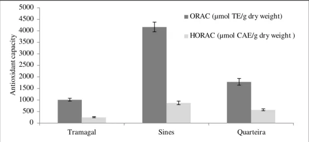
Figure 3.4 Page 30 Antioxidant capacity (ORAC and HORAC values) of Opuntia spp fruit juices varieties
Figure 3.5 Page 31 Antiproliferative effect of Portuguese Opuntia spp juices on human colon cancer cells (HT29) (incubation time: 96 hours)
Figure 3.6 Page 32 Total polyphenol content (mg GAE/g dry weight) of Sines, Quarteira and Tramagal PRCs
Figure 3.7 Page 33 Chromatograms of Opuntia PRCs from Tramagal, Sines and Quarteira (1mg/mL) at (A) 280nm, (B) 360 nm, (C) 480nm and (D) 535 nm. Legend: 1 – piscidic acid
Figure 3.8 Page 34 Antioxidant capacity (ORAC and HORAC assays) of PRCs (Sines, Tramagal and Quarteira)
Figure 3.9 Page 35 Antiproliferative activity of PRCs on HT29 cell line. (Incubation time= 72h)
Figure 3.10 Page 36 HT29 cells, in 25 cm2 flask, treated with: A) Control; B) PRC-Sines; C)
PRC- Quarteira
Figure 3.11 Page 36 Cell cycle distribution of HT29 cells incubated with PRC samples (5mg/mL) during 48 and 72 hours
Figure 3.12 Page 36 Phase contrast images of cells. A) Control (RMPI medium with solvent); B) PRC-Quarteira; C) PRC-Sines. Scale bar: 200μm
Figure 3.13 Page 38 Dried extracts obtained from Sines (S1) and Quarteira (Q1) fruit residues and its PRCs (S2 and Q2, respectively)
Figure 3.14 Page 38 Total polyphenol content of extracts derived from Sines (S1 and S2) and Quarteira (Q1 and Q2) residues
Figure 3.15 Page 39 Figure 3.15. Chromatograms profiles of S1 (50 mg/ml), S2 (10 mg/ml),
Q1(50 mg/ml) and Q2 (10 mg/ml) at A) 280 nm; B) 360 nm; C) 480 nm; D) 520 nm. Legend: 1- piscidic acid.
Figure 3.16 Page 39 Antioxidant capacity (ORAC and HORAC) of residues extracts from Sines (S1) and Quarteira (Q1) and its respective PRCs (S2 and Q2)
Figure 3.17 Page 40 Antiproliferative effect of S1 and Q1extracts on HT29 cells. (Incubation time= 24 hours)
Figure 3.18 Page 41 Antiproliferative effect of S2 and Q2on HT29 cells (Incubation time: 24 hours)
Figure 3.19 Page 42 Anticancer effect on HT29 cells after residues extracts of Sines and Quarteira and PRCs exposure for 24 hours. The results were presented in total polyphenol content of extracts (mg GAE/mL)
Figure 3.20 Page 42 Total polyphenol content of extracts obtained from Beja residue, using PLE and CSE
Figure 3.21 Page 43 Chromatograms profiles of PLE and CSE extracts (200 mg/ml) at: A) 280nm and B) 360nm. 1, 2, 3, 4, 5, 6, 7, 8, 9, 10, A, B, C e D
Figure 3.22 Page 44 Chromatograms profiles of 1, A and B extracts at: A) 280 nm and B) 360 nm
Figure 3.23 Page 44 Chromatograms profiles of 3 and C extracts at: A) 280 nm and B) 360 nm
Figure 3.24 Page 44 Chromatograms profiles of 6 and D extracts at: A) 280 nm and B) 360 nm
Figure 3.25 Page 45 Antioxidant capacity of extracts obtained from Beja residue, using PLE and CSE
Figure 3.26 Page 46 Antiproliferative effect on HT29 cell line, after 24 hours of CSE extracts treatment
Figure 3.27 Page 46 Antiproliferative effect on HT29 cell line, after 24 hours of PLE extracts treatment
Figure 3.28 Page 47 Antiproliferative effect on HT29 cell line, after 24 hours of treatment with extract 2
Figure 3.29 Page 48 Cell cycle distribution of HT29 cells incubated for 24h with different Opuntia spp. extracts
Figure 3.30 Page 48 Effect of Opuntia extracts (0.5 mg/mL) on reactive oxygen species (ROS) accumulation in HT29 cells, after 24h incubation
Figure 3.31 Page 50 Inhibitory effect of doxorubicin exposure during1 hour, in HT29 cell line and HT29 dx
Figure 3.32 Page 50 Intracellular doxorubicin accumulation in HT29 and HT29 dx
Figure 3.33 Page 51 Toxicity effect of Opuntia extracts conjugated with doxorubicin in HT29
dx cells
Figure 3.34 Page 51 Toxicity of Q1 extract (6 and 9 mg/mL) and /or doxorubicin (0-20 uM)
on HT29 and HT29dx cells.
Figure 3.35 Page 52 Toxicity of PLE-2 extract (6 and 9 mg/mL) and /or doxorubicin (0-20 µM) on HT29 and HT29dx cells
Figure 3.36 Page 52 Toxicity effect of PLE-2 extract plus doxo or using only doxo in HT29 or HT29 dx
Index of tables
Table 1.1 Page 2 Colorectal Cancer Incidence and Mortality Worldwide in 2008. Numbers are expressed in thousands
Table 1.2 Page 6 The factors that modify the risk of cancers of the colon and the rectum. Judgments are graded according to the strength of the evidence by the Panel of WCRF/AICR Continuous Update Project 2011
Table 1.3 Page 10 Mechanisms involved in the chemopreventive effect of polyphenols in colorectal cell lines
Table 2.1 Page 19 Extracts obtained from Beja residue using PLE technology
Table 3.1 Page 27 Opuntia spp. fruits varieties, color juice and phytochemical characterization
Table 3.2 Page 31 The amount of sample necessary to decrease 50% of the cellular viability (ED50) after 96 h of Opuntia juice exposure to HT29 cell line
Table 3.3 Page 33 Total polyphenol content (mg GAE/dry weight) of PRCs and juices
Table 3.4 Page 34 Anitoxidant capacity (ORAC and HORAC) of juices and its PRCs
Table 3.5 Page 35 ED50 values of PRCs in HT29 cells. (Incubation time= 72h)
Table 3.6 Page 37 Extracts obtained from Sines and Quarteira residues and its PRCs
Table 3.7 Page 40 Comparison of total polyphenols and antioxidant activity between juices, residues and respective PRCs of Sines and Quarteira Opuntia spp. fruits Table 3.8 Page 41 ED50 values in HT29 cells after 24h of residues extracts and PRCs
exposure
Table 3.9 Page 42 Extracts obtained from Beja residue using PLE and CSE
Table 3.10 Page 50 IC50 values determined for HT29 cell line and HT29 dx, after 1h drug exposure
Table 3.11 Page 53 ED50 values determined in chemosensitization activity
List of abbreviations, acronyms and symbols
Abbreviation Full form
AAPH 2‟,2‟-Azobis (2-amidinopropane) dihydrochloride
ACS American Cancer Society
AD Adsorption technology
AICR American Institute of Cancer Research
ASE Accelerated Solvent Extraction
BSA Bovine Serum Albumine
Caco-2 Caco-2 human colon carcinoma cell line
CAE Caffeic Acid Equivalents
CO2 Carbon dioxide
CSE Conventional Solvent Extraction
DCFH-DA 2‟,7‟-Dichlorofluorescin Diacetate
DNA Deoxyribonucleic acid
ED50 Median effective dose
EDTA Ethylenediamine Tetraacetic Acid
EGCG Epigallocatechin-3-gallate
EtOH Ethanol
FBS Fetal Bovine Serum
FL Disodium Fluorescein
GAE Gallic Acid Equivalents
HE Hericium erinaceus
HORAC Hydroxyl Radical Adverting Capacity
HPLC High Performance Liquid Chromatography
HT29 HT29 human colon cancer cell line
HT29 dx HT29 human colon cancer cell line drug-resistant
MDR Multidrug-Resistance
MPa MegaPascal
MTT Methylthiazolyldiphenyl tetrazolium bromide
NCI National Cancer Institute
NHE Na+/H+ exchanger
NO Nitric Oxide
ORAC Oxygen Radical Absorbance Capacity
PBS Phosphate Buffer Saline
PFE Pressurized Fluid Extraction
PLE Pressurized liquid extraction
PRC Polyphenol-Rich Concentrate
ROS Reactive Oxygen Species
SFE Supercritical Fluid Extraction
TCF Total Flavonoid
Content TPC Total Polyphenol
Content UV Ultra - violet
1.1 Introduction
1.1 Cancer worldwide
Cancer is a leading cause of mortality in human and a growing health problem all around the world. One defining feature of cancer is the rapid creation of abnormal cells that grow beyond their usual boundaries and spread to other organs, creating metastasis (Haque, et al., 2010). According to Cancerbase GLOBOCAN, in 2008, more than 12 million new cases of cancer were registered (excluding non-melanoma skin cancer) while 7.5 million people died from cancer worldwide. In 2020 it is estimated that mortality from cancer will increase to more than 10 million (Ferlay J, et al., 2010).
Since there are no particular treatment of cancer, effective preventive measures and cancer awareness among the general population is essential. Many clinical and laboratory studies support that our lifestyle and dietary factors are playing a complex multifaceted role in etiology of cancer (Haque, et al., 2010). Several organizations such as: World Health Organization (WHO), American Cancer Society (ACS), American Institute of Cancer Research (AICR) and National Cancer Institute (NCI) have established dietary guidelines to help people reduce the cancer risk (Surh, 2003).
1.1.1 Colorectal Cancer
1.1.1.1 Epidemiology, etiology and causes
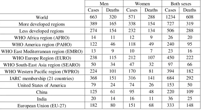
Colorectal cancer (cancer of the colon and/or rectum) is the third most common cancer in men (663 000 cases, 10.0% of the total) and the second in women (571 000 cases, 9.4% of the total) worldwide (Ferlay J, et al., 2010). In 2008, about 608 000 deaths from colorectal cancer were estimated, accounting for 8% of all cancer deaths, making it the fourth most common cause of death from cancer in world. Almost 60% of the cases occur in developed regions, but colon cancer incidence is now increasing in middle and low income countries (see table 1.1) (Ferlay J, et al., 2010).
Colon cancer is defined as any malignant neoplasm arising from the inner lining of the colonic epithelium (Rajamanickam and Agarwal, 2008). According to the 2007 Report of the World Cancer Research Fund/American Institute of Cancer Research (WCRF/AICR), approximately 95 per cent of colorectal cancers are adenocarcinomas. However other types of cancer can occur, including mucinous carcinomas and adenosquamous carcinomas. The occurrence of colon cancer is strongly related with age, 90% of the cases arising in people who are 50 years or older. Survival rates for colon cancer can vary based on a variety of factors, particularly the stage. If the cancer is detected at an early and localized stage, the five-year survival rate (the percentage of people who survive at least five years after the cancer is detected, excluding those who die from other diseases) is 90%. However, when metastasis occur, the 5-year survival rate decrease to 10% (American Cancer Society, 2008).
Etiologically, colorectal cancer may be hereditary or sporadic (Young, et al., 2005). Epidemiological studies have suggested that colon cancer is a manifestation of a number of inherited
cancer predisposition syndromes, including familial adenomatous polyposis, hereditary non-polyposis
colorectal cancer, personal or family history of colorectal cancer and/or polyps and inflammatory bowel disease (Rowley, 2005). On the other hand, sporadic colorectal cancers are due to somatic genetic mutations that occur as part of the normal cellular lifespan or because of exposure to environmental factors (Young, et al., 2005). Known causes of colon cancer include tobacco smoking, obesity and lack of exercise, infectious agents, medication, radiation, industrial chemicals, alcohol consumption and diet rich in high fat, red and processed meats. Moreover, inadequate intake of dietary fiber, fruits and vegetables are also associated with the increase of colon cancer risk (Haque, et al., 2010, Kushi, et al., 2006, Rajamanickam and Agarwal, 2008).
Table 1.1. Colorectal Cancer Incidence and Mortality Worldwide in 2008. Numbers are expressed in thousands
(adapted from Ferlay J, et al., 2010).
Men Women Both sexes Cases Deaths Cases Deaths Cases Deaths World 663 320 571 288 1234 608 More developed regions 389 165 338 154 727 319 Less developed regions 274 154 232 134 506 288 WHO Africa region (AFRO) 14 11 12 9 26 20 WHO America region (PAHO) 122 46 118 49 240 95 WHO East Mediterranean region (EMRO) 13 9 10 7 23 16 WHO Europe Region (EURO) 238 115 212 107 450 222 WHO South-East Asia region (SEARO) 50 34 47 32 97 66
WHO Western Pacific region (WPRO) 224 101 170 81 394 182 IARC membership (21 countries) 368 151 316 141 684 292 United States of America 79 24 74 26 153 50
China 125 61 95 48 220 109 India 20 14 16 11 36 25 European Union (EU-27) 182 80 151 68 333 148
1.1.1.2 Chemoteraphy and chemoprevention: a perfect combination
Conventional cancer therapies, including surgery, chemotherapy, and radiotherapy have a significant role in the overall treatment of tumors (Sarkar and Li, 2006). In particular chemotherapy has high treatment success rates in various types of cancer. However, the main drawback of this therapy consists on the multidrug-resistance (MDR) effect of cancer cell (De Boo, et al., 2009, Riganti, et al., 2005, Sarkar and Li, 2006, Schonn, et al., 2011). A variety of mechanisms by which cancer cells acquire the MDR phenotype have been described, including the reduced uptake or the increased efflux of the drug, the genetic modification of the drug's specific targets, the increased ability to repair DNA damage, the reduced capacity to enter apoptosis, the different compartmentalization and the increased rate of drug detoxification (Ferreira, et al., 2002, Gottesman, 2002). Thus, the cell resistance to anticancer drugs and the failure of chemotherapy to treat some
cancers lead to development of new therapies. The strategies of cancer treatment using combined
therapies or combined agents with distinct molecular mechanisms are considered very promising for higher efficacy, resulting in better survival (Hwang, et al., 2005, Sarkar and Li, 2006). For this reason, chemoprevention, which involves the use of specific natural products or synthetic chemical agents to reverse, suppress or prevent premalignancy before the development of invasive cancer, has appeared as a novel approach for controlling this malignant disease (Haque, et al., 2010, Rajamanickam and Agarwal, 2008, Ramos, 2008).
In terms of molecular perspective, carcinogenesis is a multistage process consisting on initiation, promotion and progression phases which involves sequential generations of cells that exhibit continuous disturbance of cellular and molecular signal cascades (Vincent and Gatenby, 2008) (figure 1.1). The initiation stage comprises the exposure or uptake of cells with a carcinogenic agent causing a genetic alteration. In the promotion stage abnormal cells persists, replicates and may originate a focus of preneoplastic cells. Finally in the last stage – progression - there is an uncontrolled growth of the cells (tumor) that involves the gradual conversion of premalignant cells to neoplastic ones with an increase of invasiveness and metastasis potential, and new blood vessel formation (angiogenesis) (Ramos, 2008). During this multistage sequence of events there are many phases for intervention to inhibit, reverse and/or delay the progress of cancer (Johnson, 2007).
Figure 1.1. Sequence of multistage carcinogenesis process (Ramos, 2008).
In recent years, dietary compounds have been recognized as cancer chemopreventive agents owing to their various health benefits and noticeable lack of toxicity and side effects (Manson, et al., 2005, Sarkar and Li, 2006). Common cancer therapies combined with these dietary compounds may exert enhanced antitumor activity through synergic action or compensation of inverse properties.
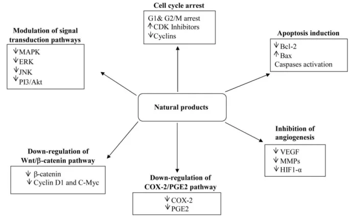
Moreover, the combination treatment may decrease the systemic toxicity caused by many chemotherapeutic drugs because lower doses could be used (Sarkar and Li, 2006). It is known that these compounds exert the anticarcinogenic activity through regulation of different cell signaling pathways (figure 1.2). Chemopreventive agents can affect all stages of carcinogenesis by inducing cell cycle arrest and apoptosis, decreasing cell proliferation and angiogenesis, inhibiting tumor cell invasion and metastasis, and modulating various signal transduction as well as COX-2/PGE2 and Wnt/β-catenin pathways, involved in colon cancer development (Dragnev, et al., 2007)
Figure 1.2. Mechanisms of action of natural products in colon cancer chemoprevention (Rajamanickam and Agarwal, 2008).
1.1.1.3 Food and nutrition on cancer chemoprevention
According to WCRF/AICR 2007 report, 30-40% of cancer could be preventable over time by appropriate food and nutrition, regular physical activity, and avoidance of obesity. On a global scale this represents over 3 to 4 million cases of cancer that can be prevented in these ways, every year (WCRF/AICR, 2007). As table 1.2 shows, colorectal cancer are greatly or mostly affected by food and nutrition. It is proven that physical activity and foods containing dietary fiber protect against colorectal cancer while foods, like red and processed meat, alcoholic drinks, body fatness and others factors increase risk of the disease. However, the findings that fruits and vegetables can protect against colon cancer are not yet conclusive, suggesting a limited-suggestive role (WCRF/AICR, 2011). Despite that, several epidemiological studies have reported an inverse correlation between decreased cancer risk and high consumption of vegetables, including cabbage, cauliflower, broccoli, brussels sprout,
tomatoes, and fruits such as, apples, grapes, and berries (Gordaliza, 2007, Vainio and Weiderpass, 2006).
The assessing of the real impact of dietary constituents on human health is difficult because in many cases the exact composition of foods and the bioavailability of active molecules are not known. The most direct evidence of beneficial effects by a particular food rich in certain compounds have come from animal models and in vitro experiments. In fact, cell culture studies constitute a valuable tool for identifying the molecular targets modulated by food compounds in cancer cells and for elucidating the molecular pathways involved in the overall disease process (Ramos, 2008).
Decrease risk Increase risk
Convincing
- Physical activity 1
- Foods containing dietary fiber
- Red meat
- Processed meat
- Alchololic drinks (men) 2 - Body fatness
- Abdominal fatness
- Adult attained height 3
Probable
- Garlic
- Milk
- Calcium
- Alcoholic drinks (woman) 2
Limited -
suggestive
- Non-starchy vegetables
- Fruits
- Foods containing folate
- Foods containing selenium
- Fish
- Foods containing vitamin D
- Selenium
- Foods containig iron
- Cheese
- Foods containing animal fats
- Foods containing sugars
Limited - no
conclusion
- Cereals (grains) and their products; potatoes; poultry; shellfish and other seafood; other dairy products; total fat; fatty acid composition; cholesterol; sugar (sucrose); coffee; tea; caffeine; total carbohydrate; starch; vitamin A; retinol; vitamin C; vitamin E; multivitamins; non-dairy sources of calcium; methionine; beta-carotene; alpha-carotene; lycopene; meal frequency; energy intake
Table 1.2. The factors that modify the risk of cancers of the colon and the rectum. Judgments are graded according
to the strength of the evidence by the Panel of WCRF/AICR Continuous Update Project 2011 (adapted
from WCRF/AICR, 2011).
1 Physical activity of all types: occupational, household, transport, and recreational. The Panel judges that the evidence for colon cancer is convincing. No conclusion was drawn for rectal cancer.
2 The judgments for men and women are different because there are fewer data for women. Increased risk is only apparent above a threshold of 30 g/day of ethanol for both sexes.
3 Adult attained heights are unlikely directly to modify the risk of cancer. It is a marker for genetic, environmental, hormonal, and also nutritional factors affecting growth during the period from preconception to completion of linear growth.
1.2 Polyphenols and health-promoting effects
“Polyphenols” (or phenolic compounds) is a generic term that refers to more than 8000 compounds widely dispersed throughout the plant kingdom (Cartea, et al., 2011). These phytochemicals are secondary metabolites and have considerable physiological and morphological functions. Polyphenols may act as phytoalexins, antifeedants, attractants for pollinators, contributors to plant pigmentation, antioxidants and protective agents against UV light, among others (Naczk and Shahidi, 2006). These bioactive properties play an important role in plant growth and reproduction and providing an efficient protection against pathogens and predators. Additionally, polyphenols contribute to the color and sensory characteristics of many fruits and vegetables (Cartea, et al., 2011, Jaganath and Crozier, 2010, Zitka, et al., 2011).
Polyphenolic compounds derived from phenylalanine and are characterized by having at least one aromatic ring with one or more hydroxyl groups attached. They are divided into several classes according to the number of phenol rings that they contain and the structural elements that bind these rings to one another (D'Archivio, et al., 2007). The main groups of polyphenols are flavonoids (eg: quercetin), phenolic acids (eg: chlorogenic acid), stilbenes (eg: resveratrol) and lignans (enterodiol). Flavonoids and phenolic acids account for 60 and 30%, respectively, of dietary polyphenols (figure
1.3) (Scalbert and Williamson, 2000).
Figure 1.3. Chemical structures of the main classes of polyphenols (adapted from Scalbert and Williamson, 2000).
Polyphenols are widespread constituents of fruits, vegetables, cereals, olive, dry legumes, chocolate and beverages, such as tea, coffee and wine (D'Archivio, et al., 2007). They have a great interest in the industry because they have many applications as food preservatives and colorants or in
production of paints, papers and cosmetic (Ignat, et al., 2011). However, in last decade, polyphenols have received much more attention for their health benefits. The most well-know property of polyphenols is their antioxidant capacity - the ability of a food constituent to enhance the endogenous antioxidant defense system (Lila, 2007). Reactive oxygen species (ROS), derived from oxidation processes, are an important part of the defense mechanisms against infection, but excessive generation of free oxygen radicals may damage the tissue. When there is an imbalance between ROS and antioxidant defense mechanisms, the ROS lead to the oxidative modification of cellular membranes or intracellular biomolecules and result in the peroxidation of membrane lipids, leading to the accumulation of lipid peroxides (Cartea, et al., 2011). Therefore, as antioxidants, polyphenols can trap ROS in the human body, protecting cells against oxidation damage and consequently limit the risk of several pathologies associated to oxidative stress (Cartea, et al., 2011, D'Archivio, et al., 2007, Lila, 2007, Ramos, 2008). In more detail, polyphenolic compounds can prevent the DNA-damage caused by free radicals or carcinogenic agents through different mechanisms: (i) direct radical scavenging (Alía, et al., 2006, Sonee, et al., 2004), (ii) chelating divalent cations involved in Fenton reaction (Nakagawa, et al., 2004) and (iii) modulation of enzymes related to oxidative stress (glutathione peroxidase, glutathione reductase, nitricoxide sinthase, lipooxygenase, xanthine oxidase, etc.) (Alía (a), et al., 2006, Alía(b), et al., 2006). Besides their antioxidant activity, several studies have revealed that polyphenols exhibit an extensive spectrum of others biological activities such as, stimulation of the immune system, antibacterial, antiviral, anti-hepatotoxic, anti-ulcer, anti-inflammatory , anti- mutagenic, and anticancer effects (figure 1.4) (Zitka, et al., 2011).
Figure 1.4. Polyphenols and their biological properties (adapted from Zitka, et al., 2011).
Dietary polyphenols can also act as pro-oxidants depending on the cell type, dose and/or time of treatment, having opposite effects on basic cell physiological processes: if as antioxidants they improve cell survival, as pro-oxidant they may induce apoptosis and block cell proliferation (Lambert, et al., 2005). Because of these several effects and properties, polyphenols may confer protection against several pathologies, such as cardiovascular disease, cancer, osteoporosis, diabetes mellitus and
neurodegenerative diseases (Parkinson`s and Alzheimer`s disease) (Scalbert, et al., 2005).
Many studies in different cell lines and animal models suggest a protective role of dietary polyphenols against colorectal cancer (table 1.3) (Araújo, et al., 2011). According to Ramos (2008), there are 6 main common chemopreventive effects that polyphenols can exert on cancer cells: (1) antioxidant effect; (2) antiproliferation and antisurvival effect; (3) induction of cell cycle arrest, (4) induction o apoptosis, (5) anti-inflammatory effect, and (6) inhibition of angiogenesis and metastasis.
It is important to note that phenolic compounds are less potent that pharmaceutical drugs but since they are ingested regularly in significant amounts as part of the diet, they may have a noticeable long-term physiological effect (Espín, et al., 2007). The average dietary intake of polyphenols varied between 0.1 and 1 g/d in occidental Europe and the United States population but only 10% are absorbed upper gastrointestinal track (Manach, et al., 2004). For that reason, when compared with other organs and tissues, the intestinal region has been pointed as a promising site for chemoprevention due to the higher exposition doses of dietary polyphenols (Manach, et al., 2004, Scalbert and Williamson, 2000). As example, after the ingestion of 250 to 500 mg of polyphenol supplements the lumen of the colon can be exposed to concentrations around 0.1 to 3 mmol/L (Dihal, et al., 2006, Scalbert and Williamson, 2000) whereas the plasma concentrations are around 1µmol/L (van der Woude, et al., 2003).
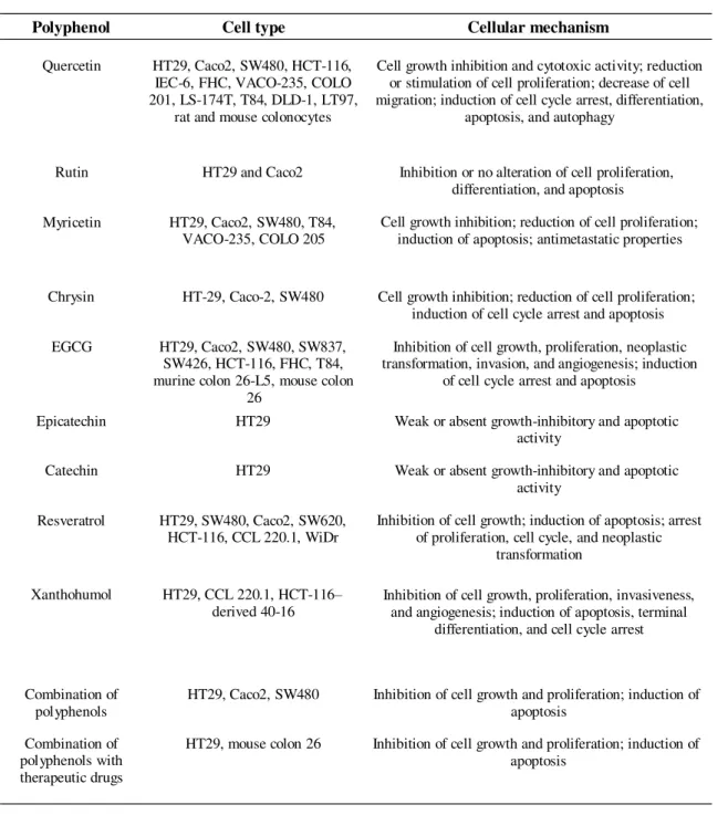
Table 1.3. Mechanisms involved in the chemopreventive effect of polyphenols in colorectal cell lines (adapted from Araújo, et al., 2011).
Polyphenol Cell type Cellular mechanism
Quercetin HT29, Caco2, SW480, HCT-116,
IEC-6, FHC, VACO-235, COLO 201, LS-174T, T84, DLD-1, LT97,
rat and mouse colonocytes
Cell growth inhibition and cytotoxic activity; reduction or stimulation of cell proliferation; decrease of cell migration; induction of cell cycle arrest, differentiation,
apoptosis, and autophagy
Rutin HT29 and Caco2 Inhibition or no alteration of cell proliferation,
differentiation, and apoptosis
Myricetin HT29, Caco2, SW480, T84,
VACO-235, COLO 205 Cell growth inhibition; reduction of cell proliferation; induction of apoptosis; antimetastatic properties
Chrysin HT-29, Caco-2, SW480 Cell growth inhibition; reduction of cell proliferation;
induction of cell cycle arrest and apoptosis
EGCG HT29, Caco2, SW480, SW837,
SW426, HCT-116, FHC, T84, murine colon 26-L5, mouse colon
26
Inhibition of cell growth, proliferation, neoplastic transformation, invasion, and angiogenesis; induction
of cell cycle arrest and apoptosis
Epicatechin HT29 Weak or absent growth-inhibitory and apoptotic
activity
Catechin HT29 Weak or absent growth-inhibitory and apoptotic
activity
Resveratrol HT29, SW480, Caco2, SW620,
HCT-116, CCL 220.1, WiDr Inhibition of cell growth; induction of apoptosis; arrest of proliferation, cell cycle, and neoplastic
transformation
Xanthohumol HT29, CCL 220.1, HCT-116–
derived 40-16 Inhibition of cell growth, proliferation, invasiveness, and angiogenesis; induction of apoptosis, terminal
differentiation, and cell cycle arrest
Combination of
polyphenols HT29, Caco2, SW480 Inhibition of cell growth and proliferation; induction of apoptosis
Combination of polyphenols with therapeutic drugs
HT29, mouse colon 26 Inhibition of cell growth and proliferation; induction of
apoptosis
1.3 Opuntia spp.
The prickly pear cactus (Opuntia spp.) or cactus pear belongs to the Cactaceae family
(Feugang, et al., 2006) and is a long-domesticated crop critically important in agricultural economies throughout the arid and semiarid parts of the world, where water can be a limitation for cultivation (Griffith, 2004). It is widely distributed in Europe, Southwestern United States, Northern Mexico, much of Latin America, South Africa and the Mediterranean countries (Utkarsha, et al., 2010).
Two parts of the plant have been used for food: the „„nopal‟‟ or cladodes (stems) and the fruits
or the prickly pears (figure 1.5). In particular, cladodes are consumed in Mexican regions as a constituent of salads (Medina, et al., 2007) while Opuntia spp. fruits are known to be fresh and sweet
fruits that can be eaten fresh, dried or preserved in jams, syrups or processed into candy-like products
(Galati, et al., 2003, Medina, et al., 2007).
Figure 1.5. Images of Opuntia spp. cladodes and fruits.
Opuntia fruits are fleshy and elongated berries, varying in shape, size and color (orange, yellow, red, purple, green, white) and have a consistent number of hard seeds (Piga, 2004). This fruit was ignored by the scientific community until the beginning of the 1980s when several lab reports and studies demonstrated their biological activity. Recently, investigations on the chemical components and the nutritional value of Opuntia spp. have attracted attention in the food, nutritional and pharmacological field (Feugang, et al., 2006, Hernández-Pérez, et al., 2005, Piga, 2004, Saenz, 2000, Stintzing, et al., 2001). Nowadays it is known that Opuntia spp. fruits are sources of several bioactive and nutritionally valuable compounds (Cayupán, et al., 2011, Utkarsha, et al., 2010) and their concentrations are dependent on the climate, cultivation site and respective fruit variety (Feugang, et al., 2006). Opuntia fruits are rich in polyphenolic compounds, namely flavonoids such as quercetin, kaempferol, isorhamnetin (Tesoriere (a), et al., 2005) and betalains, (Stintzing, et al., 2001). Betalains are nitrogen-containing vacuolar pigments and can be yellow-orange (betaxanthins) or red-violet
(betacyanins). Recently, betalains have received much more interest due to their possible use as natural food colorants in industry (Azeredo, 2009) . Opuntia fruits also contain vitamins E, K1 and C
(Ramadan and Morsel, 2003, Stintzing, et al., 2001), amino acids, especially proline and taurine (Stintzing, et al., 2001, Tesoriere, et al., 2005), minerals, namely calcium, potassium and magnesium (Gurrieri, et al., 2000, Lee, et al., 2005, Piga, 2004, Stintzing, et al., 2001) and sugars, such as glucose and fructose, (Galati, et al., 2003, Piga, 2004).
As numerous fruits and vegetables, Opuntia fruits have been reported to be beneficial to health. Recent studies reported that Opuntia fruits may prevent oxidative stress by acting as scavengers of free radicals (Butera, et al., 2002, Chavez-Santoscoy, et al., 2009, Galati, et al., 2003, Kuti, 2004, Tesoriere, et al., 2004). In particular, betalains have shown to be more potent antioxidants than ascorbic acid (Stintzing, et al., 2005). The anticancer effect of Opuntia fruits extracts has also been reported in vitro using ovarian, cervical and bladder cancer cells and in vivo using a nude mice ovarian cancer model (Zou, et al., 2005). The antiproliferative effect of Opuntia was also reported in human cancer cells of gliobastomas (Hahm, et al., 2010) and leukemia (Sreekanth, et al., 2007). Additionally, Opuntia fruits have shown others beneficial effects such as antiulcerogenic (Galati, et al., 2003), antinflammatory and analgesic (Loro, et al., 1999).
Due to the great number of potentially active nutrients and their multifunctional prop erties
Opuntia spp. fruits can be considered as perfect candidates for the production of health-promoting food and food supplements.
1.4 Nutraceuticals and functional foods
In the past few years, there has been an increase demand by consumers for health-promoting food products and two new concepts appear in the market: nutraceuticals and functional food.
In 1989, a new hybrid term between nutrients and pharmaceuticals, "nutraceuticals", has been coined by Stephen DeFelice, MD, founder and chairman of the Foundation for Innovation in Medicine (Brower, 1998). Nutraceuticals can be found in presentations similar to drugs (pills, extracts, tablets, etc) (Espín, et al., 2007) and is defined as diet supplements that deliver a concentrated form of a presumed bioactive agent from a food (Zeisel, 1999).
Functional foods represents a type of food that when consumed regularly exert a specific health-beneficial effect (i.e., a healthier status or a lower risk of disease) beyond their nutritional properties, and this effect must be scientifically proven (Espín, et al., 2007).
Some of the most common bioactive ingredients found in the nutraceutical and functional food market are polyphenols such as anthocyanins, proanthocyanidins, flavonols, stilbenes, hydroxycinnamates, coumarins, ellagic acid and ellagitannins, isoflavones, lignans, etc. (Espín, et al., 2007). Most of these compounds are isolated from natural sources using several extraction techniques.
1.4.1 Extraction methods
In recent years, extraction, isolation and purification of phytochemicals from natural sources, provide business opportunities and offers additional environmental and economic incentives for food industry. In order to obtain such valuable compounds, extraction techniques have been widely investigated. For that, it is of critical importance to select efficient extraction procedure/method and maintain the stability of phenolic compounds. Conventional solvent extractions (CSE) are the most commonly used procedures to prepare extracts from plant materials due to their ease of use, efficiency, and wide applicability (Dai and Mumper, 2010). The yield of chemical extraction depends on several factors such as the type of solvents, extraction time and temperature, sample/solvent ratio as well as on the chemical composition and physical characteristics of the samples (Dai and Mumper, 2010, Xu and Chang, 2007). Organic solvents, such as methanol, ethanol, acetone, ethyl acetate and their combinations with water have been widely used for the extraction of phenolics from plant materials (Xu and Chang, 2007). However, due to the low selectivity and extraction yields (unwanted substances such as sugar, organic acids and/or fats may be also recovered) of CSE additional steps may be required to successful isolate the bioactive ingredients (Dai and Mumper, 2010). Additionally, CSE employed large amounts of toxic solvents (Dai and Mumper, 2010) and longer extraction times Therefore nmerous others extraction methods have been proposed such as: adsorption technology (AD) (Soto, et al., 2011) microwave, ultrasound-assisted extractions, and techniques based on use of compressed fluids such as: supercritical fluid extraction (SFE) and pressurized liquid extraction (PLE) (Dai and Mumper, 2010).
The adsorption technology (AD) is attractive for its relative simplicity of design, operation and scale up, high capacity and favorable rate, insensitivity to toxic substances, ease of regeneration and low cost. Additionally, it avoids using toxic solvents and minimizes degradation (Soto, et al., 2011). Adsorption is a process of accumulation of molecules from a bulk solution onto the external and internal surfaces of the adsorbent. This process involves various chemical interactions such as hydrophobic, electrostatic attraction and hydrogen bonding. The sorption capacity of the adsorbents strongly depends on the surface area, contact time, polarity, concentration and the degree of hydrophobicity in the adsorption system (Barkakatia, et al., 2010). There are several types of adsorbents that can be used, including activated carbons, minerals and resins (Soto, et al., 2011). In particular, the use of resins (synthetic polymeric adsorbents with hydrophilic or hydrophobic nature) has many advantages, such as: chemically inert, durable and stable, high adsorption capacity, efficiency, selectivity and ease of regeneration, with relatively low cost and limited toxicity.
Other technique with growing interest for extraction of phytochemicals is pressurized liquid extraction (PLE) (or accelerated solvent extraction (ASE) or pressurized fluid extraction (PFE)) , and it partly derives from supercritical fluid extraction (Camel, 2001) . In this technology carbon dioxide can be used in combination with water and/or an alcohol (forming a gas-expanded liquid) to extract pytochemicals (Seabra, et al., 2010). The combined use of high pressures (3.3-20.3 MPa) and
temperatures (40-200°C) provides faster and efficient extractions that require small amounts of classic solvents (Dai and Mumper, 2010). Moreover high pressure extraction has other advantages such as the fact that native enzymes, which degrade phenolic compounds, are inhibited by extraction pressure increasing and CO2 addition. Besides that supercritical fluid processed materials do not require
additional sterilization steps (Seabra, et al., 2010). In recent years, PLE has been successfully applied to the extraction of phenolic compounds from different plant materials such as grape seeds and skin (Piñeiro, et al., 2006), apples (Alonso-Salces, et al., 2001) spinach (Howard and Pandjaitan, 2008), and eggplants (Luthria and Mukhopadhyay, 2006).
1.5 Aim and rational of the thesis
This thesis focus on the evaluation of the bioactive effect of several Portuguese Opuntia fruits derived from two different species (O. ficus-indica and O. robusta) with potential application in colon cancer therapy. To achieve this, an integrated approach was developed by: i) investigating functional properties of Opuntia spp. fruit juices, and ii) developing and/or evaluating the anticancer activity of Opuntia spp. natural extracts.
Within this context, the work was divided into three parts, as esquematly presented in figure 1.6.
Figure 1.6. Structure of the thesis.
In part 1, five Opuntia fruit juices were evaluated in terms of phenolic compounds and bioactivity in an effort to distinguish promising functional beverages. In part 2, the development of
Opuntia natural extracts were performed by processing the most effective Opuntia juices and the most interesting fruit juice residues with clean technologies. All extracts were screened for their antiproliferative effect in HT29 cells and the most promising ones were further selected to be fully characterized in Part 3. In this part, the analysis of cell cycle distribution and ROS generation were
performed, in order to better understand the mechanisms underlying HT29 cell death. Additionally, a drug-resistant cell line (HT29 dx) was created to evaluate the chemosensitization effect of natural
extracts.
2. Experimental procedure
2.1 Chemicals
EtOH 96% (AGA, Lisbon, Portugal), distillated water (ITQB, Lisbon), food grade macroporous resin Amberlite XAD-16 (Sigma-Aldrich, St Louis, USA) and were used for extraction experiments.
For phytochemical characterization: sodium carbonate (Na2CO3) were purchased from Sigma-
Aldrich (St Quentin Fallavier, France), Folin Ciocalteau reagent was acquired from Panreac (Barcelona, Spain) and gallic acid was purchased from Fluka (Germany). Sodium nitrite (NaNO2)
were from Riedel-de-Haën (Seelze, Germany), aluminium chloride (AlCl3) and sodium hydroxide
(NaOH) was obtained from Sigma-Aldrich, in Germany. (+)-Catechin hydrate was from Sigma
(Japan).
Chemicals used for antioxidant activity assays were: 2‟,2‟-Azobis (2- amidinopropane)
dihydrochloride (AAPH), 6-hydroxy-2,5,7,8- tetramethylchroman-2-carboxylic acid (Trolox), caffeic acid (C9H8O4), cobalt floride tetrahydrate (CoF2), hydrogen peroxide (H2O2) and picolinic acid
(C6H5NO2) were purchased from Sigma-Aldrich (St Quentin Fallavier, France). Disodium fluorescein
(FL) was from TCI Europe (Antwerp, Belgium). Sodium chloride (NaCl), potassium chloride (KCl) and monopotassium phosphate (KH2PO4) were from Sigma-Aldrich (St Quentin Fallavier, France) and
sodium phosphate dibasic dehydrate (Na2HPO4⋅2H2O) from Riedel-de-Haën (Seelze, Germany) were
used for phosphate buffer solution preparation (PBS).
All cell culture media and supplements, namely fetal bovine serum (FBS), glutamine, RPMI 1640 and trypsin/EDTA were obtained from Invitrogen (Gibco, Invitrogen Corporation, Paisley, UK). Moreover, chemicals used for cell-based assays were: MTS tetrazolium compound (3-(4,5- dimethylthiazol-2-yl)-5-(3-carboxymethoxyphenyl)-2-(4-sulfophenyl)-2H-tetrazolium) from Promega Corporation (Madison, USA), dimethyl sulphoxide (DMSO) (99.5%, Panreac, Barcelona, Spain),
ribonuclease A (Calbiochem, Darmstadt, Germany), 2‟,7‟- dichlorofluorescin diacetate (DCFH-DA),
methylthiazolyldiphenyl - tetrazolium bromide (MTT), Triton X (C14H22O(C2H4O)n), propidium iodide
(C27H34I2N4), doxorubicin hydrochloride (C27H29NO11.HCl) , bovine serum albumin (BSA) and
Bradford reagent all from Sigma-Aldrich (St Quentin Fallavier, France). PBS for cells was purchased from Sigma-Aldrich (St. Louis, USA) and CyQUANT Cell Proliferation Assay Kit was from Invitrogen (Carlsbad, California, USA).
2.2 Opuntia spp. samples and fruit juices preparation
Opuntia spp. fruits were collected in five different regions of Portugal, named Tramagal, Beja, Marvão, Sines and Quarteira. The plants are grown without any agronomical inputs and fruits were harvested by hand during October 2010.
Firstly, for sample preparation, spikes were removed with a brush and prickly pears were processed in a food processor (UFESA, LC5005, China) to collect the fruit juice and the
corresponding residue, which are mainly composed by hard seeds and part of the peel. Fruits juices were centrifuged (Avanti J26 XPI, Beckman Coulter®, California, USA) at 9000 rpm for 10 minutes and the supernatant was collected and filtrated in a 0.22 μm filter (Sarstedt, Nümbrecht, Germany). The resulting juices and residues were separated and preserved under frozen storage (-20ºC) until analyses.
2.3 Opuntia spp. extracts
2.3.1 Opuntia spp. extracts from fruit juices: adsorption technology (AD)
Opuntia juices were used for production of polyphenols-rich concentrates (PRCs) through adsorption technology. For that a food grade macroporous resin Amberlite XAD-16 was used. This resin is allowed to food applications by the U.S. Food and Drug Administration Code of Federal Regulation Title 21 (Scordino, et al., 2003). Before the experimental procedure it was necessary the resin preconditionation. This was realized by an extensive wash of the resin with distilled water to remove salts and impurities as described by Serra (2010). Then the resin was dried for 24 h at 70ºC and cooled in desiccators and further immersed in ethanol (96%). After 12 h later ethanol was replaced by distilled water through washing. Once prepared to use, Opuntia spp. juices were put in contact with resin XAD-16 inside flaks (equivalent to 30 mg of polyphenols/g of resin). These flaks were protected from light and submitted to an agitation at 200 rpm for 4 h. Afterwards the supernatant were removed and resins were washed three times with distilled water in order to remove water soluble constituents (i.e. sugars, organic acids and minerals). Polyphenols were recovered from resins in the elution step with ethanol (96%). The ethanolic fractions were vacuum filtered through one layer of filter pa per (Filter-lab, Barcelona, Spain) and concentrated in a rotary evaporator (BUCHI Rotavapor R-210, Flawil, Switzerland) under reduced pressure at 40ºC (BUCHI Heating Bath B-491, Flawil, Switzerland). Finally, extracts were transferred into a water phase and freeze dried (Modulyo freeze dried, Eduards, Sussex, UK) at -20ºC, in the absence of light, during 48h. The resulting PRCs were kept in a cool, dry and dark environment until analyses. The final extracts were designated as PRCs according with sample origin (Quarteira, Sines and Tramageal).
2.3.2 Opuntia spp. extracts from fruit juices residues: Conventional solvent
extraction (CSE)
Opuntia fruits residues from Sines, Quarteira and Beja were extracted using CSE under the conditions described below.
Opuntia fruits residues from Sines and Quarteira were extracted in the dark with EtOH:H2O
(50:50 v/v) solution (1:20, w/v), for 2 h at room temperature in constant agitation (IKA® dual-speed mixer RW 20.n, Aldrich, St. Luis, USA). The extracts were then centrifuged at 9000 rpm for 10 minutes and the supernatants were concentrated in a rotary evaporator (BUCHI Rotavapor R -210,
Flawil, Switzerland) in a water bath at 40ºC and under reduced pressure, in order to remove ethanolic
fraction. The final extracts were designated as Q1 (from Quarteira residue) and S1 (from Sines residue).
Opuntia residue from Beja was extracted in a 1:20 (w/v), for 2 hours, in the dark, at room temperature in constant agitation (IKA® dual-speed mixer RW 20.n, Aldrich, St. Luis, USA) using different solutions: i) 100% EtOH; ii) 100% H2O; iii) 60%EtOH:40%H2O and iv) 70%EtOH:30%H2O.
The extracts were then filtered, centrifuged at 9000 rpm for 10 minutes and the supernatants wer e concentrated in a rotary evaporator in a water bath at 40ºC under reduced pressure. Finally, extracts were freeze dried (Modulyo freeze dried, Eduards, Sussex, UK) at -20ºC, in the absence of light, during 48h. The final extracts were designated as A (100% EtOH), B (100% H2O) C
(60%EtOH:40%H2O) and D (70%EtOH:30%H2O).
2.3.3 Opuntia spp. extracts from fruit juices residues: adsorption technology (AD)
Conventional extracts obtained from Quarteira and Sines fruit juice residues (Q1 and S1) were submitted to adsorption technology using the adsorbent Amberlite XAD-16 resin. The experimental procedure was the same as described for fruit juices (section 2.3.1). The final extracts were designated as Q2 and S2.
(PLE)
2.3.4 Opuntia spp. extracts from fruit juices residues: Pressurized liquid extraction
Pressurized solvent extracts from Opuntia ficus-indica fruit residue from Beja were kindly provided by Dr. Hermínio de Sousa and his laboratory team group (Laboratory of Polymer Processing and Supercritical Tecnology, Universidade de Coimbra, Portugal). Ten extracts obtained at 313K and 20MPa and using diverse CO2/EtOH/H2O mixtures were provided (table 2.1).
Table 2.1. Extracts obtained from Beja residue using PLE technology
Sample ID Molar fracctions EtOH CO2 H2O
1 1 0 0 2 0.4 0.6 0 3 0.4 0 0.6 4 0.7 0.3 0 5 0.4 0.3 0.3 6 0.7 0 0.3 7 0.8 0.1 0.1 8 0.5 0.4 0.1 9 0.5 0.1 0.4 10 0.6 0.2 0.2
2.4 Phytochemical characterization
2.4.1. Polyphenols
Total phenolic content by Folin Ciocalteau method
Total concentration of phenolic compounds present in Opuntia spp. extracts were determined according to the Folin Ciocalteau colorimetric method (Singleton and Rossi, 1965) as described by Serra (2010). Briefly, 20μL of the appropriate dilutions of Opuntia samples were added to 1580μL of distilled water and oxidized with 100 μL of Folin Ciocalteau reagent. The reaction was neutralized with 300 μL of sodium carbonate solution (Na2CO3) and incubated at 40ºC for 30 minutes. The
absorbance of samples were measured at 765 nm in a spectrometer (Genesys10uv, Thermo Spectronic, New York, USA) and gallic acid was used as standard (0-800 mg/L) for the calibration curve. The results were expressed as means of triplicates (mg of gallic acid equivalents per gram of extract - mgGAE/g).
HPLC analysis
HPLC analysis of Opuntia spp. juices were performed by Analytical Group of IBET (Oeiras, Portugal) coordinated by Dr. Rosário Bronze. Briefly the analysis were performed on a Waters®
Alliance 2695 (Waters) equipped with a quaternary pump, solvent degasser, auto sampler and column oven, coupled to a Photodiode Array Detector Waters 996 PDA (Waters). A pre-column (RP-18, 5µm)
and reversed phase column (RP-18 Synergy, 2.5 m Max-RP from Phenomenex) with oven at at 35ºC were used for separation. The gradient mobile phase consisted of 0.5% formic acid p.a in ultra pure water (A): LC-MS grade acetonitrile (B) at a flow rate of 0.30 mL/min. Total polyphenol content of fruit juices were assayed using acid gallic as standard. Solutions of 5, 20, 40, 60, 80 and 100 ppm of gallic acid were used for calibration curve. Results were expressed as mg GAE/ L of juice fruit.
Identification of polyphenol compounds was done by HPLC/MS. A triple quadrupole mass
spectrometer MicroMass® Quattro micro (Micromass, Waters) outfitted with electrospray ionization
source (ESI) was used in tandem at temperature of 120ºC and capillary voltage of 3.0 kV. The compounds were ionized in both negative and positive ion mode and spectra of the column elute were recorded in the range m/z 100-1000. High purity nitrogen (N2) was used both as drying gas and as a
nebulising gas. Ultrahigh-purity Argon (Ar) was used as collision gas at a pressure of 1×10⁻⁴ mBar.
MassLynx software was used for data acquisition and processing.
2.4.2 Flavonoid content
The measurement of flavonoid content was performed by the AlCl3 complexation method
described by (Zhishen, et al., 1999) modified for the reader microplate (Powerwave XS Microplate Spectrophotometer, Biotek Instruments, Winooski, USA) according to Tavares, et al., 2010 . To each
well of a 96-well microplate was added 125 μL of distilled water, 25 μL of Opuntia spp. fruit juice and
7.5 μL of NaNO2 5% (w/v). The mixture was allowed to stay at room temperature and after 6 minutes
were added 15 μL of AlCl3 10% (w/v). After 5 min of incubation, 100μL of NaOH (1M) was added
and the solution of each well was mixed by pippetting up and down. The absorbance was measured at 510 nm. (+)-Catechin hydrate, minimum 98% (w/w) was used as standard, and the results are expressed as mg catechin equivalents per L of juice (mg CE/L).
2.5. Antioxidant activity
2.5.1 Oxygen radical absorbance capacity (ORAC)
ORAC assay was carried out by following the method previously described (Huang, et al., 2002) modified for the FL800 microplate fluorescent reader as described by Serra et al., 2010. This assay measures the ability of the antioxidant species present in the sample to inhibit the oxidation of disodium fluorescein (FL) catalyzed by peroxyl radicals generated from AAPH.
Briefly, in a 96-well microplate were added 25 µL of the appropriate sample dilution and 150 µL of disodium fluorescein (2×10⁻⁷ mM). The microplate was put in a fluorescent reader and allowed
to incubat at 37ºC, for 10 minutes. The reaction was started with 25 µL of AAPH (153 mM) added through the injector. Fluorescence emitted by the reduced form of FL was measured in an FL800 microplate fluorescent reader (Bio-Tek Instruments, Winooski, VT, USA) and recorded every 1 min at the emission wavelength of 530±25 nm and excitation wavelength of 485±20 nm for a period of 30 min. Phosphate buffer (75 mM, pH=7.4) was used to prepare AAPH and FL solutions and was used as blank. Solutions of 5, 10, 20, 40, and 50 μmol/L of Trolox were used as control standards. All samples, including the blank and the controls, were analyzed as triplicates. Final ORAC values were calculated by a regression equation between the Trolox concentration and the net area under the FL decay curve and were expressed as Trolox Equivalents per litre of Opuntia juice or gram of extract (μmol TE/L of juice or μmol TE/g of extract).
2.5.2 Hydroxyl Radical Adverting Capacity (HORAC)
HORAC assay was based on a previously reported method (Ou, et al., 2002), modified for the FL800 microplate fluorescence reader (Bio-Tek Instruments, Winooski, VT, USA) as described by Serra et al., 2010. This assay evaluates the hydroxyl radical prevention capacity of a sample using fluorescein (FL) as the probe. The hydroxyl radical was generated by a Co(II)-mediated Fenton like reaction and, similarly to ORAC assay, the fluorescence decay curve of FL was used to quantify the
HORAC value. Briefly, 10 µL of appropriate dilutions of samples were added to 180 µL of FL (4x10⁻³
µM) in a 96 well microplate. After 10 minutes of incubation at 37ºC were added 10µL of H2O2 (0.55
M). The reaction was started with 10 µL of CoF2 added through the injector. Fluorescence emitted by
the reduced form of FL was measured and recorded every 1 minute during 35 minutes. The FL800 microplate fluorescence reader was used with fluorescence filters for an excitation wavelength of