Article
ISSN 0102-695X
doi: 10.1590/S0102-695X2011005000090
Received 25 Aug 2010 Accepted 3 Jan 2011 Available online 20 May 2011
myocardial infarction in rats
Sita Sharan Patel,
*,1Neelesh Kumar Verma,
1Beauty Rathore,
2Govind Nayak,
2Akhlesh Kumar Singhai,
2Priya Singh
31Cardiovascular Research Division, Himalayan Pharmacy Institute, India, 2Division of Pharmacology, Lakshmi Narain College of Pharmacy, India, 3Department of Phytochemistry & Pharmacognosy, V.N.S. Institute of
Pharmacy, India.
Abstract: The present study was designed to evaluate the cardioprotective potential of aqueous flower extract of Bombax ceiba L., Malvaceae (BC), on the basis of biochemical and histopathological parameters in Adriamycin (Adr) induced myocardial infarction in rats and to compare with vitamin E, a known cardioprotective antioxidant. Male Wister rats were used as in vivo model for the study. BC was administered orally to Wister rats at different doses (150 mg/kg, 300 mg/kg and 450 mg/kg, b.w.) for six days/week for four weeks. Thereafter, all the groups except saline were administered Adr (20 mg/kg, i.p.). There was a significant decrease in myocardial superoxide dismutase, catalase and reduced glutathione in animals treated with Adr. Concurrently marked increase in extent of lipid peroxidation was reported. Co-treatment of BC/vitamin E and Adr resulted in an increase in the cardiac antioxidant enzymes and reduction in lipid peroxidation as compared to Adr-treated animals. Adr showed significant decrease (p<0.001) in the level of cardiac marker enzymes [Lactate dehydrogenase (LDH) and Serum glutamic oxaloacetic transaminase (SGOT)] in heart homogenate with corresponding increase in their level in serum. In BC/vitamin E treated groups significant increase (p<0.001) of LDH in heart homogenate and decrease of SGOT and LDH in serum were observed. Microscopic studies in Adr-treated animals revealed mitochondrial swelling, leukocyte infiltration, lipid inclusions and myofibrillar loss whereas the pre-treatment with BC/vitamin E led to a lesser degree of Adr-induced histological alterations. These findings suggest that aqueous flower extract of BC has protective effect against Adr-induced cardiotoxicity and may have potential as a cardioprotective agent.
Keywords:
adriamycin
Bombax ceiba
cardiovascular disease catalase
vitamin E
Introduction
Cardiovascular disease (CVD) is now the most common cause of death worldwide. Before 1900, infectious diseases and malnutrition were the most common cause of death throughout the world, and CVD was responsible for less than 10% of all deaths. Today CVD accounts for ~30% of death worldwide, including nearly 40% in high-income countries and about 28% in low- and middle-income countries (Gaziano & Gaziano, 2008). Myocardial infarction is the acute condition of necrosis of the myocardium that occurs as a result of imbalance between coronary blood supply and myocardial demand. It is well recognized that ischemic tissue generates oxygen-derived free radicals and other reactive species which bring about oxidative damage of membrane lipids, proteins, carbohydrates and DNA,
Cardioprotective effect of Bombax ceiba lowers against acute adriamycin-induced myocardial infarction in rats
Sita Sharan Patelet al.
catalase (CAT) and reduced glutathione (GSH).
A large proportion of the Indian population for their physical and psychological health needs depend on traditional system of medicine. Medicinal plants have become the focus of intense study in terms of conservation as to whether their traditional uses are supported by actual pharmacological effects or merely based on folklore. Herbal medicines are free from side effects and less costly when compared to synthetic drugs. Bombax ceiba L., Malvaceae (BC), is an important medicinal plant of tropical and subtropical India. Its medicinal usage has been reported in the traditional systems of medicine such as Ayurveda, Siddha and Unani. Bombax ceiba described as a cotton tree and it is used extensively for treatment
of some diseases like inlammation (Buckingham, 1992),
algesia, hepatotoxicity (Saleem et al., 2003) and CVD i.e. hypertension, as well as well known for anti-angiogenic and in addition, BC is also known to be endowed with potent free-radical scavenging activity (Vieira et al., 2009). The effect of BC on Adr-induced cardiotoxicity is still unclear; hence the present work includes the
cardioprotective activity of aqueous lower extract of BC
against Adr-induced myocardial infarction in rats.
Materials and Methods
Plant material
The lowers of Bombax ceiba L., Malvaceae, were collected in the month of October from Eastern part of India, (Sikkim Himalayas) Majhitar, East Sikkim, India. The Herbarium specimen (No. 167) of plant was deposited in the Department of Pharmacognosy and it
was identiied by Dr. J. P. Mohanty, the Head Department
of Pharmacognosy, Himalayan Pharmacy Institute, East Sikkim.
Extraction
After collection and identiication the lowers
were dried in shade and powdered (no. 60 mesh) and 100 g of the dried powder was soxhlet extracted successively with petroleum ether, chloroform, methanol and water. The weight of aqueous extract after drying was calculated as 4.77 g.
Experimental animals
Adult albino rats (Wister strain, 200-250 g) were used. They were housed in standard environmental conditions and fed with rodent diet and water ad libitum. All animal experiments were carried out in accordance with the guidelines of Committee for the Purpose of Control and Supervision on Experiments on Animals (CPCSEA, 2003). The institutional animal ethical
committee has given approval for conducting animal experiments (HPI/08/60/IAEC/0043).
Cardioprotective activity
To study the effect of aqueous extract of BC against Adr-induced cardiotoxicity, six groups of seven animals in each were taken and treated as follows, maximum tolerated dose of BC were administered: Group I: Normal saline (0.75 ml/animal), orally 6 days/ week for 4 weeks.
Group II: Saline (0.75 ml/animal) + Adr (20 mg/kg), single i.p. injection after 4 weeks.
Group III: BC (150 mg/kg), orally for 4 weeks + Adr single i.p. injection after 4 weeks.
Group IV: BC (300 mg/kg), orally for 4 weeks + Adr single i.p. injection after 4 weeks.
Group V: BC (450 mg/kg), orally for 4 weeks + Adr single i.p. injection after 4 weeks.
Group VI: Vitamin E (100 mg/kg), orally for 4 weeks + Adr single i.p. injection after 4 weeks.
The animals were sacriiced after 48 h of Adr
administration under pentobarbital sodium (50 mg/ kg, i.p.) anesthesia and hearts were excised out for the estimation of biochemical parameters and histological studies.
Biochemical assays
Frozen tissue sample of the rat hearts were weighed and homogenized (Homogenizer REMI RQM-122, Remi Instrument, India) (1:10, w/v) in 100 mmol/L phosphate buffer (pH 7.4) containing 0.05% sodium azide in an ice bath. The homogenate was centrifuged at 5000 rpm for 10 min. The supernatant was frozen at -78 °C in aliquots until used for biochemical assays.
Protein estimation
The levels of total proteins were determined in heart homogenates of experimental animals by using the Bradford (1976) method.
Lipid peroxidation
Thiobarbituric acid reactive substances (TBARS) levels in the heart homogenates and serum
were determined by modiied method of Okhawa et
double-H2O2 + 2GSH →2H2O + GSSG (oxidized glutathione)
GSH-Px in the tissue homogenate oxidizes glutathione and simultaneously, H2O2 is reduced to water. This reaction is arrested at 10 min using trichloroacetic acid and the remaining glutathione is reacted with DTNB solution to result in a colored compound, which is measured spectrophotometrically at 420 nm.
Cardiac biomarkers
Lactate dehydrogenase (LDH) and Serum glutamic oxaloacetic transaminase (SGOT) activities in heart homogenate and serum were assayed by using Star 21 plus Biochemistry Auto Analyser (Cuesta Care Inc., Atascadero, USA).
Histopathological examination
The heart tissues (one from each) obtained from all experimental groups were washed immediately with
saline and then ixed in 10% buffered neutral formalin solution. After ixation, the heart tissues were processed embedding in parafin. Then, the tissues were sectioned
and stained with haematoxylin and eosin (H & E) and examined under high power microscope (x 400) and photomicrographs were taken.
Statistical analysis
The results were subjected to one way analysis of variance (ANOVA) followed by Bonferroni test p<0.05
were considered signiicant.
Results and discussion
The current study entails the cardioprotective
potential of the aqueous lower extract of BC against acute Adr-induced cardiotoxicity for the irst time.
BC is a plant, well known for its cardioprotective properties in the traditional Indian system of medicine. In the present study cardioprotective effect of chronic oral administration of BC against Adr-induced acute cardiotoxicity were evaluated in male Wister rats. The major chemical constituents present in the aqueous
extract of BC (tannins, lavonoids and glycosides) may
be responsible for the potent antioxidant activities (Vieira et al., 2009).
The existing experimental evidences suggest that Adr-induced oxidative stress is due to the generation of free radicals in the heart tissue (Hardina et al., 2000; Naidu et al., 2002). The principle ROS generated are superoxide radicals and hydroxyl radicals, which have the potential to cause damage to various intracellular components. Cardiac muscle is particularly susceptible distilled water and 5.0 mL of n-butanol:pyridine (15:1,
v/v) mixture were added to the tubes and centrifuged at 5000 rpm for 10 min. The absorbance of organic layer was measured at 540 nm. Malonyldialdehyde (MDA), an end product of lipid peroxidation forms pink color adducts with TBARS. The extent of lipid peroxidation was expressed as µM of MDA/g heart tissue.
Glutathione estimation
Myocardial GSH was estimated according to
the modiied method of Ellman (1959). The heart tissues
were homogenized with 10% TCA buffer and centrifuged at 3000 rpm for 10 min at 4 °C. The reaction mixture contained 0.1 mL of supernatant, 2.0 mL of 0.3M phosphate buffer (pH 8.4), 0.4 mL of double-distilled water and 0.5 mL of DTNB [5,5-dithiobis(2-nitrobenzoic acid)]. The reaction mixture was incubated for 10 min and the absorbance was measured at 412 nm within 15 min. The concentration of GSH was expressed as µg/g of heart tissue.
Antioxidant enzyme assays in heart homogenates
SOD levels in the myocardial tissue of rats were
determined according to the modiied method of Kakkar et al. (1984). Briely heart tissues were homogenized in
0.25 M Tris sucrose buffer pH 7.4 and centrifuged at 10,000 rpm for 15 min at 4 °C. Supernatant (600 µL) was added to the solution containing 1.2 mL of sodium pyrophosphate buffer (0.052 M, pH 8.3), 0.1 mL of phenazine methosulphate solution (186 µM) and 0.3 mL of nitro blue tetrazolium solution (300 µM). Reaction was initiated by the addition of 0.2 mL of nicotinamide adenine dinucleotide-reduced disodium salt (NADH) solution (780 µM). This reaction mixture was incubated for 90 s at room temperature and then stopped by the addition of 1 mL glacial acetic acid. Absorbance of reaction mixture was read spectrophotometrically at 560 nm. SOD activity was expressed as U/mg protein.
Levels of CAT were estimated by the modiied
method of Aebi (1984). Hearts were homogenized at 4 °C in 50 mM potassium phosphate buffer (pH 7.4) and centrifuged at 5000 rpm for 10 min. Ethanol equal to 0.01 mL/mL of supernatant was added and incubated
for 30 min in ice. Triton 100X was added to the inal
concentration of 1%. Supernatant (50 µL) was added to a cuvette containing 1.95 mL of 50 mM phosphate buffer (pH 7.0). Then 1.0 mL of 30 mM hydrogen peroxide was added and rate of decomposition of hydrogen peroxide was measured spectrophotometrically at 240 nm. CAT activity was expressed as U/mg protein.
Cardioprotective effect of Bombax ceiba lowers against acute adriamycin-induced myocardial infarction in rats
Sita Sharan Patelet al.
to free-radical injury, because it contains low levels of free-radical detoxifying enzymes/molecules like SOD, GSH and CAT (Takacs et al., 1992). Furthermore, Adr
also has high afinity for the phospholipid component
of mitochondrial membrane in cardiac myocyte, leading to accumulation of Adr in the heart tissue (Takacs et al., 1992). It was observed that 20 mg/kg (Singh et al., 2008) dose of Adr-induced moderate lesions in the myocardium
and signiicantly altered various biochemical parameters
resulting myocardial infarction. Therefore, the cardioprotective activity of BC was evaluated against this dose.
Myocardial lipid peroxidation was signiicantly
increased (p<0.001) in Adr-treated animals as compared to normal animals. Pre-treatment with BC (300 mg/
kg and 450 mg/kg) and vitamin E showed signiicant
(p<0.001) reduction of lipid peroxidation as compared to GII (Table 1). Adr-induced myocardial lesions have been well documented in patients as well as in experimental animals (Ytrehus & Hegstad, 1991; Lenaz & Page, 1976; Doroshow, 1991). Studies have shown the Adr cardiotoxicity to proceed via production of free radicals.
Lipid peroxidation has been identiied as one of the basic
deteriorative reactions in cellular mechanisms during free radicals induced myocardial injury. The increased levels of malondialdehyde (MDA) indicate excessive formation of free radicals by Adr and activation of the lipid peroxidative process, resulting in irreversible damage to heart in animals subjected to Adr stress. BC
treatment signiicantly decreased the MDA levels by
preventing formation of lipid peroxides from fatty acids.
Myocardial GSH levels were signiicantly
reduced (p<0.001) in Adr-treated animals as compared to untreated animals. Pre-treatment with BC showed
signiicant increase (p<0.001) in GSH levels at the doses of 300 mg/kg and 450 mg/kg as compared to Adr treated
group. Treatment of animals with dose of 150 mg/kg of
BC led to insigniicant alteration in the levels of GSH
(Table 1).
Reduced glutathione is one of the most abundant non-enzymatic antioxidant bio-molecule present in the body (Meister, 1984). Together with GSH-Px, glutathione
reductase (GR) and CAT–SOD couple, it eficiently
scavenges free radical species such as H2O2, superoxide anions and alkoxy radicals.
As a substrate for antioxidant enzymes GSH-Px and glutathione transferase (GST), it protects cellular constituents from the damaging effects of ROS and peroxides formed during metabolism. Decreased GSH levels in Adr intoxicated rats may be due to its enhanced utilization for augmenting the activities of GSH-Px and GST.
Glutathione levels depleted by Adr-induced
damage were signiicantly (p<0.001) elevated by BC (300 and 450 mg/kg) and vitamin E pre-treatment. It may be understood that increased levels of GSH could be because of its enhanced synthesis or due to improved GR activity in presence of BC.
Adr treatment to Wister rats causes signiicant
decrease (p<0.001) in SOD activity in the myocardium as compared to control. Pre-treatment with BC (300
mg/kg and 450 mg/kg) signiicantly increased the SOD
activity (p<0.001) as compared to Adr-treated animals.
No signiicant increase in SOD activity was noticed at
150 mg/kg dose of BC (Table 1).
Adr-induced myocardial necrosis produced
a signiicant depletion in activities of antioxidant
enzymes such as CAT (p<0.001) and GSH-Px (p<0.001) compared to normal animals. BC (300 and 450 mg/kg) and vitamin E pre-treatment to myocardial necrotic rats
signiicantly restored the activities of CAT (p<0.001) and GSH-Px (p<0.001). BC 150 mg/kg, however, could only
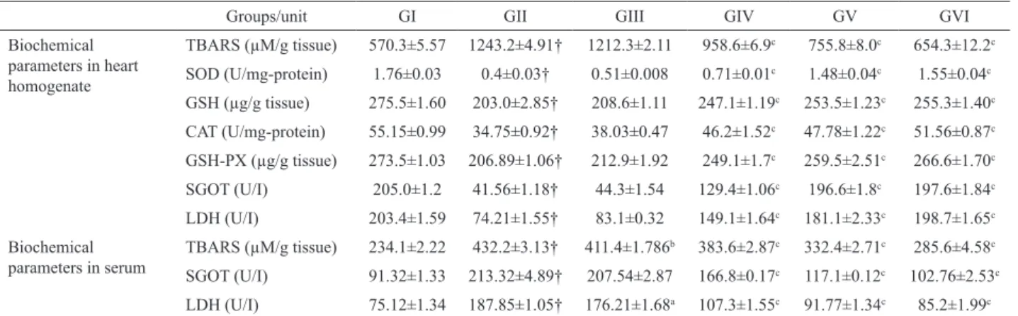
Table 1. Biochemical parameters in different experimental groups
Groups/unit GI GII GIII GIV GV GVI
Biochemical parameters in heart homogenate
TBARS (µM/g tissue) 570.3±5.57 1243.2±4.91† 1212.3±2.11 958.6±6.9c 755.8±8.0c 654.3±12.2c
SOD (U/mg-protein) 1.76±0.03 0.4±0.03† 0.51±0.008 0.71±0.01c 1.48±0.04c 1.55±0.04c
GSH (µg/g tissue) 275.5±1.60 203.0±2.85† 208.6±1.11 247.1±1.19c 253.5±1.23c 255.3±1.40c
CAT (U/mg-protein) 55.15±0.99 34.75±0.92† 38.03±0.47 46.2±1.52c 47.78±1.22c 51.56±0.87c
GSH-PX (µg/g tissue) 273.5±1.03 206.89±1.06† 212.9±1.92 249.1±1.7c 259.5±2.51c 266.6±1.70c
SGOT (U/I) 205.0±1.2 41.56±1.18† 44.3±1.54 129.4±1.06c 196.6±1.8c 197.6±1.84c
LDH (U/I) 203.4±1.59 74.21±1.55† 83.1±0.32 149.1±1.64c 181.1±2.33c 198.7±1.65c
Biochemical parameters in serum
TBARS (µM/g tissue) 234.1±2.22 432.2±3.13† 411.4±1.786b 383.6±2.87c 332.4±2.71c 285.6±4.58c
SGOT (U/I) 91.32±1.33 213.32±4.89† 207.54±2.87 166.8±0.17c 117.1±0.12c 102.76±2.53c
LDH (U/I) 75.12±1.34 187.85±1.05† 176.21±1.68a 107.3±1.55c 91.77±1.34c 85.2±1.99c
n=6; †p<0.001 versus GI; ap<0.05, bp<0.01, cp<0.001 versus GII; Values are obtained by one way ANOVA followed by Bonferroni tests; GI: Normal
restore the Adr depleted activities of CAT and GSH-Px
insignii cantly.
SOD, CAT and GSH-Px constitute a mutually
supportive enzyme system of the i rst line cellular
defense against oxidative injury, decomposing O2 and H2O2 before their interaction to form the more harmful hydroxyl radical (Li et al., 1988).
In the present study, SOD activity decreased
signii cantly in the Adr group of animals may be due to
an excessive formation of superoxide anions. A decrease in SOD activity can result in the decreased removal of superoxide anions, which can be harmful to the myocardium (Sharma et al., 2001). The activities of H2O2 scavenging enzymes CAT and GSH-Px also decreased
signii cantly after Adr treatment. The decline in these
enzyme levels may be explained by the fact that excessive superoxide anions may inactivate SOD, thus, resulting in an inactivation of the H2O2 scavenging enzymes. Pre-treatment with BC/vitamin E to Adr challenged rats heart effectively prevented the decrease in SOD, CAT and GSH-Px activities, which may be correlated directly to the scavenging of radicals by BC resulting in protection of these enzymes (Tosaki et al., 1994; Panda & Naik, 2008).
Adr showed signii cant (p<0.001) decrease in the level of cardiac marker enzymes (SGOT and LDH) in heart homogenate with a corresponding increase in their levels in the serum when compared with normal control. Increase in the activity of these enzymes in serum could be due to leakage of these enzymes from the heart as a result of free radicals induced necrosis (Peer et al.,
2008). In vitamin E and BC (300 and 450 mg/kg) treated
groups signii cant (p<0.001) increase of LDH in heart homogenate and decrease of SGOT and LDH in serum were observed.
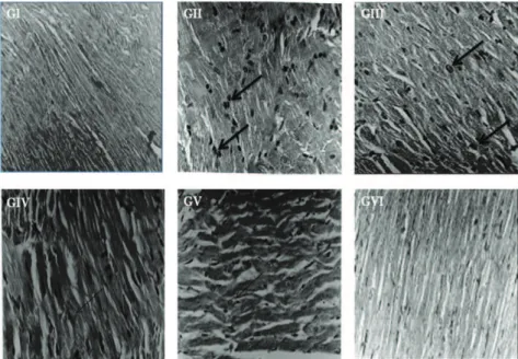
Cardiotoxicity induced by Adr was further assessed using H&E stain. The heart of control group showed regular cell distribution and normal myocardium morphology. Histology of the rat heart from Adr-treated animals revealed the cytoplasmic vacuole formation,
mitochondrial swelling, leukocyte ini ltration and myoi brillar loss, which is a typical i nding in Adr-induced cardiomyopathy. Myocardial infarction was signii cantly
reduced in animals those received BC/vitamin E treatment (Figure 1). BC (300 and 450 mg/kg, p.o.) and vitamin E (100 mg/kg, p.o.) maintained all biochemical and histopathological parameters near normal as compared to Adr group, indicating cardioprotective activity of BC.
Conclusion
In conclusion, BC showed cardioprotective effect against Adr-induced myocardial infarction and it may be due to its antioxidant effect. Therefore further studies are required to prove the potential of this plant.
Acknowledgement
Authors acknowledge to All India Council for Technical Education, New Delhi & Himalayan Pharmacy
Institute, East Sikkim for their i nancial support to
carry-out this research work (AICTE/F. No: 8023/BOR/RID/ RPS-206/2007).
Figure 1. Effect of aqueous extract of Bombax ceiba L., Malvaceae, l owers on myocardial morphology: (GI) Control rat heart
showed normal structure. (GII) Rat treated with Adr alone showed cytoplasmic vacuole formation, mitochondrial swelling, leukocyte
ini ltration, edema and myoi brillar loss. (GIII) BC 150 mg/kg+Adr could not relieve the damage. (GIV) BC 300 & (GV) 450 mg/kg+Adr
Cardioprotective effect of Bombax ceiba lowers against acute adriamycin-induced myocardial infarction in rats
Sita Sharan Patelet al.
References
Aebi H 1984. Catalase in vitro. In: Packer L, Orlando FL (org.)
Methods in enzymology. New York: Academic Press, p 121-126.
Bradford MM 1976. A rapid and sensitive method for the quantitation of microgram quantities of protein utilizing the principle of protein-dye binding. Anal Biochem 7: 248-254.
Buckingham J 1992. Dictionary of natural products. London:
Champan and Hall scientiic data division.
CPCSEA 2003. Guidelines for laboratory animal facility. Indian
J Pharmacol 35: 257-274.
Doroshow JH 1991. Doxorubicin-induced cardiotoxicity. New
Engl J Med 324: 843-845.
Ellman GL 1959. Tissue sulphydryl groups. Arch Biochem Biophys 82: 70-77.
Gaziano TA, Gaziano JM 2008. Epidemiology of cardiovascular
disease. In: Fauci AS, Braunwald E, Kasper DL, Hauser
SL, Longo DL, Jameson JL, Loscalzo J (org.) Principles of internal medicine. New York: McGraw Hill, p 1375-1379.
Hardina R, Gersl V, Klimtova I, Simunek T, Machackova
J, Adamcova M 2000. Anthracycline induced cardiotoxicity. Acta Medica 43: 75-82.
Kakkar P, Das B, Viswanatham PN 1984. A modiied
spectrophotometric assay of super oxide dismutase.
Indian J Biochem Biophys 21: 130-132.
Lenaz LN, Page JA 1976. Cardiotoxicity of adriamycin and relateld anthracyclines. Cancer Treat Rev 3: 111-120. Li JL, Stantman FW, Lardy HA 1988. Antioxidant enzyme
systems in rat liver and skeletal muscle. Arch Biochem Biophys 263: 150-160.
Meister A 1984. New aspects of glutathione biochemistry and transport selective alterations of glutathione metabolism.
Nutr Rev 42: 397-400.
Naidu MUR, Vijay Kumar K, Krishna Mohan I, Sundaram C,
Singh S 2002. Protective effect of Gingko biloba extract against doxorubicin-induced cardiotoxicity in mice.
Indian J Exp Biol 40: 894-900.
Okhawa H, Qohishi N, Yagi K 1979. Assay of lipid peroxides
in animal tissues by thiobarbituric acid reaction. Anal Biochem 95: 351-358.
Olson RD, Mushlin PS 1990. Doxorubicin cardiotoxicity: Analysis of prevailing hypothesis. Fed Am Soc Exp Biol J 4: 3076-3086.
Panda VS, Naik SR 2008. Cardioprotective activity of Ginkgo biloba phytosomes in isoproterenol-induced myocardial necrosis in rats: A biochemical and histoarchitectural evaluation. Exp Toxicol Pathol 60: 397-404.
Peer PA, Trivedi PC, Nigade PB, Ghaisas MM, Deshpande AD 2008. Cardioprotective effect of Azadirachta indica A. Juss. on isoprenaline induced myocardial infarction in rats. Int J Cardiol 126: 123-126.
Rotruck JT, Pope AL, Ganther HE, Hofeman DG, Hoeksta WG 1973. Selenium: biochemical role as a component of glutathione peroxidase. Science 179: 588-590.
Saleem R, Ahmad SI, Ahmad M, Faizi Z, Rehman S, Ali M, Faizi S 2003. Hypotensive activity and toxicology of constituents from Bombax ceiba stem bark. Biol Pharm Bull 26: 41-46.
Sharma M, Kishore K, Gupta SK, Joshi S, Arya DS 2001.
Cardioprotective potential of Ocimum sanctum in isoproterenol induced myocardial infarction in rats. Mol Cell Biochem 225: 75-83.
Singh G, Singh AT, Abraham A, Bhat B, Mukherjee A, Verma
R, Agarwal SK, Jha S, Mukherjee R, Burman AC
2008. Protective effects of Terminalia arjuna against Doxorubicin-induced cardiotoxicity. J Ethnopharmacol 117: 123-129.
Sinha BK, Politi PM 1990. Anthracyclines. Cancer Chemother
11: 45-57.
Takacs IE, Matkovics B, Varga SI, Homolay P, Feer G, Seres T 1992. Study of the myocardial antioxidant defence in various species. Pharmacol Res 25: 177-178.
Tosaki A, Engelman DT, Pali T, Engelman RM, Droy Lefaix MT 1994. Ginkgo biloba Extract (EGb-761) improves postischemic function in isolated preconditioned working rat hearts. Coronary Artery Dis 5: 443-450. Vieira TO, Said A, Aboutab E, Azzam M, Creczynski P, Tania
B 2009. Antioxidant activity of methanolic extract of
Bombax ceiba. Redox Rep 14: 41-46.
Ytrehus KT, Hegstad AC 1991. Lipid peroxidation and
membrane damage of the heart. Acta Physiol Scand 324: 843-845.
*Correspondence
Sita Sharan Patel
Cardiovascular Research Division, Himalayan Pharmacy Institute Majhitar, East Sikkim, 737136 India