ISSN 0102-695X DOI: 10.1590/S0102-695X2013005000054 Received 3 May 2013 Accepted 10 Jul 2013 Available online 9 Aug 2013
pharmacognostic quality control
Juliana Youssef,
1Patrícia M. Döll-Boscardin,
*,2Paulo V. Farago,
2Márcia R. Duarte,
3Jane M. Budel
21Laboratório de Farmacognosia, Faculdades Integradas do Brasil, Brazil,
2Departamento de Ciências Farmacêuticas, Universidade Estadual de Ponta Grossa,
Brazil,
3Departamento de Farmácia, Universidade Federal do Paraná, Brazil.
Abstract: Gochnatia polymorpha (Less.) Cabrera, Asteraceae, is popularly known as cambará and cambara-de-folha-grande in Brazil. It is used in traditional medicine to treat respiratory and gastrointestinal disorders. Pharmacological studies revealed
anti-inl ammatory, antispasmodic, antibacterial and antiviral activities. The goal of this
paper was to carry out morphological and anatomical studies in order to describe the aerial parts of G. polymorpha. The botanical material was collected, i xed, and prepared
according to usual light and scanning electron microtechniques. The leaves are simple, oblong-lanceolate to elliptical-lanceolate in form with mucronate acute apex, rounded
base, entire or slightly toothed margin, and short petiole. In transection, the epidermis
is uniseriate along the leaf blade. A subepidermal layer next to the adaxial side is present. Anomocytic stomata are seen only on the abaxial surface. Capitate glandular trichomes and T-shaped non-glandular trichomes occur on the leaves. The mesophyll
is dorsiventral and minor collateral vascular bundles are enclosed by a sheath of
thick-walled parenchymatic cells. The midrib is biconvex and the petiole has a circular shape. The epidermis of the stem consists of a single layer of cells with glandular and non-glandular trichomes. The vascular cylinder shows typical structure and perivascular i ber caps are next to the phloem.
Keywords: Asteraceae
Gochnatia polymorpha
medicinal plant morpho-anatomy
Introduction
Gochnatia Kunth belongs to Asteraceae and shows about seventy species occurring in America from
Mexico to Argentina and in Asia. This genus consists of
trees or shrubs that particularly grow in Brazil where there are 22 species distributed in Rio Grande do Sul, Paraná, São Paulo, Goiás, Minas Gerais, Bahia, and Ceará (Freire et al., 2002). Chemical studies have reported the presence of sesquiterpenes lactones, diterpenes, triterpenes,
l avonoids, coumarins, and essential oil in species of
Gochnatia (Catalan et al., 1996; Silva et al., 2011). Gochnatia polymorpha (Less.) Cabrera is a representative of Mutisieae and is popularly known as cambará (Rossato & Kolb, 2010) and cambará-de-folha-grande (Lorenzi, 2002) in Brazil. It is a medium sized tree (Schlemper et al, 2011) with a twisted and branched trunk of sympodial growth. It can reach 10 m in high (Lorenzi, 2002) and is distributed in the regions of neotropical savanna, mainly in Southeastern Brazil (Lorenzi, 2002; Rossato & Kolb, 2010).
It has been used in folk medicine for treating respiratory problems such as colds and coughs and for avoiding gastrointestinal diseases (Alice et al., 1995; Mors et al., 2000; Stefanello et al., 2006b; Schlemper et al., 2011). Pharmacological studies have demonstrated that G. polymorpha showed anti-inl ammatory (Moreira et al., 2000), antimutagenic (Horn & Vargas, 2008), antispasmodic (Schlemper et al., 2011), and antimicrobial activities (Stefanello et al., 2006b).
Various terpene compounds were isolated from
l owers of G. polymorpha such as: lupeol, lupeol acetate,
lupeol palmitate, taraxasterol, taraxasteryl acetate, pseudotaraxasterol, pseudotaraxasterol acetate, α-amyrin, α-amyril palmitate, β-amyrin, and β-amyril palmitate (Silva et al., 2011). The essential oil from l owers showed
(E)-nerolidol, eugenol, and phenylacetaldehyde as major constituents while the volatile oil from roots presented a
higher content of β-bisabolene and bisabolol (Stefanello et al., 2006a). Two ent-kaurenediterpenes were also isolated from the aerial parts of G. polymorpha (Sacilotto et al., 1997).
species is remarkable required for pharmacognostic purposes. In that sense, morpho-anatomical studies are essential tools to provide low cost and reliable data. In general, medicinal plants are mainly sold as fragments or powders and morpho-anatomical descriptions can be used
as the irst parameters for their quality control.
Considering the lack of available data on the morphology and anatomy of G. polymorpha, the goal of the present work was to carry out the macro- and microscopic
identiication of its aerial parts as a contribution to the
pharmacognostic studies involving Gochnatia.
Materials and Methods
The aerial parts of Gochnatia polymorpha (Less.)
Cabrera, Asteraceae, were collected in São Maximiano
Farm located in Guaíba, Rio Grande do Sul (30° 10' S and
51° 20' W, 27 m high), in December 2010. The species was identiied by the voucher ICN 996231 lodged at the
herbarium from the Instituto de Ciências Naturais at the Universidade Federal do Rio Grande do Sul.
The plant material was ixed in FAA 70 (Johansen,
1940) and kept in 70% ethanol solution (v/v) (Berlyn
& Miksch, 1976). This material was sectioned by hand
or using rotary microtome to obtain semipermanent and permanent slides for microscopic studies, respectively.
Transverse and longitudinal sections were stained either
with toluidine blue (O’Brien et al., 1964) or astra blue and basic fuchsine (Roeser, 1972).
For microchemical tests, transections of the
previously ixed material were prepared by freehand. The
following standard solutions were used for microchemical
Figure 1.Gochnatia polymorpha (Less.) Cabrera, Asteraceae. A. General aspect; B. Aspect of aerial parts; C. Numerous trichomes on
tests: hydrochloric phloroglucin for ligniied elements
(Foster, 1949), Sudan III for lipophilic compounds (Sass, 1951), ferric chloride for phenolic substances (Johansen, 1940), and lugol to detect starch (Berlyn & Miksch, 1976). Photos were taken using light microscope with different
magniications.
For the analysis of scanning electron microscopy
(SEM) (Souza, 1998), the samples were ixed in FAA 70,
dehydrated in a graded ethanol series and CO2 critical
point drying apparatus (Bal-Tec CPD-030), coated with gold (Balzers SCD-030) and examined using the Jeol
JSM- 6360LV microscope.
Results
Morphological analysis of Gochnatia polymorpha (Figure 1A) reveals simple leaves varying from oblong-lanceolate to elliptical-oblong-lanceolate in form and measuring 8-12 cm in length and 3-5 cm in width, with alternate
phyllotaxy, mucronate acute apex, rounded base, entire
or slightly toothed margin, pinnate venation and short
petiole. The adaxial surface is bright green as compared to the gray abaxial side (Figure 1B). In addition, the leaves
show coriaceous consistency.
In surface view, the leaf blade shows that the
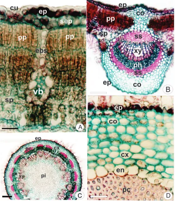
Figure 2. Gochnatia polymorpha (Less.) Cabrera, Asteraceae. Transection. A. Leaf blade with cuticule (cu), epidermis (ep),
anticlinal walls of epidermal cells are straight and relatively thin on both sides. Anomocytic stomata are only observed
on the abaxial surface. The stomata are at the same level
of other cells of epidermis or slightly raised above the surface.
In transection, the epidermis is uniseriate along
the leaf blade. An adaxial subepidermal cell layer is
observed (Figure 1D) which can represent a hypodermis.
However no ontogenetic study was performed to conirm this hypotesis. The epidermis is coated with a thick and smooth cuticle (Figures 1D, 2A) on both sides. The cuticle is slightly thicker on the adaxial surface.
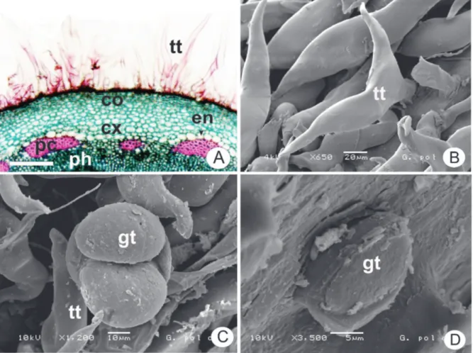
Several non-glandular trichomes occur only
on the abaxial surface (Figure 1C) and some glandular
trichomes inserted in small depressions are found on both
sides (Figures 1D, 2A). The non-glandular trichomes are branched with T-shaped apical cell (Figures 1C, 3B). The
capitate glandular trichomes are stalked or sessile and the head consists of one or two cells (Figures 1D, 2A, 3C, 3D).
The mesophyll reveals dorsiventral organization
comprising 2-3 layers of palisade parenchyma and 5-6 layers of spongy parenchyma (Figures 2A, B). Minor
collateral vascular bundles are embedded in the mesophyll and they are enclosed by a sheath of thick-walled
parenchymatic cells and ibers. This sheath also presents extensions towards the epidermis (Figures 1D, 2A).
The midrib shows a biconvex shape in transection, being slight convex on the adaxial surface. The uniseriate epidermis is covered by a thick and smooth cuticle. There are 8-10 layers of angular collenchyma on the adaxial side and 5-6 layers on abaxial side. In the ground parenchyma,
G. polymorpha exhibits a single collateral vascular bundle
which is surrounded by scleriied sheath (Figure 2B). The petiole is circular in transection and its
epidermis has the same characteristics previously
described for the leaf blade. The vascular system shows
an interrupted arch shape with two collateral bundles in
an adaxial position. The vascular system is surrounded by scleriied cells. Scleriied cells are common in the petiole cortex.
The stem presents an incipient secondary growth and has a circular shape in transection (Figure 2C). The
epidermis consists of a single layer of cells (Figure 2D) with glandular and non-glandular trichomes (Figures 3B,
C, D) similar to those found in the leaf. The collenchyma
Figure 3. Gochnatia polymorpha (Less.) Cabrera, Asteraceae. A. Stem detail with non-glandular trichome (tt), collenchyma (co),
forms a continuous ring and contains chloroplasts (Figure 2D). An endodermis containing amyloplasts bounds the
cortex internally (Figures 2D). The vascular cylinder
shows a typical structure (Figure 3A) and a vascular
cambium is evident. Perivascular iber caps are next to the phloem (Figures 2C, 3A). The pith cells show pitting and are slightly ligniied. In transection, elongated pith cells are veriied close to the perimedular zone while they are
circular in the central zone (Fig. 2C).
Discussion
Gochnatia spp. have simple leaf morphology but its leaves demonstrate great variation in shape from linear
to suborbicular. The venation is predominantly pinnate, although in taxa such as Gochnatia arequipensis Sandwith, Gochnatia glutinosa Klatt, Gochnatia rotundifolia Less. the leaves are three-veined (Freire et al., 2002). In this study, the leaf morphology of Gochnatia polymorpha (Less.) Cabrera is in accordance with the previous reports of Alice et al. (1995) and Freire et al. (2002) for this species.
An adaxial hypodermis is observed in a few
number of Gochnatia species whereas it is absent in G. amplexifolia (Gardner) Cabrera, G. cardenasii S.F. Blake, G. discoidea (Less.) Cabrera, G. foliolosa (D. Don) D. Don
& Arn. ex Hook., G. glutinosa and G. vernonioides Kunth. Occasionally, a descontinuous hypodermis is reported, e.g. G. argentina Cabrera, G. discolor Baker and G. orbiculata (Malme) Cabrera (Freire et al., 2002). In this investigation, the term subepidermal layer is used since no ontogenetic approach was performed for elucidating whether this layer has its origin from epidermis or mesophyll.
The anatomical characters observed in the
leaf blade, mainly the presence of a subepidermal layer, correspond to that described by Rossato & Kolb (2010; 2012) for G. polymorpha. However, these authors demonstrate that this species can show up to three layers
of adaxial hypodermis in higher luminosity environments.
According to Metcalfe & Chalk (1950), the presence of stomata in surface view has diagnostic value for Asteraceae. In addition, several genera of Asteraceae can present anomocytic (predominantly) and anisocytic stomata as observed in Calea (Farago et al., 2006; Budel et al., 2006), Baccharis (Oliveira et al., 2011; Souza et al., 2011), Lucilia (Duarte et al., 2011), Mikania (Budel et al., 2009; Gasparetto et al., 2010). In this work, anomocytic
stomata are veriied in G. polymorpha on the abaxial surface. However, G. barrosii Cabrera shows anomocytic stomata on both sides (Rossato & Kolb, 2012).
Various secretory structures such as ducts, cavities and glandular trichomes can occur on leaves of Asteraceae (Castro et al., 1997; Budel et al., 2013). However, ducts and cavities are not present in G. polymorpha. The glandular trichomes of many Asteraceae species are multicellular, biseriate, stalked
or sessile (Fahn, 1988). The glandular trichomes of
G. polymorpha present all these usual characteristcs of Asteraceae. In particular, the glandular trichomes of G. polymorpha demonstrate a head consisting of one or two cells as reported by Bieras & Sajo (2009).
The non-glandular trichomes are diverse in
anatomy, morphology, and microstructure, but they
are mainly classiied by the morphology, resulting in descriptive terms such as stellate, T-shaped, dendritic, and
spiral (Werker, 2000). In this study, G. polymorpha presents
T-shaped non-glandular trichomes. Simple and T-shaped
non-glandular trichomes were previously described for G. polymorpha by Alice et al. (1995) and Bieras & Sajo (2009). Stellate non-glandular trichomes are found in G. barrosii (Bieras & Sajo, 2009).
In particular, the two apical cells of the non-glandular trichomes of G. polymorpha are sharp-pointed.
This characteristic is widely reported in several genera
of Asteraceae as Acanthospermum (Martins et al., 2006), Ageratum (Oliveira et al, 1993, Tavares et al, 2000; Procopio et al., 2003), Baccharis (Budel & Duarte, 2007; Budel & Duarte, 2008a; Budel & Duarte, 2008b; Budel et al., 2012), Bidens (Procopio et al., 2003), Calea (Budel et al., 2006), Conyza, Galinsoga (Procopio et al., 2003) and Gochnatia (Bieras & Sajo, 2009).
Several species of Asteraceae show mesophyll distinguished in palisade and spongy parenchyma (Budel et al., 2005; Budel & Duarte, 2010; Oliveira et al., 2011; Souza et al., 2011). In this sense, G. polymorpha exhibits
a dorsiventral mesophyll as expected. Similar structural
arrangement was observed for this species in the paper of Rossato & Kolb (2010; 2012). Bieras & Sajo (2009) also
veriied a dorsiventral mesophyll for G. barrosii.
The minor collateral vascular bundles of leaves
of G. polymorpha are enclosed by a sheath of
thick-walled parenchymatic cells and ibers. This sheath also presents extensions towards the epidermis as indicated
for this species by Rossato & Kolb (2012). According to
Oliveira et al. (1993), the presence of a iber cap in the
vascular system is a remarkable attribute concerning the diagnosis of herbal drugs. In this investigation, the stem of G. polymorpha demonstrates perivascular iber caps
next to the phloem. In addition, ibers around the vascular
bundles are a characteristic of Mutisieae which suggests a phylogenetic relationship (Rossato & Kolb, 2010).
The endodermis is formed by a layer of cells
that bounds internally the cortical region. In Asteraceae,
the endodermis can exhibit Casparian strips which is
Conclusion
Considering the previously investigated macro- and microscopic aspects of the aerial vegetative organs of Gochnatia polymorpha (Less.) Cabrera, the
morpho-anatomical characters of uniseriate epidermis, adaxial
subepidermal cell layer, anomocytic stomata on the
abaxial surface, capitate glandular trichomes, T-shaped
non-glandular trichomes, dorsiventral mesophyll, and
perivascular iber caps next to the phloem in the stem
should be taken into account as quality control parameters for its pharmacognostic study.
Acknowledgements
The authors thank Dr. Nelson Ivo Matzenbacher for plant identiication and Centro de Microscopia
Eletrônica of Federal University of Paraná by performing the electron micrographs.
Authors’ contributions
JY contributed in collecting the plant sample and
its identiication, running the laboratory work, analyzing
of data and drafting the paper. PMDB and PVF contributed in performing the scanning electron microscopy (SEM) analysis and critical reading of the manuscript. MRD contributed to the critical reading of the manuscript. JMB supervised the laboratory work and contributed in
plant identiication and herborizing procedure. All the authors have read the inal manuscript and approved the
submission.
References
Alice CB, Siqueira NCS, Mentz LA, Silva G, Jose KFD 1995.
Popular use of medicinal plants: atlas pharmacognostic. Canoas: Publisher of Ulbra.
Berlyn GP, Miksch JP 1976. Botanical microtechnique and cytochemistry. Ames: Iowa State University.
Bieras AC, Sajo MG 2009. Leaf structure of the Cerrado (Brazilian savanna) woody plants. Trees-Struct Funct 23: 451-471.
Budel JM, Duarte MR, Santos CAM, Farago PV, Matzenbacher NI 2005. O progresso da pesquisa sobre o gênero Baccharis, Asteraceae: I - Estudos botânicos. Rev Bras Farmacogn 15: 268-271.
Budel JM, Duarte MR, Farago PV, Takeda IJM 2006. Caracteres
anatômicos de folha e caule de Calea unilora Less., Asteraceae. Rev Bras Farmacogn 16: 53-60.
Budel JM, Duarte MR 2007. Caracteres morfoanatômicos de partes vegetativas aéreas de Baccharis coridifolia DC. (Asteraceae-Astereae). Lat Am J Pharm 26: 723-731. Budel JM, Duarte MR 2008a. Estudo farmacobotânico de folha e
caule de Baccharis uncinella DC., Asteraceae. Lat Am J
Pharm 28: 740-746.
Budel JM, Duarte MR 2008b. Estudo farmacobotânico de partes vegetativas aéreas de Baccharis anomala DC., Asteraceae. Rev Bras Farmacogn 18: 761-768.
Budel JM, Duarte, MR, Kosciuv I, Morais TB, Ferrari, LP 2009.
Contribuição ao estudo farmacognóstico de Mikania laevigata Sch. Bip. ex Baker (guaco), visando o controle
de qualidade da matéria-prima. Rev Bras Farmacogn 19: 545-552.
Budel JM, Duarte MR 2010. Macro and microscopic characters of the aerial vegetative organs of carqueja: Baccharis usterii Heering. Braz Arch Biol Technol 53: 123-131. Budel JM, Duarte MR, Döll-Boscardin PM, Farago PV,
Matzenbacher NI, Sartoratto A, Maia BHLNS 2012 Composition of essential oils and secretory structures of
Baccharis anomala, B. megapotamica and B. ochracea. J Essen Oil Res 24: 19-24.
Budel JM, Farago PV, Duarte MR 2013 Pharmacobotanical study of Baccharis cognata DC. (Asteraceae: Astereae). Lat Am J Pharm 32: 550-554.
Castro MM, Leitão-Filho HF, Miller WR 1997. Use of secretory
structures in the identiication of genera of Asteraceae
from the cerrado vegetation. Braz J Botany 20: 163-174.
Catalan CAN, Borkosky SA, Joseph-Nathan P 1996. The
secondary metabolite chemistry of the subtribe Gochnatiinae (tribe Mutisieae, family Compositae).
Biochem Syst Ecol 24: 659-718.
Duarte MR, Budel JM, Matzenbacher NI, Menarim DO 2011. Microscopic diagnosis of the leaf and stem of Lucilia nitens Less., Asteraceae. Lat Am J Pharm 30: 2070-2075.
Esau K 1976. Anatomy of seed plants. New York: Edgard Blucher.
Fahn A 1988. Secretory tissues in vascular plants. New Phytologist 108: 229-257.
Farago PV, Budel JM, Duarte MR, Jurgensen I, Takeda IJM
2006. Anatomia da folha e do caule de Calea longifolia
(Asteraceae). Lat Am J Pharm 25: 512-517.
Freire SE, Kalina U, Sancho G 2002. Gochnatia (Asteraceae, Mutisieae) and the Gochnatia complex: Implications
from taxonomic morphology. Ann Mo Bot Gard 89: 524-550.
Foster AS 1949. Practical plant anatomy. 2. ed. Princeton: D. Van Nostrand.
Gasparetto JC, Campos FR, Budel JM, Pontarolo, R 2010.
Mikania glomerata Spreng. e M. laevigata Sch.
Bip. ex Baker, Asteraceae: estudos agronômicos,
genéticos, morfoanatômicos, químicos, farmacológicos,
toxicológicos e uso nos programas de itoterapia do
Brasil. Rev Bras Farmacogn 20: 627-640.
Horn RC, Vargas VM 2008. Mutagenicity and antimutagenicity of used in popular medicine in the Salmonella/ microsome assay. Toxicology in vitro 22: 1043-1049. Johansen NDA 1940. Plant microtechnique. New York: McGraw
Lorenzi H 2002. Brazilian trees: automatic identiication and
cultivation of woody plants in Brazil. 4th ed. Institute
Plantarum, New Odessa, p. 384.
Martins LRR, Mourão KSM, Albiero ALM, Cortez DAG, Dias-Filho BP, Nakamura CV 2006. Estudo morfoanatômico preliminar do caule e da folha de Acanthospermum australe (Loel.) Kuntze (Asteraceae-Heliantheae). Rev Bras Farmacogn 16: 42-52.
Metcalfe CR, Chalk L 1950. Anatomy of dicotyledons: leaves,
stem, and woods in relation to taxonomy with notes on economic uses. Oxford: Clarendon Press.
Moreira AS, Spritzer V, Schapoval EE, Schenkel EP 2000.
Anti-inlammatory activity of extracts and fractions from the
leaves of Gocnatia polymorpha. Phytother Res 14: 638-640.
Mors WB, Rizzini CT, Pereira NA 2000. Medicinal plants of Brazil. Michigan: Reference Publications.
O'Brien TP, Feder N, McCully ME 1964. Polychromatic staining
of plant cell walls by toluidine blue O. Protoplasm 59: 368-373.
Oliveira F, Lucia M, Garcia LO 1993. Caracterização
farmacognóstica da droga e do extrato luído de mentrasto
- Ageratum conyzoides L. Lecta 11: 63-100.
Oliveira AMA, Santos, VLP, Franco, CRC, Farago PV, Duarte, MR, Budel JM 2011. Comparative morpho-anatomical study of Baccharis curitybensis Heering ex Malme and Baccharis spicata (Lam.) Baill. Lat Am J Pharm 30: 1560-1566.
Procopio SO, Silva EAM, Silva AA, Ferreira EA 2003. Anatomia foliar de plantas daninhas do Brasil. Viçosa: Ed. UFV, v. I.
Roeser KR 1972. Die nadel und der schwarzkiefer massenprodukt kunstwerk-der natur. Mikrokosmos 61: 33-36.
Rossato DR, Kolb RM 2010. Gochnatia polymorpha (Less.) Cabrera (Asteraceae) mudanças na estrutura da folha
devido a diferenças nas condições de luz e edáicas. Acta Bot Bras 24: 605-612.
Rossato DR, Kolb RM 2012. Structural and functional leaf traits of two Gochnatia species from distinct growth forms in a sclerophyll forest site in Southeastern Brazil. Acta Bot Bras 26: 849-856.
Sass JE 1951. Botanical microtechnique. 2nd ed. Ames: Iowa
State College.
Sacilotto ACBC, Vichnewski W, Herz W 1997. Ent-kaurenediterpenes from Gochnatia polymorpha var.
polymorpha. Phytochemistry 44: 659-661.
Schlemper V, Freitas SA, Schlemper SRM 2011. Efeitos
antiespasmódico do extrato hidroalcoólico de Gochnatia polymorpha sp. loccosa no íleo cobaia. J Med Plant Res 5: 288-294.
Silva LB, Strapasson RLB, Riva D, Salvador MJ, Stefanello MEA
2011. Triterpenos das lores de Gochnatia polymorpha
subsp. loccosa. Rev Bras Farmacogn 21: 556-559. Stefanello MEA, Cervi AC, Wisniewshi JRA, Simionatto EL
2006a. Óleo essencial de Gochnatia polymorpha (Less.) Cabrera ssp. loccosa. Quim Nova 29: 999-1002.
Stefanello MEA, Salvador MJ, Ito IY, Macari PAT 2006b. Avaliação da atividade antimicrobiana e citotóxica de extratos de Gochnatia polymorpha ssp. loccosa. Rev
Bras Farmacogn 16: 525-530.
Souza W 1998. Técnicas básicas de microscopia eletrônica
aplicadas às Ciências Biológicas. Rio de Janeiro: Sociedade Brasileira de Microscopia Eletrônica. Souza CA, Farago PV, Duarte MR, Budel JM 2011.
Pharmacobotanical study of Baccharis singularis (Vell.) G.M. Barroso, Asteraceae. Lat Am J Pharm 30: 311-317.
Tavares ES, Gil VR, Viana VRC 2000. Anatomia do eixo
vegetativo de Ageratum conyzoides L. (Asteraceae). Rev Bras Farm 81: 25-28.
Werker E 2000. Trichome diversity and development. Adv Bot Res 31: 1-35.
*Correspondence
Patrícia Mathias Döll-Boscardin
Departamento de Ciências Farmaceuticas, Universidade Estadual de Ponta Grossa
Av. Carlos Cavalcanti, 4748, 84030-900 Ponta Grossa-PR, Brazil