ISSN 0102-695X
DOI: 10.1590/S0102-695X2013005000057 Received 11 May 2013
Accepted 26 Jul 2013 Available online 20 Aug 2013
(
Cinchona
spp.) in Brazilian traditional
medicine. Part I:
Polyouratea hexasperma
Nádia S. Somavilla,
1,2Gustavo P. Cosenza,
3,4Christopher W.
Fagg,
5Maria G. L. Brandão
*,3,41Laboratório de Anatomia Vegetal, Universidade de Brasília, Brazil,
2Departamento de Botânica, Universidade Federal de Juiz de Fora, Brazil,
3Dataplamt, Museu de História Natural e Jardim Botânico, Universidade Federal de
Minas Gerais, Brazil,
4Faculdade de Farmácia, Universidade Federal de Minas Gerais, Brazil,
5Faculdade de Ceilândia and Departamento de Botânica, Universidade de Brasília,
Brazil.
Abstract: This research is part of a larger study of the Brazilian species that are commonly referred to as quinas and are usually used as substitute of Cinchona
species. The purpose of the present study was the botanical characterization of the whole and powdered stem bark of Polyouratea hexasperma (A. St.-Hil.) Tiegh., Ochnaceae, by morphological and anatomical description, and the analysis of its chemical profile. The external texture of the bark, the whitened pit in the inner bark and the presence of cristarque cells, as well as the shape and arrangement of other lignified cells, are the most important macroscopic and microscopic features for the characterization of the bark. Chlorogenic and cafeic acids were detected in the chemical analysis and can also be used in the identification of the bark.
Keywords:
bark anatomy chemical analysis cristarque cells sclereids phenolic acids
Introduction
Quina (or china) is the traditional name originally assigned to Cinchona species, specii cally Cinchona calisaya Wedd. and C. succirubra Pav. ex Klotzsch, Rubiaceae. These plants are native to Peru, and their barks are sources of quinine and other alkaloids that have important medical applications. Quinine has been used to treat humans with malaria for over 350 years and is still in use, especially in cases of chloroquine-resistant Plasmodium falciparum (Kaur et al. 2009; Dondorp & Nosten, 2009). Quinine is also commercially used as
a l avoring agent in tonic water due to its bitter l avor
(Brasic, 1999).
Species of the Cinchona genus that produce quinine do not naturally grow in Brazil, and the interest in finding natural substitutes for Cinchona barks stimulated the search for Cinchona species in the Brazilian territories in the 18th and 19th centuries (Dean,
1996; Ribeiro, 2005). A consequence of these searches was the appearance of several substitutes for Cinchona plants that also received the name quina. In 1799, for example, the Brazilian naturalist Frei M. C. Vellozo wrote a book entitled Quinographia Portugueza, in
which he describes data on almost thirty substitutes for species of Cinchona which produces quinine (Vellozo, 1799). The German naturalist K. F. von Martius, in 1854, also registered several substitute species in his book Systema Materia Medica Vegetal (Martius, 1854). In 1916, Waldemar Peckolt, publish a monography with the description of fifteen species used as quinas in different regions of Brazil (Peckolt, 1916). In his travel notebook, the French naturalist A. Saint-Hilaire registered several species of quina used by the Brazilian population (Brandão et al., 2012), and in his book, Usual Plants of Brazilians (Saint-Hilaire, 2009), he reported the presence of seven substitutes for Cinchona spp. Barks from the quina-mineira (Remijia ferruginea (A. St.-Hil.) DC.) and quina-do-campo (Strychnos pseudoquina A. St.-Hil.) species were already widely used as tonics and febrifuges in conventional medicine. Monographs for these barks were included in the first edition of the Brazilian Pharmacopoeia (Brandão et al., 2009). Despite not being currently used in the preparation of industrialized medicine (Brandão et al., 2010), the barks of these quina plants are still being sold in popular markets all over the country (Brandão et al., 2013).
In recent years, our research group has focused on the restoration of information and samples of the plants used in traditional Brazilian medicine in the past centuries. A database including images and data on such plants is available at www.dataplamt.org.br. The present study is part of a larger project in which we are retrieving and evaluating the bitter native species known as quina. Analytical methods for the characterization of vegetal drugs from these species are scarce or do not exist, and
this lack of methods may make their identiication, as well as their quality control, dificult. The inal objective of this
project is to gather information to better understand these remedies from native Brazilian species and to promote their better use and conservation. In this study, we show
the results from the morpho-anatomic and chemical proile
analysis of the bark of the quina species Polyouratea hexasperma (A. St.-Hil.) Tiegh., Ochnaceae.
Materials and Methods
Plant material
The samples of Polyouratea hexasperma (A. St.-Hil.) Tiegh., Ochnaceae, bark were collected from a small tree on the University of Brasília campus (DF, Brazil), located at 15.46.12.9S, 47.52.06W at an altitude of 1040 m. The voucher was deposited in the University of Brasília herbarium (Fagg, C.W. 2191 UB), and the vegetal drug (dried bark) was deposited in the Dataplamt-UFMG (DAT-105).
Morpho-anatomical analysis
Soon after the samples were collected, the samples
were ixed in a solution of formalin, acetic acid, and 70%
ethanol (FAA), in a proportion of 1:1:18 per volume (Johansen, 1940), rinsed in distilled water, and stored in
70% ethanol. These samples were dehydrated in an ethanol
series, embedded in Leica HistoResin, and sectioned using a rotary microtome Leica RM2145 (Leica Microsystems,
Wetzlar, Germany). The 3-7 μm sections were stained with 0.05% toluidine blue solution (O’Brien et al., 1964).
The slides were mounted using Verniz vitral incolor 500®
(Paiva et al., 2006). Free-hand sections were obtained with
the aid of a Ranvier’s microtome and were submitted to
various histochemical tests; these included ferric chloride (Johansen, 1940) and potassium dicromate (Gabe, 1968) staining to detect phenolic compounds, acid phoroglucin staining for lignin detection (Sass, 1951), lugol staining
for starch (Kraus & Arduin, 1997), Draggendorff’s reagent (Yoder & Malhberg, 1976) and Wagner’s reagent (Furr &
Mahlberg, 1981) tests for alkaloids and Sudan III (Sass, 1951) and Sudan IV staining for lipid detection (Gerlach, 1984). Part of each sample was submitted to the maceration process for dissociation and tissue component analysis. For
this analysis, the samples were placed in Franklin solution and maintained in a kiln at 60 ºC for 72 h (Kraus & Arduin, 1997). After this process, the macerate was washed with distilled water to completely remove the Franklin solution
and was stored in 50% ethanol. The macerates were stained with 1% ethanolic safranin. The slides were analyzed and
described using an Olympus CX31 optical microscope and photographed using a digital Olympus C-7070 camera with wide zoom.
Chromatographic proile for phenolic and alkaloids in TLC
Preparation of fractions enriched in phenolic substances
Briely, 1 g of dried P. hexasperma bark was
extracted under relux conditions with 20 mL 70% ethanol for 30 min. The sample was then iltered through ilter
paper and concentrated and dried using a rotary evaporator. The resulting residue was diluted with 50 mL water and extracted three times with 30 mL ethyl acetate. The ethyl acetate fraction was concentrated to dryness and dissolved in 1 mL methanol for subsequent analysis.
Preparation of alkaloid fraction
Briely, 3 g of dried P. hexasperma bark was
extracted under relux conditions with 20 mL 0.1 M HCl
for 30 min. The solution was alkalized with NH4OH to pH 9.0 and extracted three times with 20 mL ethyl ether. The organic phase was concentrated to dryness, and the resulting residue was dissolved in 2 mL methanol for subsequent analysis.
Chromatographic analysis
Results and Discussion
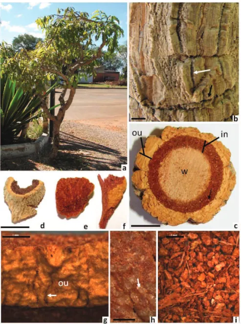
Poliouratea hexasperma (A. St.-Hil.) Tiegh., Ochnaceae, is a small tree that grows between two and three meters in height. This species is characteristically found in the Brazilian Cerrado (Figure 1A), but it also occurs in Bolivia and Paraguay (Killeen et al., 1998).
A. de Saint-Hilaire irst observed the use of this species
as a substitute for Cinchona spp. in the 19th century. He
irst described this species in 1824 and named it with the
basionym Gomphia hexasperma A. St.-Hil. The bark of this species has been used to heal wounds caused by insect
bites in both animals and humans; the bark has also been used as an astringent (Martius, 1854; Saint-Hilaire, 2009). On the other hand, Peckolt (1916) not cited this species in your study of Brazilian quinas.
Externally, the bark of P. hexasperma is corky and ranges in color from grey to yellowish grey. The
bark has sparse deep discontinuous issures, which can be
longitudinal or transversal (Figure 1B). The bark can also have grey-green lichens. When cut on the transverse plane, the bark can be divided into an outer bark and an inner
bark, according to Trockenbrodt (1990); this deinition
considers all the tissues outside of the vascular cambium
Figure 1. Polyouratea hexasperma (A. St.-Hil.) Tiegh. A. Tree. B. Externally aspect of bark showed longitudinal and transversal
issures (arrows white and black, respectively). C. Cross-section showed outer (ou), inner(in) bark and wood (w). Arrow points the whitened dots. D-F. Dry fragments of bark (plant drug). G. Detail of outer bark (ou) without cork. Arrows points the issure. H. Detail of whitened dots (arrow) of inner bark. I. Powder of the bark with ibrous pieces. Scale bars: 1 cm (B-F), 2 mm (G, I) and 0,5 mm
as bark, regardless of its speciic structure. According to
this same author, the terms outer bark and inner bark are recommended, and the term bast, which is sometimes used to identify the inner bark, should be avoided. The outer bark is creamy-brown in color, and the inner bark is reddish-brown with white dots (Figure 1C). Dry fragments (plant drug) can be represented by complete bark or parts (Figure 1D-F). When the plant drug is devoid of cork, the outer bark is yellowish in color and appears wrinkled, with
small transverse issures (Figure 1F-G). The white dots observed on the inner bark are translucent when magniied
(Figure 1H). The powder is reddish in color, with little
yellow pieces and ibrous pieces (Figure 1I).
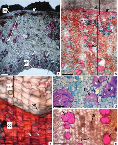
When cross-sectioned, the outer bark is formed by the periderm with phellem, phellogen and phelloderm cells (Figure 2A,B). The phellogen has a subepidermal origin according to ontogeny analyse in young stem (data not shown). The phellem is very regular and formed by several layers of juxtaposed rectangular cells, with slightly thickened walls, followed by one layer of phellogen (Figure 2C). In the phelloderm, the cells developed thick
walls (Figure 2A,B), ligniied and formed a continuous ring
just below the phellogen (Figure 2B). The deposition of lignin in these cells was thicker in the inner and anticlinal walls, thereby giving this structure the shape of the letter “U” (Figure 2C, arrows). These cells are called cristarque
Figure 2. Microscopy characteristics in transversal section of the bark of the Polyouratea hexasperma (A. St. Hi.) Tiegh. A. General
view of the young bark showed the periderm (pe) with phelloderm without ligniication (arrow), cortex (c) and secondary phloem (ph). Thin arrow showed the ibers of the primary phloem. B. Part of the outer bark (ob) showed the phellem (asterisk) and phelloderm with ligniied cell wall (arrow) and part of the inner bark showed the cortex (c) with clusteres of sclereids. C. Magniication of square
in (b). Cork with rectangular cells (co), phellogen (pg) and cristarque cells (cc) of the phelloderm showed thickening on the inner
and anticlinal walls (arrows). D. Cortex region with stone cells (stars) and bundle of primary phloem ibers (i). E. Secondary phloem
cells and usually have a crystal inside the lumen (Rao et al., 1967; Dickinson, 2000). Nevertheless, the presence of the crystals was not detected in the lumen of these cells in P. hexasperma. The presence of the cristarque cells is a diagnostic characteristic of the Ochnaceae family and a few others families (Dickinson, 2000). Phelloderm cells with thickened walls are common in tropical plants (Roth, 1981), and these cells can provide protection to the inner tissue when the cork is lost (Yamamoto, 1989).
The inner bark is formed by the cortex and the secondary phloem (Figure 2A). In the cortex, there are a large number of parenchyma cells interspersed with clusters of sclereids, as well as single sclereids and groups
of sclerenchyma ibers (Figure 2B and 2D). These ibers
are reminiscent of the primary phloem (Figure 2A). In the secondary phloem, lengthened sclereids were found close to the vascular cambium; these sclereids were found in isolation as well as in clusters (Figure 2E). Idioblasts containing druses (Figure 2E and 3B) and lipidic substances in the shape of oil droplets (Figure 3B) or adhered to druses were observed mostly in the parenchyma cells of the secondary phloem. Cells with phenolic compounds and idioblasts containing starch grains (Figure 2E) were distributed throughout the cortex and the secondary phloem, especially in the parenchyma cells of the cortex. The presence of phenolic compounds in these regions of
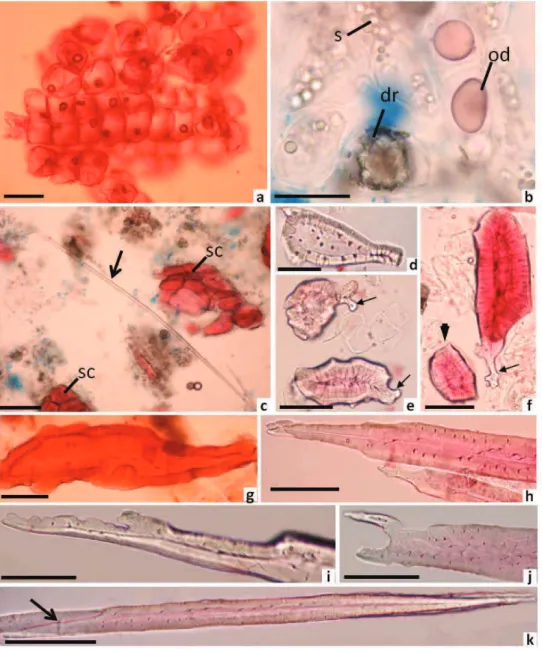
Figure 3. Macerate of Polyouratea hexasperma (A. St. Hi.) Tiegh. A. Cork cells. B. Idioblasts with druses (dr), starch grains (s)
and oil droplets (od). C. Part of primary phloem iber (arrow) and cluster of sclereids (sc). D. Stone cell. E. Sclereids with adorned
the bark is common in plants from the Cerrado (Costa et al., 1997), and these compounds usually help protect the plant from ultraviolet B radiation and insect herbivory in the leaves (Izaguirre et al., 2007). However, the function of these compounds in the bark remains unknown.
After the maceration process, the following cell
types were identiied: clustered or isolated square-shape
cork cells (Figure 3A), idioblasts containing druses and oil droplets (Figure 3B), sieve tube members with sieved
areas and sieve plates, parenchyma cells and ligniied cells. The ligniied cells showed variation in the cells types, such as narrow and elongated primary phloem ibers
(Figure 3C), and sclereids of diverse shape; these sclereids included stone cells or brachysclereids (Figure 3D, F), short sclereids with or without adorned ends (Figure 3E,F), sclereids with irregular forms (Figure 3G) and lengthened sclereids with intrusive growth and adornments at the end of the cells (Figure 3H-K). Short sclereids with and without adorned ends had polylamellate walls with branched pits
(Figure 3D-G). Phloem ibers and lengthened sclereids
(Figure 3H-K) did not possess polylamellate walls, but the lengthened sclereids did possess branched pits. These pits
were not visible in the phloem ibers.
Fibers and sclereids in the secondary phloem and cortex are common features and are found in most barks (Esau, 1977; Parameswaran, 1980; Trockenbrodt, 1990; Junikka, 1994). However, the characterization of the cells types, such as their shape, location and source, are important (Parameswaran, 1980; Evert, 2006) and could assist in the diagnosis of vegetal drugs.
Previous phytochemical studies have shown the presence of terpenoids, isoflavonoids, flavonoid glycosides, biisoflavanones and biflavones in the roots, leaves and stem barks of P. hexasperma. The biflavone are considered to be chemical markers of the genus Polyouratea (Moreira et al., 1994; 1999; Daniel et al., 2005; Carvalho et al., 2008; Fernandes, 2008; Suzart et al, 2007). The biflavone 7-O-methyl-agathisflavone has been shown to inhibit DNA topoisomerase and therefore possesses anticancer activity (Grynberg et al., 2002; Daniel et al., 2007). Flavonoids were only weakly observed in our study, being rutin and quercetin absent. Conversely, chlorogenic acids among other caffeic acids were clearly observed in the TLC analysis and the presence of these substances can be useful for characterization of the bark. Alkaloids were also weakly observed in Rf of 0.60, above the bands for quinine, quinidine, cinchonine and cinchonidine, which were used as standards. In spite of TLC analyzes reveal the presence of these substances in the bark, histochemical tests were negative for alkaloids, hindering its localization in tissues.
Conclusion
This study reports the morpho-anatomical and chemical characteristics of the bark of the quina substitute Polyouratea hexasperma (A. St.-Hil.) Tiegh. The presence of cristarque cells in the phelloderm and the adorned sclereids are anatomical characters of diagnostic value. Furthermore, the presence of chlorogenic acids may
be useful for identiication of the extracts of the bark in
addition to already existing chemical markers. Wherefore,
these characteristics could contribute to the identiication
of this species and help in quality control analyses.
Acknowledgements
The authors are grateful to FAPEMIG (PPM-2010) and CNPq for their fellowships (150523/2011-4,
150453/2012) and inancial support (563311/2010-0,
563563/2010-9).
Authors’ Contribution
NSS contributed in anatomy and histochemical studies. MGLB is the coordinator of the research and GPC has done the chromatographic analyses. CWF contributed
in collecting plant material, identiication and herbarium confection. All the authors have read the inal manuscript
and approved the submission.
References
Brandão MGL, Cosenza GP, Grael CFF, Netto NL, Monte-Mór RLM 2009. Traditional uses of American plant species from the 1st edition of Brazilian Oficial Pharmacopoeia.
Rev Bras Farmacogn 19: 478-487.
Brandão MGL, Cosenza GP, Stanislau AM, Fernandes GW 2010. Influence of Brazilian herbal regulations on the use and conservation of native medicinal plants.
Environ Monitor Assess 164: 369-377.
Brandão MGL, Pignal M, Romaniuc S, Grael CFF, Fagg CW
2012. Useful Brazilian plants listed in the ield books
of the French naturalist Auguste de Saint-Hilaire (1779-1853). J Ethnopharmacol 143: 488-500.
Brandão MGL, Cosenza GP, Pereira FL, Vasconcelos AS, Fagg CW 2013.Change in the trade in native medicinal plants in Brazilian public markets. Environ Monit Assessment.
DOI 10.1007/s10661-013-3081-y.
Brasic JM 1999. Should people with nocturnal leg cramps drink tonic water and bitter lemon? Psychol Rep 84: 355-367. Carvalho MG, Suzart LR, Cavatti LC, Kaplan MAC 2008.
New lavonoids and other constituents from Ouratea hexasperma. J Braz Chem Soc 19: 1423-1428.
385-399.
Daniel JFS, Carvalho MG, Cardoso RS, Agra MF, Eberlin MN
2005. Other lavonoids from Ouratea hexasperma. J Braz Chem Soc 16: 634-638.
Daniel JFS, Alves CCF, Grivicich I, da Rocha AB, Carvalho MG
2007. Antitumor activity of bilavonoids from Ouratea
and Luxemburgia on human cancer cell lines. Indian J Pharmacol 39: 184-186.
Dean A 1996. A ferro e fogo: a história e a devastação da Mata Atlântica brasileira. Cia das Letras: Rio de Janeiro. Dickinson WC 2000. Integrative plant anatomy. Academic Press,
San Diego.
Dondorp AM, Nosten F, Yi P 2009. Artemisinin resistance in
Plasmodium falciparum malaria New Engl J Med 361: 455-467.
Esau K 1977. Anatomy of seed plants. 2ed. John Wiley, New York.
Evert RF 2006. Esau’s plant anatomy: merystems, cells, and tissues of the plant body: theirs structure, function and development. 3ed. Jon Willey & Sons Inc., New Jersey. Fernades RD 2008. Estudo químico e atividades biológicas de
Ouratea hexasperma var. planchonii Engl. (Ochnaceae). 118p. Dissertação de Mestrado. Programa de Pós-graduação em Química. Universidade Federal Rural do Rio de Janeiro.
Furr M, Mahlberg PG 1981. Histochemical analyses of lacticifers and glandular trichomes in Cannabis sativa. J Nat Prod 44: 153-159.
Gabe M 1968. Techniques histologiques. Masson & Cie, Paris. Gerlach D 1984. Botanische Mikrotechnik. Thieme, Stuttgart. Grynberg NF, Carvalho MG, Velandir JR, Oliveira MC, Moreira
IC, Braz-Filho R, Echevarria A 2002. DNA polymerase
inhibitors: bilavonoids from Ouratea species. Braz J Med Biol Res 35: 819-822.
Izaguirre MM, Mazza CA, Svatos A, Baldwin IT, Ballare CL 2007. Solar ultraviolet-B radiation and insect herbivory trigger partially overlapping phenolic responses in
Nicotiana attenuata and Nicotiana longilora. Ann Bot
99: 103-109.
Johansen DA 1940. Plant Microtechnique. Macgraw-Hill Book Company, New York.
Junikka L 1994. Survey of English macroscopy bark terminology. IAWA Journal 15: 3-45.
Kaur K, Jain M, Kaur T, Jain R 2009. Antimalarials from nature.
Bioorg Med Chem17: 3229-3256.
Killeen TJ 1998. A checklist of the vascular plants of Parque Nacional Noel Kempff Mercado and surrounding areas. In: Killeen TJ, Schulenberg TS (Eds). A biological assessment of Parque Nacional Noel Kempff Mercado, Bolivia. RAP Working Papers 10, Conservation International, Washington, D.C.
Kraus JE, Arduin M 1997. Manual básico de métodos em morfologia vegetal. Rio de Janeiro, EDUR.
Martius KF 1854. Systema de Materia Medica Vegetal. Rio de Janeiro: Eduardo e Henrique Laemmert.
Moreira IC, Sobrinho DC, Carvalho MG, Braz-Filho R 1994.
Isolavanone dimers hexaspermone A, B and C from
Ouratea hexasperma. Phytochemistry 35: 1567-1572. Moreira IC, Carvalho MG, Bastos ABFO, Braz-Filho R
1999. A lavones dimer from Ouratea hexasperma. Phytochemistry 51: 833-838.
O'Brien TP, Feder N, McCully ME 1964. Polychromatic staining of plant cell walls by toluidine blue. Protoplasma 59: 368-373.
Paiva JGA, Fank-de-Carvalho SM, Magalhães MP, Graciano-Ribeiro D 2006. Verniz vitral incolor 500®: uma alternativa
de meio de montagem economicamente viável. Acta Bot Bras 20: 257-264.
Parameswaran N 1980. Some remarks on the nomenclature of
ibers, sclereids and ibre-sclereids in the secondary
phloem of trees. IAWA Bulletin 1: 130-132.
Peckolt WL 1916. Monograia das falsas quinas brasileiras. Rio de Janeiro.
Rao AR, Malaviya M, Menon VK 1967. On the cauline sclereids of three species of Ochna. Proc Indian Acad Sci, Plant Sci 66: 19-24.
Ribeiro RF 2005. Florestas Anãs do Sertão. O Cerrado da Historia de Minas Gerais. Belo Horizonte, Autêntica.
Roth I 1981. Structural patterns of tropical barks. Gebruder Borntraeger, Berlin-Stuttgart. p 81-111.
Saint-Hilaire A 2009. Plantas usuais dos Brasileiros. Código, Dataplamt. Belo Horizonte.
Sass JE 1951. Botanical Microtechnique. Iowa State University Press, Ames.
Suzart LR, Daniel JFS, Carvalho MG 2007. Biodiversidade
lavonoídica e aspectos farmacológicos em espécies dos
gêneros Ouratea e Luxemburgia (Ochnaceae). Quim Nova 30: 984-987.
Trockenbrodt M 1990. Survey and discussion of the terminology used in bark anatomy. IAWA Bulletin 11: 141-166. Vellozo FMC 1799. Quinographia Portugueza. Ou Coleção de
várias memórias sobre 22 espécies de quinas tendentes
ao seu descobrimento nos vastos domínios do Brazil. Lisboa, Impressora da Santa Igreja Patriarcal.
Yamamoto K 1989. Morfologia, anatomia e sistemática do gênero Ouratea Aublet (Ochnaceae): levantamento preliminar das características de importância taxonômica
e avaliação das classiicações vigentes. Campinas,
175p. Dissertação de Mestrado, Instituto de Biologia da Universidade Estadual de Campinas.
Yoder LR, Mahlberg PG 1976. Reactions of alkaloid and histochemical indicators in laticifers and specialized parenchyma cells of Catharanthus roseus
(Apocynaceae). Am J Bot 63: 1167-1173.
*Correspondence
Maria das Graças Lins Brandão
Av. Gustavo da Silveira, 1035, 31080-010 Belo Horizonte-MG, Brazil