Original article (short paper)
Electromyographic assessment of trunk and
shoulder muscles during a Pilates pull-up exercise
Isabel C.N. Sacco Eduardo T.T. Mori Bergson C. Queiroz
University of São Paulo, Brazil
Nadia Marconi
University of Brasília, Brazil
Ivye L. R. Pereira
University of São Paulo, Brazil
Abstract—This study compares surface electromyographic activity of the internal oblique, rectus abdominis, multiidus,
iliocostalis, anterior deltoids during the pull-up on a lower and on a higher dificulty level. We assessed nine adults with
previous experience in Pilates. The root mean square (RMS) values were normalized by maximum isometric
contrac-tion for each participant. During the ascent phase, the low spring posicontrac-tion showed a signiicantly higher RMS than the
high spring position of 8.9% for deltoid, 17.2% for internal oblique, 22.3% for rectus abdominis, 4.1% for iliocostalis,
and 5.6% for multiidus, and in the descent phase, the RMS in the lower spring exceeded signiicantly the high spring
position in 1.6% for the deltoid, 10% for internal oblique, 31.4% for rectus abdominis and 11.4% for iliocostalis. There was no predominance of abdominal muscles over the shoulder muscle in any spring position. The pull-up exercise can be a useful choice for the core and anterior deltoid muscles strengthening.
Keywords: biomechanics, electromyography, exercise therapy, abdominal wall
Resumo—“Avaliação eletromiográica de músculos do tronco e do ombro durante um exercício Pilates de pull-up.” Este
estudo compara a atividade eletromiográica de superfície dos músculos oblíquo interno, reto abdominal, multíidos, iliocostal e deltóide anterior durante o pull-up em dois níveis de diiculdade (mola alta e mola baixa). Foram avaliadas
nove adultos com experiência anterior em Pilates. Os valores RMS foram normalizados pela contração isométrica máxima.
Durante a fase de subida, a posição de mola baixa mostrou RMS signiicativamente maiores em relação a alta de 8,9% para deltóide, 17,2% para o oblíquo interno, 22,3% para o reto abdominal, 4,1% para iliocostal, e 5,6% para o multíido, e na fase de descida, em 1,6% para o deltóide, 10% para oblíquo interno, 31,4% para o reto abdominal e 11,4% para o iliocostal. Não houve predomínio dos músculos abdominais sob o músculo do ombro em qualquer posição de mola. O exercício de pull-up pode ser ferramenta útil para o fortalecimento da musculatura do core e do músculo deltóide anterior. Palavras-chave: biomecânica, eletromiograia, terapia por exercício e parede abdominal
Resumen—“Evaluación electromiográica de los músculos del tronco y del hombro durante un ejercicio Pilates de pull-up.” Este estudio compara la EMG supericial de los músculos recto del abdomen, oblicuo interno, multiidos,
ilio-costal y deltoides anterior durante el ejercicio pull-up en dos niveles de diicultad. Se evaluaron a nueve adultos
experimentados en Pilates. Los valores de RMS se normalizaron por la contracción isométrica máxima. Durante la
fase de ascenso, la posición baja del resorte mostró valores signiicativamente majores de RMS que la posición alta de
8,9% para lo deltoides, 17,2% para oblicuo interno, 22,3% para recto abdominal, 4,1% para ilio-costalis, y 5,6% para
multiidos. En la fase de descenso, el RMS, en el muelle inferior, excede signiicativamente la posición alta del resorte
en 1,6% para el deltoides, 10% para oblicuo interno, 31,4% para recto abdominal y 11,4% para ilio-costalis. No hubo
predominio de los músculos abdominales bajo los deltoides anteriores. Pull-up puede ser una herramienta útil para el trabajo del core y para la fortiicación del deltoides anterior.
Introduction
The Pilates method was created by Joseph Pilates and consists of stretching, strengthening, and proprioception exercises with the entire attention of practitioners focused on muscle control,
postu-re, and breathing (Johnson, Larsen, Ozawa, Wilson, & Kennedy, 2006; Muscolino & Cipriani, 2004a; Stolze et al., 2012; Wells, Kolt, & Bialocerkowski, 2012). The main focus of the method is on trunk musculature; therefore, it can be classiied as a program of “core stabilization”(Amorim, Sousa, & Santos, 2011; Akuthota & Nadler, 2004; Barr, Griggs, & Cadby, 2007; Stolze, Allison, & Childs, 2012). Pilates method has been considered as an im
-portant tool for rehabilitation and injury prevention (Anderson & Spector, 2000, Lim, Poh, Low, & Wong, 2012) and also has been the subject of recent trends in Brazilian scientiic investigations (Bertolla, Baroni, Leal Junior, & Oltramari, 2007; Queiroz et al.,
2010; Rocha-e-Silva, 2009; Sacco et al., 2005).
Trunk lexion exercises, such as the pull-up, may compro -mise the ability of the lumbar extensor muscles to bear the shear forces on the lumbar column. Despite some evidences correlating these exercises with increased susceptibility to disc
injury, it is possible to increase the spine stability and thereby reduce the risk of damage to the structures of the column, by controlling the strength of the abdominal and back extensors muscles (Brown & McGill, 2008). In one more physiological
sense the “Pilates’ core stabilization” is related to coordinated
and simultaneous activation of the trunk and hip muscles, which is associated to reduction the risk of disc injury (Lim et al., 2012; Van Dieen, Cholewicki, & Radebold, 2003; Wong, Leong, Chan, Luk, & Lu, 2004).
For this reason, the Pilates’ pull-up exercise could be considered as a good therapeutic and itness option when the main goal is workout abdominal, back extensors muscles, and shoulder muscles in a closed-kinetic chain in opposition to
the elastic loads attached to the chair equipment. During the exercise performance, the practitioner also has the challenge
to control shoulder and trunk muscles simultaneous and dyna
-mically (Akuthota & Nadler, 2004; Isacowitz, 2006; Pilates,
2000). Despite being recommended for shoulder girdle muscles strengthening (Isacowitz, 2006), the main focus of the pull-up exercise is to strengthen the core that comprises the coordinated
and simultaneous activation of the trunk and hip muscles, and
which is one of the fundamental concepts of Pilates (Gallagher
& Kryzanoswska, 2000a; Muscolino & Cipriani, 2004b; Pilates,
2000). The main principle is the tightening of the muscular centre of the body or “powerhouse,” located between the pelvic
loor and the ribcage during exercises. Probably, this is one of the
main reasons why Pilates is suitable to promote core stability,
control and strength (Wells et al., 2012).
Because of its complexity, the pull-up exercise can be
used in a inal phase of sports rehabilitation and improvement
of the performance of athletes and dancers. A biomechanical investigation of the pull-up exercise would lead to a better com-prehension of its potential to strengthen the core and shoulder girdle muscles and to understand the relationship between the
activation of trunk lexors and extensors muscles. Identiication of muscles working patterns is a main concern among resear
-chers and clinicians (Marques, Hallal, & Gonçalves, 2012) and
these deeper insights will allow us to discuss more properly the potential advantages and disadvantages of this exercise for the core stabilization and for the lumbar region, thereby con-tributing to the grounding both of rehabilitation and physical conditioning practices.
This study aimed to compare the surface electromyographic
activity of the internal oblique, rectus abdominis, multiidus,
iliocostalis, and anterior deltoid during the pull-up on two levels
of dificulty, modiied by the different anchoring positions of
the springs. This study addressed the following hypotheses: (i) the different anchoring of spring positions would generate
distinct patterns of trunk and shoulder muscles activation, (ii) the trunk muscles would activate predominantly over shoulder muscles, regardless of the level of exercise dificulty and phase of movement, (iii) among the trunk muscles there would be a predominant activity of lexors muscles, particularly the internal
oblique, over the extensors muscles regardless of the level of
exercise dificulty and phase of movement.
Methods
Experimental approach to the problem
The present study characterized the EMG activity of anterior
trunk muscles (internal oblique and rectus abdominis), posterior trunk muscle (multiidus and iliocostalis) and a shoulder girdle
muscle (anterior deltoid) during the pull-up exercise performed
in two dificulty levels of dificulty by nine experienced healthy adults. Within-muscle EMG comparison were made to evaluate the existence of co-contraction (trunk lexor and extensor) and the importance of trunk and shoulder muscle activation accor -ding to the phase of the exercise (ascent or descent phase). The spring anchoring change might bring important variation in the intensity of exercise inferred from EMG activity.
Participants
Nine healthy adults (5 men and 4 women, 28 ± 5 years; 1.72
± 0.07m; 68 ± 12 kg) volunteered for this study. All assessed in -dividuals had previous Pilates training (at least 6 months, 2 times
a week, 48 sessions minimum). Participants were excluded from
the study if they had a history of recent (in the last two years,
with a duration of seven days) lower back pain, musculoskeletal or neuromuscular injury, previous abdominal surgeries, scoliosis,
important lower limbs asymmetry, or postural deviations. All procedures were approved by the Local Ethics Commit-tee (protocol no. 079/10), and the participants gave their written informed consent.
Task
During the pull-up the volunteers were instructed to perform
the equipment with wrists in extension while upper limbs; trunk and hips were in lexion (Figure 1). During the upward phase, the
volunteer lifted the body weight upwards moving the shoulders
in extension and the trunk segments and hip in lexion while the
lower limbs are extended with the feet in contact with the pedal. In the upward phase, springs progressively reduce their lengths. The opposite combination of movements is observed during the downward phase, while springs stretch. During both movement
phases, the ankles are kept in plantar lexion. The force provided
by this pedal could be modulated by the number, the strength, and the positioning of the springs (Isacowitz, 2006; Queiroz et
al., 2010; Gallagher & Kryzanowska, 2000b).
The pull-up exercise was performed in two levels of dificul -ty (high and low springs) in a random order (by simple drawing) for each participant, in four series of three cycles, with a one-minute interval to rest. This approach provided a total of 12
cycles in each level of dificulty for analysis. The performance
speed was controlled by verbal commands during the upward and downward movements of the lower limbs, following the
cadence marked by a digital metronome to 100 bpm. All parti -cipants were experts in the Pilates method and experienced in responding to verbal rhythmic commands.
Procedures
During the pull-up exercise, the electromyographic signal of four muscles (internal oblique, rectus abdominis, iliocostalis,
multiidus) in the trunk and one in the arm (anterior deltoid),
were unilaterally recorded on the right side of the body. This exercise started with the participant in the position 1 (as shown
in Figure 1), with lower limbs extended and feet touching the
equipment’s pedal. The pedals were connected to springs that
were used in different positions classiied as low and high. The trunk was in lexion; the pelvis was retroverted and the hands
were holding the top of the chair.
The exercise was performed by raising the body using the strength of the abdominals and upper limbs with an increase in
the lexion of the trunk segment (Figure 1). Two springs pro -vided assistant force on the pedal to raise the body. This force was modulated by changing the chair´s spring position in order to modify the exercise’s demand.
The elastic constant (k) of each spring has been found as
280 N/m. At the beginning of the movement, the spring, which
was ixed in the high position, at 35° from the horizontal plan,
provided a vertical support of 209 N and in the low spring
position, at 25° from the horizontal plan, provided a support of 150 N (Figure 1).
An 8-channel EMG system (DE 2.2L Bagnoli model; Delsys, Boston, MA, EUA) was used to measure electromyography (EMG). Ag/AgCl bipolar electrodes (10 mm) were placed over each muscle with an interelectrode distance of 20 mm center to
the center. EMG signals were sampled at 1kHz and ampliied 2000 times; the common mode rejection ratio was 92 dB (at
60/10Hz) and the input impedance of the system was 1015Ω.
After skin preparation, which consisted of shaving and cleaning with rubbing alcohol and gauze, the electrodes were ixed with
transpore® (3M) adhesive tape on the right side of the body.
The placement of electrodes on the iliocostalis, multiidus
and anterior deltoid muscles followed the recommendations of
the project Surface EMG for a non-invasive assessment of mus
-cles (SENIAM, 2005). For the rectus abdominis, we followed the recommendations of Grenier and McGill (Grenier & McGill,
2007), and for the internal oblique, we followed Escamilla,
McTaggart, Fricklas, and DeWit (2006). A reference electrode
was placed over the right clavicle.
For normalization purposes, before the performance of
the exercises, four seconds of electromyographic data were recorded for each muscle while the participants performed maximum voluntary isometric contractions (MVIC) against manual resistance. The highest mean value during 500 ms from the two central seconds’ window from two trials of each
muscle was chosen as the representative MVIC. For testing the MVICs of the iliocostalis and lumbar multiidus muscles,
trunk extension was performed in a prone position, with the
lower limbs restrained and maximum resistance applied to the
upper back (SENIAM, 2005). For testing the MVIC of the rec
-tus abdominis muscles, the upper trunk was maximally lexed with maximum resistance applied to the shoulders in the trunk extension direction with knees lexed 90o and feet restrained
(Escamilla et al., 2006). For testing the MVIC of the internal oblique muscles, the trunk was maximally lexed and rotated
to the left and to the right side with maximum resistance at the shoulders in the opposite direction of rotation in a supine
position with knees lexed 90o and feet restrained (Escamilla et al., 2006). And for testing the MVIC of the anterior deltoid muscles, the participant was sitting with the shoulder
abduc-tion in slight lexion, arms hanging vertically and the palm
pointing inwards. The maximum resistance applied against the antero-medial surface of the arm in the direction of adduction and slight extension (SENIAM, 2005).
Tracking data from the right greater trochanter was used to
synchronize the phases (upward and downward) of the Pull-up
cycles. An Optotrak Motion Analysis System – OPTOTRAK 3020 (Northern Digital, Waterloo, Ontario, Canada) sampled at 100 Hz the LEDs (light emitting diodes) ixed at the right greater trochanter. All EMG data were sampled at 1 kHz using the Optotrak software and a synchronization unit (ODAUII)
with a 12-bit acquisition board.
Data processing
After the offset removal from the raw EMG—if existent, the
signal was digitally zero lag bandpass iltered (Butterworth, 2nd order 10-400 Hz) and normalized by the MIVC EMG signal.
Each movement phase (upward and downward), determined by
kinematic data of the trochanter, was represented by its avera -ge root mean square (RMS) of the whole phase duration. The average RMS of 12 cycles (12 ascent phases and 12 descent phases) was calculated for each muscle and spring.
Statistical analyses
The mean values of the normalized muscles activities were compared between the two phases (ascent x descent) in each spring position and between spring positions within each movement phase by two-sided paired t-tests. The ive muscles were compared at each movement phase and spring position by ANOVAs for repeated measures followed by Newman-Keuls post-hoc tests. We adopted an alpha error of 0.05 and statistics were performed using Statistica software (v.7 StatSoft, Inc.). Given the sample size evaluated, an alpha of 5%, a moderate effect size (f=0.45), the statistical power was 0.852 for the F test used.
Results
The percentage differences presented refer to the ratio of the highest value obtained by the absolute difference between the values.
Comparisons between springs
In the ascent phase, the EMG activity observed in the low
spring position (150 N assistance force) signiicantly outper
-Muscle Spring position Ascent phase Descent phase p¹
Internal Oblique (% MVIC)
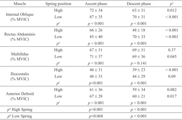
High 72 ± 34 63 ± 31 0.012
Low 87 ± 35 70 ± 31 < 0.001
p¹ p < 0.001 p < 0.001
Rectus Abdominis (% MVIC)
High 66 ± 26 48 ± 18 < 0.001
Low 85 ± 40 70 ± 33 < 0.001
p¹ p < 0.001 p < 0.001
Multiidus (% MVIC)
High 67 ± 31 69 ± 31 0.37
Low 71 ± 37 69 ± 36 0.045
p¹ p < 0.001 p = 0.141
Iliocostalis (% MVIC)
High 46 ± 31 39 ± 23 < 0.001
Low 48 ± 33 44 ± 29 0.09
p¹ p<0.001 p < 0.001
Anterior Deltoid (% MVIC)
High 61 ± 36 59 ± 34 0.082
Low 67 ± 28 60 ± 21 0.017
p¹ p < 0.001 p < 0.001
p² High Spring p<0.001 p < 0.001
p² Low Spring p=0.068 p < 0.001
Table 1. Mean (± standard deviation) values of normalized RMS (by the maximal voluntary contraction – MVIC) of the muscles evaluated, p values of comparisons between the phases of the movement (ascent x descent) and between spring positions (high x low), and p values of comparisons among muscles in each phase and spring.
predominantly over shoulder muscles since the anterior deltoid showed higher activity than rectus abdominis and iliocostalis
in the descent phase on the high spring position. Furthermore, during the ascent phase on the low spring position, no signiicant difference between trunk and shoulder muscles was identiied.
In the third hypothesis, there were no predominance activity
of lexors muscles over the extensors muscles as was assumed,
since in the ascent phase on the high spring position, there was
an increase in the internal oblique activity over multiidus, but
in the descent phase on the high spring position, there was an
increase in the multiidus activity over internal oblique. In the
ascent phase on the high spring position, there was also an
increase in the multiidus muscle over anterior deltoid and the
iliocostalis muscle. However, in the descent phase on the high spring position, the anterior deltoid showed higher activity than rectus abdominis and iliocostalis, as we said, despite showing
less activity than multiidus. In the descent phase in the low spring position, multiidus showed higher activity compared
to the anterior deltoid.
In the comparisons between the ascent and descent phases for each muscle, in the high spring position in the ascent phase,
we observed higher RMS values, with signiicantly difference,
for the internal oblique, rectus abdominis and iliocostalis. In contrast, in the low spring position in the ascent phase, the
in-ternal oblique, rectus abdominis, multiidus and anterior deltoid
muscles showed higher EMG values.
We can suggest by the comparisons among the trunk lexors
muscles that internal oblique and rectus abdominis are equally
important for the trunk lexion in this exercise in spite of the level of dificulty. However, the higher activation of the internal
oblique for both spring positions and movement phases indica-tes the crucial importance of this muscle to maintain the pelvis
retroversion while the trunk is lexed, as Queiroz et al. (2010)
pointed out in other Pilates exercises; while the rectus abdominis
acts, beyond the function of the trunk lexor, as an anchor for the
internal oblique action, as also proposed by McGill et al. (1996).
During the pull-up exercise, the trunk lexion and the pelvic retroversion modiies the lever arms, considering the shoulder joint as the axis, the in two ways: [1] the center of body mass
gets near the pull-up main movement axis in the shoulder,
de-creasing the resistance arm to the shoulder and [2] this forward
movement of the center of mass also decreases the resistance arm to the pedal axis, increasing the ratio between the force arm
(pedal axis – pedal extremity where the foot rests) and resistance
arm. The resultant internal forces, in addition to the spring assis-tance force, allow the body to go up. In this case, it is possible
to propose the importance of the trunk lexor muscles (internal
oblique and rectus abdominis) to bring forward the center of
mass in the ascent phase for the high spring position. For the
low spring position, where less assistance is provided, one may need the additional action of the anterior deltoid to modify the
lever arm, besides the trunk muscles support.
The multiidus muscle showed the highest activity (67%
MVIC) compared to the other extensor assessed (iliocostalis 46% MVIC) in the ascent phase in the high spring position.
McGill, Hughson, and Parks (2000) identiied that lumbar lexion compromises the ability of the force generation of the
formed the high spring position (assistance force of 209 N) in 8.9% for anterior deltoid (p<0.001), 17.2% for internal oblique (p<0.001), 22.3% for rectus abdominis (p<0.001), 4.1% for ilio-costalis (p<0.001) and 5.6% for multiidus muscles (p<0.001). In the descent phase, the EMG signals observed in the lower
spring exceeded signiicantly the EMG values observed in the
high spring position in 1.6% for the anterior deltoid (p<0.001), 10% for internal oblique (p<0.001), 31.4% for rectus abdominis (p<0.001) and 11.4% for iliocostalis muscle (p<0.001), but not
for the multiidus.
Comparison among muscles
In the high spring position, during the ascent phase, the in-ternal oblique muscle showed a 6.9% higher activity compared
to the multiidus muscle (p=0.004), while the multiidus muscle showed an 8.9% greater activity (p=0.002) compared to the an-terior deltoid, and 31.3% greater (p=0.009) than the iliocostalis
muscle. For the descent phase, in the high spring position, the multiidus showed an 8.7% higher activity (p=0.026) than the internal oblique, and 14.5% higher (p<0.001) than the anterior deltoid muscle. The anterior deltoid was more active in 18.6% (p=0.003) than the rectus abdominis, and in 33.8% (p=0.004) than the iliocostalis muscle.
In the low spring position, the muscles differ from each other
only during the descent phase. The multiidus presented a 13%
higher activity (p=0.048) than the anterior deltoid.
Comparison between phases of movement (ascent and descent)
In the high spring position, the internal oblique (12.5%, p=0.012), rectus abdominis (27.3%, p=<0.001), and iliocostalis (15.2%, p=<0.001) muscles showed signiicantly greater electrical activity in the ascent phase over the descent phase. In the low spring position, the internal oblique (19.5%, p=<0.001), rectus abdominis (17.6%, p=<0.001), multiidus (2.8%, p=0.045) and anterior deltoid (10.4%, p=0.017) muscles showed signiicantly greater electrical activity in the ascent phase over the descent phase.
Discussion
The general results allowed us to conirm the irst hypothesis
whether the different anchoring of spring positions would
gene-rate distinct patterns of trunk and shoulder muscles activation,
since we observed distinct levels of muscle activity for each level of spring anchoring.
In the ascent phase with the low spring position, which provided less assistance (150 N on the starting position), we observed greater EMG activity of all assessed muscles com-pared to the high spring position, and in the descent phase, anterior deltoid, internal oblique, and rectus abdominis and iliocostalis showed greater activation compared to the high
iliocostalis muscle due to the obliquity of its ibers. This me -chanical disadvantage may reduce the ability of this muscle to
control intervertebral anterior shear forces while the multiidus function would be less affected by lumbar lexion. Therefore, the higher activity of the multiidus could relect its pivotal
role in lumbar vertebrae positioning and in controlling the
in-tervertebral forces. This key role inferred by our EMG results is also consistent with the description that Wilke, Wolf, Claes, Arand, and Wiesend (1995) and Bojadsen, Silva, Rodrigues,
and Amadio (2000) made in their studies, highlighting the role
of the multiidus as the major lumbar extensor and stabilizer. However, even in exercises that require no trunk lexion and
are performed isometrically (Holding test), there was a
predo-minant action of the multiidus muscle (78% MVIC) compared to the iliocostalis (65% MVIC) (Ng, Richardson, & Jull, 1997).
Moreover, in the descent phase with the high spring position,
the activity of multiidus overcomes the internal oblique, which was the most active muscle in the ascent phase among the ive assessed muscles, suggesting that the multiidus muscle plays
not only an important role in the spinal stabilization but also in
the eccentric trunk control.
According to Granata and Marras (2000) and Kavcic,
Gre-nier, and McGill (2004b), the co-contraction of both lexors and extensors muscles during the trunk lexion increases the
spine’s capability to support critical overloads and increase
trunk stability; however, it increases the compression forces
in the lumbar spine. The study of co-contraction in several patterns of exercises can guide the planning of training and
rehabilitation activities (Marques, Hallal, & Gonçalves,
2012). In the pull-up exercise, we observed values of MVIC between 48% (rectus abdominis) and 87% (internal oblique)
for the lexor muscles, and values between 39% (iliocostalis) and 71% (multiidus) for the extensor muscles. Kavcic et
al. (2004b) have found that values of MVIC around 57% for internal oblique, 46% for rectus abdominis and 25% for
multiidus, provided enough co-contractions for stabilization purposes. All these indings together with our results suggest that the activity magnitude between lexors and extensors in the pull-up exercise can provide suficient spine stability when used in training practice. However, it is important to take into
consideration the probable increase in the lumbar spine loads (Kavcic et al., 2004b) not only due to the exercise itself, but
due to the position of trunk lexion, which has been suggested
as a main factor for intervertebral disc herniation (Callaghan
& McGill, 2001). Therefore, one should be careful to indicate
this exercise in cases of lumbar dysfunctions.
Since the multiidus, internal oblique, rectus abdominis, and
anterior deltoid muscles generated EMG activities generally above 60% of MVIC, we suggest that the pull-up exercise is
an effective option for strengthening purposes(Anderson &
Spector, 2000), with either the high or low spring positions. Our
results conirm the pull-up effectiveness as a tool for strengthe
-ning the core muscles, as stated by Kryzanowska and Gallagher
(2000a) and Isacowitz (2006). The iliocostalis showed values between 39% and 48% MVIC, these activation magnitudes are
suficient to gain strength, which for Ekstrom et al. (2007) is
at least 45% of MVIC.
Practical applications
The pull-up exercise is effective as an exercise for stren-gthening and conditioning all “power house” muscles, and the anterior deltoid plays an important role in controlling ec-centrically the lowering of the body in the descent phase. The practitioner of this exercise must have a previous appropriate physical preparation for its performance since it demands high muscle activation to perform it while stabilize the lumbar spine. In order to progressively enhance the load and the level of mus-cular activity, it is highly recommended to change the anchoring position of the springs. The low spring position homogenizes
and increases muscle activity, especially the trunk muscles.
References
Akuthota, V., & Nadler, S. F. (2004). Core strengthening. Archives of Physical Medicine and Rehabilitation,85(3 Suppl 1), S86-92. Amorim, T. P., Sousa, F. M., Santos, & Rodrigues, J.A. dos. (2011).
Inluence of Pilates training on muscular strength and lexibility in dancers. Motriz: Revista de Educação Física, 17, 660-666. Anderson, B. D., & Spector, A. (2000). Introduction to Pilates-based
rehabilitation. Orthopaedic Physical Therapy Clinics of North America, 9(3), 395-410.
Barr, K. P., Griggs, M., & Cadby, T. (2007). Lumbar stabilization: a review of core concepts and current literature, part 2. American Journal of Physical Medicine & Rehabilitation, 86(1), 72-80. Bertolla, F., Baroni, B.M., Leal Junior, E.C.P., & Oltramari, J.D. (2007).
Effects of a training program using the Pilates method in lexibility of sub-20 indoor soccer athletes. Revista Brasileira de Medicina do Esporte, 13, 222-226.
Bojadsen, T. W., Silva, E. S., Rodrigues, A. J., & Amadio, A. C. (2000). Comparative study of Mm. Multiidi in lumbar and thoracic spine. Journal of Electromyography and Kinesiology, 10(3), 143-149. Brown, S. H., & McGill, S. M. (2008). How the inherent stiffness of the
in vivo human trunk varies with changing magnitudes of muscular activation. Clinical Biomechanics (Bristol, Avon), 23(1), 15-22. Callaghan, J.P., & McGill, S.M. (2001). Intervertebral disc herniation:
studies on a porcine model exposed to highly repetitive lexion/ extension motion with compressive force. Clinical Biomechanics (Bristol, Avon), 16, 28-37.
Ekstrom, R. A., Donatelli, R. A., & Carp, K. C. (2007). Electromyo -graphic analysis of core trunk, hip, and thigh muscles during 9 rehabilitation exercises. Journal of Orthopaedic & Sports Physical Therapy, 37(12), 754-762.
Escamilla, R. F., McTaggart, M. S., Fricklas, E. J., DeWitt, R., Kelleher, P., Taylor, M.K, . . .& Moorman, C.T. (2006). An electromyographic analysis of commercial and common abdominal exercises: impli-cations for rehabilitation and training. Journal of Orthopaedic & Sports Physical Therapy, 36(2), 45-57.
Gallagher, S. P., & Kryzanoswska, R. (2000a). The Pilates method of body conditioning. Philadelphia: BainBridgeBooks.
Gallagher, S.P., & Kryzanowska, R. (2000b). The complete writings of Joseph H Pilates. Philadelphia: BainBridgeBooks.
Granata, K. P., & Marras, W. S. (2000). Cost-beneit of muscle cocontrac -tion in protecting against spinal instability. Spine, 25(11), 1398-1404. Grenier, S. G., & McGill, S. M. (2007). Quantiication of lumbar sta
-bility by using 2 different abdominal activation strategies. Archives of Physical Medicine and Rehabilitation, 88(1), 54-62.
Isacowitz, I. (2006). Pilates. Champaign, IL - Florida.
(2006). The effects of Pilates-based exercise on dynamic balance in healthy adults. Journal of Bodywork and Movement Therapies, article in press.
Lim, E. C. W., Poh, R. L. C., Low, A. Y., & Wong, W. P . (2012). Ef -fects of pilates-based exercise on pain and disability in individuals with persistent nonspeciic low back pain_ a systematic review with meta-analysis. Journal of Orthopaedic & Sports Physical Therapy,41(2),70-80.
Kavcic, N., Grenier, S., & McGill, S. M. (2004b). Quantifying tissue loads and spine stability while performing commonly prescribed low back stabilization exercises. Spine, 29(20), 2319-2329. Marques, N.R., Hallal, C.Z., & Gonçalves, M. (2012). Padrão de
co-ativação dos músculos do tronco durante exercícios com haste oscilatória. Motriz: Revista de Educação Física, 18, 245-252. McGill, S., Juker, D., & Kropf, P. (1996). Appropriately placed surface
EMG electrodes relect deep muscle activity (psoas, quadratus lumborum, abdominal wall) in the lumbar spine. Journal of Bio-mechanics, 29(11), 1503-1507.
McGill, S. M., Hughson, R. L., & Parks, K. (2000). Changes in lumbar lordosis modify the role of the extensor muscles. Clinical Biome-chanics(Bristol, Avon), 15(10), 777-780.
Muscolino, J.E., & Cipriani, S. (2004a). Pilates and the ‘powerhou -se’ - I. Journal of Bodywork and Movement Therapies, 8, 15-24. Muscolino, J.E., & Cipriani, S. (2004b). Pilates and the “powerhouse’
- II. Journal of Bodywork and Movement Therapies, 4(122-130). Ng, J. K., Richardson, C. A., & Jull, G. A. (1997). Electromyographic amplitude and frequency changes in the iliocostalis lumborum and multiidus muscles during a trunk holding test. Physical Therapy, 77(9), 954-961.
Pilates, J.H. (2000). The complete writings of Joseph H. Pilates. Phi-ladelphia: BainBridgeBooks.
Queiroz, B. C., Cagliari, M. F., Amorim, C. F., & Sacco, I. C. (2010). Muscle activation during four Pilates core stability exercises in quadruped position. Archives of Physical Medicine and Rehabili-tation, 91(1), 86-92.
Rocha-e-Silva, M. (2009). Recent trends in Brazilian medical research. An overview. Clinics (Sao Paulo), 64(10), 1007-1013.
Sacco, I.C.N., Andrade, M.S., Souza, P.S., Nisiyama, M., Cantuária, A.L., Maeda, F.Y.I., & Pikel, M. (2005). Método Pilates em revista: aspectos biomecânicos de movimentos especíicos para reestrutu -ração postural - estudos de caso. Revista Brasileira de Fisiotearpia e Movimento,13(4), 65-78.
SENIAM. Project 2005. Retrieved 07.agosto.2010, from http://www. seniam.org
Stolze, L., Allison, S. C., & Childs, J. D. (2012). Derivation of a preli -minary clinical prediction rule for identifying a subgroup of patients with low back pain likely to beneit from pilates-based exercise. Journal of Orthopaedic & Sports Physical Therapy, 42(5), 425-436. Van Dieen, J. H., Cholewicki, J., & Radebold, A. (2003). Trunk muscle
recruitment patterns in patients with low back pain enhance the stability of the lumbar spine. Spine, 28(8), 834-841.
Wells, C., Kolt, G. S., & Bialocerkowski, A. (2012). Deining Pilates exercise: a systematic review. Complementary Therapies in Me-dicine, 20(4), 253-262.
Wilke, H. J., Wolf, S., Claes, L. E., Arand, M., & Wiesend, A. (1995). Stability increase of the lumbar spine with different muscle groups. A biomechanical in vitro study. Spine, 20(2), 192-198.
Wong, K. W., Leong, J. C., Chan, M. K., Luk, K. D., & Lu, W. W. (2004). The lexion-extension proile of lumbar spine in 100 healthy volunteers. Spine, 29(15), 1636-1641.
Authors’ note
Isabel C.N. Sacco, Eduardo T.T. Mori, Bergson C. Queiroz, and Ivye L. R. Pereira are afiliated with the Physical Therapy, Speech and Occupational Therapy Department, School of Medicine, University of São Paulo, SP, Brazil.
Nadia Marconi is afiliated with the Physical Therapy Department, University of Brasília, DF, Brazil.
Corresponding author:
Dr. Isabel Sacco
Movement and Posture Biomechanics Laboratory; Physiotherapy, Speech Therapy and Occupational Therapy Department. USP Rua Cipotânea, 51, Cidade Universitária, São Paulo, SP 056360-160, Brazil Phone: 55 11 30918426.
Email: [email protected] www.usp.br/labimph.
Acknowledgements
The authors are grateful to CNPq (National Council for Scientiic and Technological Development) for the scholarship awarded to Mori, to Olavo Luppi Silva for supporting the EMG processing, and to Nove de Julho University, where data were collected. There is no commercial relationship, which may lead to a conlict of interests, whether inancial or personal, with third parties or persons.
Manuscript received on September 15, 2013 Manuscript accepted on March 19, 2014