61
Jurno ME et al. Síndrome de desmielinização osmótica: relato de caso
Radiol Bras. 2012 Jan/Fev;45(1):61–62
Osmotic demyelination syndrome: report of a case
with favorable outcome
*
Síndrome de desmielinização osmótica: relato de caso com evolução favorável
Mauro Eduardo Jurno1, Marina Horta Azevedo de Castro2, Mariana de Assis Lage2, João Henrique Dupin3, Antonio José Fonseca de Paula4, Gustavo de Vasconcelos Bello5
The authors report a case of a patient with favorable outcome after diagnosis of osmotic demyelination syndrome (central pontine and extrapontine myelinolysis) confirmed by magnetic resonance imaging.
Keywords: Osmotic demyelination syndrome; Osmotic myelinolysis; Central pontine and extrapontine myelinolysis.
Os autores relatam um caso de paciente apresentando evolução favorável após confirmado diagnóstico de síndrome de desmielinização osmótica (mielinólise pontina central e extrapontina) através de exame de ressonância magnética.
Unitermos: Síndrome de desmielinização osmótica; Mielinólise osmótica; Mielinólise pontina e extrapontina.
Abstract
Resumo
* Study developed at Hospital Regional de Barbacena – FHEMIG, Barbacena, MG, Brazil.
1. PhD, Coordinator of the Medical Practice Residency Pro-gram at Hospital Regional de Barbacena – FHEMIG, Professor at Faculdade de Medicina de Barbacena, Barbacena, MG, Bra-zil.
2. MDs, Medical Practice Residents, Hospital Regional de Barbacena – FHEMIG, Barbacena, MG, Brazil.
3. MD, Resident of Psychiatry, Centro Hospitalar Psiquiátrico de Barbacena – FHEMIG, Barbacena, MG, Brazil.
4. MD, General Clinician and Oncologist, Preceptor of Med-ical Practice, Hospital Regional de Barbacena – FHEMIG, Bar-bacena, MG, Brazil.
5. MD, Radiologist, Titular Member of Colégio Brasileiro de Radiologia e Diagnóstico por Imagem (CBR), Barbacena, MG, Brazil.
Mailing Address: Dr. Mauro Eduardo Jurno. Rua Fernando Laguardia, 45, Santa Tereza II (Geraldo Xavier). Barbacena, MG, Brazil, 36201-118. E-mail: [email protected]
Received January 17, 2011. Accepted after revision July 25, 2011.
Jurno ME, Castro MHA, Lage MA, Dupin JH, Paula AJF, Bello GV. Osmotic demyelination syndrome: report of a case with favorable outcome. Radiol Bras. 2012 Jan/Fev;45(1):61–62.
0100-3984 © Colégio Brasileiro de Radiologia e Diagnóstico por Imagem
CASE REPORT
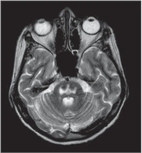
pons, sparing the corticospinal tract, with subtle involvement of the basal nuclei and the lateral aspect of the thalami, with bilat-eral areas of necrosis areas at the globus pallidus (Figures 1, 2 and 3). No abnormal contrast medium uptake was observed, and the radiologist concluded that the images confirmed signs of osmotic demyelination, necrosis in the globus pallidus and signs of decrease in the infra- and supratentorial volumes.
The patient progressed with improve-ment in swallowing, allowing the progres-sion from enteral to oral feeding. Improve-ment was also observed in limb spasticity, with a good response to the administration of introduction of a muscle relaxant drug. After one month, with progressive improve-ment of the clinical picture, the patient was able to communicate verbally (unable to raise the voice, still presenting dysarthria). MRI was repeated, with the same image pattern demonstrated by the previous study. Within two months, the patient had recov-ered from the previously described deficits.
DISCUSSION
Osmotic demyelination syndrome is a demyelinating disorder of the brain prima-rily affecting the pontine region, sometimes compromising extrapontine regions(1). In
spite of the relevance of the association of this syndrome with metabolic disorders, such a condition. Upon admission the use
of neuroleptic drugs was discontinued. Laboratory tests demonstrated severe hydroelectrolytic disorder: hyponatremia (sodium: 123 mEq/l; RV: 135 to 145 mEq/ l) and hypokalemia (potassium: 2.6 mEq/ l; RV: 3.5 to 5.2 mEq/l). Electrolytes re-placement was initiated, and sodium con-centration in blood was 147 mEq/l in the first 48 hours after admission.
Adynamia and prostration were ob-served. After psychiatric evaluation, clonazepan and mirtazapine were adminis-tered, but the clinical picture worsened. The patient presented severe prostration, drowsiness, hyporesponsiveness, eyes opening only upon stimulation, nonspecific motor response (withdrawal reflex) and incomprehensive verbal response (Glasgow coma scale: 9), and then was transferred to the intensive care unit. At admission to such unit, physical evaluation demon-strated isochoric and photoresponsive pu-pils, absence of nuchal rigidity, right-sided Babinski sign and right upper limb paresis. The patient progressed to a spastic paraly-sis and mutism.
Alterations were not observed at brain computed tomography. As the low level of consciousness of the patient persisted even after normalization of her potassium and sodium levels, MRI was requested, which demonstrated signal hyperintensity on T2-weighted sequences, compromising the INTRODUCTION
The present case report refers to a pa-tient with favorable outcome after osmotic demyelination syndrome confirmed by magnetic resonance imaging (MRI).
CASE REPORT
62
Jurno ME et al. Síndrome de desmielinização osmótica: relato de caso
Radiol Bras. 2012 Jan/Fev;45(1):61–62 particularly the rapid correction of
hy-ponatremia, this entity may occur in pa-tients with no sign of hydroelectrolyte im-balance(2). In such cases, the patients
present other risk factors such as a history of alcohol or drug abuse, malnutrition, hepatic disorders, cancer and Addison’s disease(3,4). Generally, mutism and
dysar-thria are the first symptoms of this syn-drome. Alterations such as lethargy and affective disorders are also commonly ob-served and may be confused with psychi-atric disorders. The progression of such patients is variable, ranging from complete recovery to death. In general, recovery is slow and gradual. The mortality rate asso-ciated with severe hyponatremia is between 40% and 50%(5,6).
In the present case, hyponatremia may have resulted from the utilization of
anti-depressant drugs or from the previous vom-iting episodes. It is important to prevent osmotic demyelination syndrome by means of an appropriate approach for patients with hyponatremia. Some authors consider it impossible to establish a totally risk free level of sodium correction; but in cases of symptomatic hyponatremia, sodium re-placement must not exceed 10–12 mEq within 24 hours.
The MRI report describes findings com-patible with osmotic demyelination syn-drome(7,8).
REFERENCES
1. Grafton ST, Bahls FH, Bell KR. Acquired focal dystonia following recovery from central pontine myelinolysis. J Neurol Neurosurg Psychiatry. 1988;51:1354–5.
2. Pietrini V, Mozzani F, Crafa P, et al. Central pon-tine and extraponpon-tine myelinolysis despite careful correction of hyponatremia: clinical and
neuro-Figure 1. Magnetic resonance imaging, sagittal plane: image of the pontine region.
Figure 2. Magnetic resonance imaging, axial plane: extrapontine lesions.
Figure 3. Magnetic resonance imaging, axial plane: pontine lesion image.
pathological findings of a case. Neurol Sci. 2010; 31:227–30.
3. Sugimoto T, Murata T, Omori M, et al. Central pon-tine myelinolysis associated with hypokalaemia in anorexia nervosa. J Neurol Neurosurg Psychiatry. 2003;74:353–5.
4. Martin RJ. Central pontine and extrapontine my-elinolysis: the osmotic demyelination syndromes. J Neurol Neurosurg Psychiatry. 2004;75 Suppl 3:iii22–8.
5. Thompson PD, Miller D, Gledhill RF, et al. Mag-netic resonance imaging in central pontine myeli-nolysis. J Neurol Neurosurg Psychiatry. 1989;52: 675–7.
6. Germiniani FMB, Roriz M, Nabhan SK, et al. Mielinólise pontina central e extra-pontina em paciente alcoolista sem distúrbios hidro-eletrolíti-cos: relato de caso. Arq Neuropsiquiatr. 2002;60: 1030–3.
7. Abbott R, Silber E, Felber J, et al. Osmotic demy-elination syndrome. BMJ. 2005;331:829–30. 8. Howard SA, Barletta JA, Klufas RA, et al. Best