Arq Bras Oftalmol. 2008;71(2):265-8
Glaucoma agudo bilateral em um paciente com dengue: relato de caso
Trabalho realizado no Instituto da Visão da Santa Casa de Sobral - Sobral (CE) - Brasil.
1Pós-Graduando do Departamento de Oftalmologia da
Universidade Estadual de Campinas - UNICAMP - Cam-pinas (SP) - Brasil; Oftalmologista da Santa Casa de Sobral - Sobral (CE) - Brasil.
2Médico Clínico-Infectologista da Santa Casa de Sobral
- Sobral (CE) - Brasil.
3Farmacêutica, Especialista em Saúde da Família pela
Escola de Saúde da Família Visconde de Sabóia - Sobral (CE) - Brasil.
Endereço para correspondência: Paulo de Tarso Ponte Pierre Filho. Av. Gerardo Rangel, 801 Apto.1001 -Sobral (CE) CEP 62041-380
E-mail: [email protected] Recebido para publicação em 15.05.2007 Última versão recebida em 14.08.2007 Aprovação em 02.09.2007
Nota Editorial: Depois de concluída a análise do artigo sob sigilo editorial e com a anuência do Dr. Enyr Saran Arcieri sobre a divulgação de seu nome como revisor, agradecemos sua participação neste processo.
Paulo de Tarso Ponte Pierre Filho1
Jurandir Pontes Carvalho Filho2
Érika Teles Linhares Pierre3
Bilateral acute angle closure glaucoma in a patient
with dengue fever: case report
Keywords:Dengue/complications; Glaucoma; Intraocular pressure/etiology; Brazil; Case reports [Publication type]
Ocular complications in dengue fever are uncommon but may result in visual loss. The authors report the first documented case of a patient with dengue fever who presented with simultaneous bilateral acute angle closure glaucoma. The disease was confirmed by specific serological tests. Despite the treatment, severe visual impairment occurred in this case.
ABSTRACT
RELATOS DE CASOS
INTRODUCTION
Dengue fever (DF), the most important mosquito-borne viral disease affecting humans, occurs in over 100 countries, with an estimated 100 million cases per year and more than 2.5 billion people at risk worldwide(1). It
has emerged as a global public health problem in the past several decades and is now endemic in most tropical and subtropical areas of Central and South America, Southest Asia, the Western Pacific, Africa, and the Eastern Mediterranean(1-2).
Classic DF is a self-limiting, influenza-like illness caused by any of the four serotypes (DEN 1-4) of dengue virus. Dengue hemorrhagic fever (DHF) is a more severe form of the disease characterized by multisystem hemor-rhagic manifestations, thrombocytopenia, increased vascular permeability, and plasma leakage. The early phase of DHF is indistinguishable from DF. The death rate for untreated DHF can be as high as 10-20% in places where emergency supportive treatment with intravenous fluids and platelet repla-cement is not readily accessible(3).
In Brazil, more than a million cases were reported since the reintro-duction of dengue in 1982(4). The state of Ceará in the Northeast region of
Brazil has had successive dengue epidemics that involved dengue virus serotypes DEN-1, DEN-2, and DEN-3. In 2006, there were 25,569 confirmed cases of dengue in this state and 172 fulfilled the World Health Organization criteria for DHF(5). Usually, epidemics are more intense in cities, which is
attributed to favorable epidemiological characteristics of rapidly growing urban agglomerations such as crowding, poor housing, as well as the existence of many putative breeding sites such as disposable containers, bottles and used tires(6).
266Bilateral acute angle closure glaucoma in a patient with dengue fever: case report
Arq Bras Oftalmol. 2008;71(2):265-8
Ophthalmic complications associated with DF and DHF have not been classically described. Within the ophthalmic community, this complication is being observed more fre-quently in recent times. However, only a few isolated case reports have been published(8-13). We describe a patient with
DF who developed bilateral acute angle closure glaucoma (AACG).
CASE REPORT
A previously healthy 67-year-old woman was admitted to the hospital with sudden loss of vision, intense ocular pain, lacrimation, photophobia, and redness of both eyes. She re-ported a 3-day history of fever, headache, nausea, and vo-miting. Her previous medical and ocular history was unremar-kable. Her physical examination revealed fever of 38ºC and mild dehydration. No rash, jaundice or neck stiffness was noted. Blood test revealed a hemoglobin rate 12.9 g/dL, leu-kopenia (4.0 x 10⁹/L), and thrombocytopenia (100 x 109/L).
Alanine aminotransferase (ALT)/aspartate aminotransferase (AST) were 125/65 IU/L. Blood sugar, creatinine, electrolytes were normal.
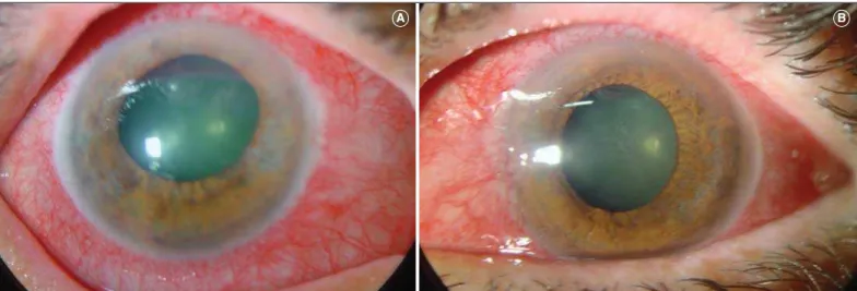
In view of the severity of her visual symptoms she was referred to the Emergency Ophthalmology Department. Oph-thalmological examination revealed that both eyes had visual acuity of hand motions, and elevated intraocular pressure (IOP) (60 mmHg in the right eye (RE) and 54 mmHg in the left eye (LE)), by applanation. Both eyes were injected with cor-neal edema, shallow anterior chamber, iris atrophy, and fixed moderately dilated pupils (Figure 1). Both crystalline lenses exhibited nuclear sclerosis 2+ and glaucomflecken, gonios-copy showed closed angles. No angle structures could be seen beyond Schwalbe’s line, even on indentation, however, there were no goniosynechiae. Fundoscopy was not clear. The axial length was 22.13 mm and 22.12 mm in the RE and LE, respectively. B-Scan ultrasonography showed normal
poste-rior segments in both eyes. A diagnosis of bilateral AACG was made and she was treated with 20% intravenous mannitol followed by topical 2% pilocarpine q.i.d., 0.5% timolol b.i.d., and 1% prednisolone acetate q.i.d. to both eyes. The IOP reduced to 22 and 20 mmHg in right and left eyes respectively and bilateral laser iridotomies were performed.
On the following day her symptoms were present but improving. Computed tomography of the brain was normal. Blood tests showed a worsening leukopenia (3 x10⁹/L) as well
as thrombocytopenia (80 x 109/L). During hospital stay, she
also was given intravenous and oral fluids and paracetamol. Five days later she was almost asymptomatic with a platelet count of 108 x 109/L and was discharged. Specific serological
tests (IgG and IgM) confirmed the dengue infection. She was advised to continue on timolol, pilocarpine and predni-solone eye drops for further 14 days. The patient’s ocular findings improved gradually over the subsequent month. However, her visual symptoms had still not completely disap-peared and visual acuity was 20/200 in both eyes. Her RE IOP was 20 mmHg and left was 19 mmHg without any treatment. The eye fundus exploration without dilatation did not exhibit significant alterations.
DISCUSSION
DF and DHF continue to be a global challenge. There is no specific treatment available nor any immediate prospect of a vaccine, and the mosquito control measures in most of the hyperendemic areas are inadequate(14).
Ophthalmic complications in dengue infections are rare, but there has been an increasing number of cases reported in recent literature(8-14). The main ocular findings include
sub-conjunctival hemorrhages, macular edema and blot hemor-rhages. Less common features include exudative retinal deta-chment, cotton wool spots, and anterior uveitis. A majority of patients were reported to have residual visual impairment(13-14).
Figure 1 - Conjunctival hyperemia, corneal edema, iris atrophy, and fixed moderately dilated pupils are seen in the right (A) and left (B) eyes
267
Bilateral acute angle closure glaucoma in a patient with dengue fever: case report
Arq Bras Oftalmol. 2008;71(2):265-8
The pathogenesis of ocular complications in dengue fever is not fully understood. Thrombocytopenia in severe dengue may predispose towards hemorrhage. However, clinical pre-sentation and behavior of these complications can suggest an immunogenic etiology rather than an infective one(15). Other
postulates include viral mutations, viral virulence and host susceptibility. Viral genetic mutations have been demonstra-ted to occur within the various serotypes, however the biolo-gic effects induced by these mutations are as yet uncharac-terized(15-16). More research is still needed to evaluate the
patho-genesis of these ocular manifestations.
Bistis (1929, apudRichardson(17)) published an article in
1929 concerning the epidemic of dengue in Athens, in which he said that iritis, glaucoma, and retrobulbar neuritis had been mentioned as complications. During the same epidemic Anar-gyros (1929, apud Richardson(17)) reported five cases with
ocular complications. In one, glaucoma of the left eye appea-red on the fourth and last day of the fever, simultaneously with the appearance of the exanthem, and two days later there was a less severe attack of glaucoma in the right eye. Iridec-tomy was done, first in the left, then in the right eye. Another patient, aged fifty-four years, had two years previously lost the sight of the right eye from glaucoma. The eye had remai-ned painless until convalescence from dengue took place, when the conjunctiva became injected and there was intense pain. The eye was enucleated and the vitreous body was found to be full of blood clots.
Gabrielides (1929, apudRichardson(17)) observed two
ca-ses of angle closure glaucoma during dengue infection in wo-men aged sixty and fifty-four years respectively. In the first case symptoms appeared on the third day; in the second case the symptoms, with the complete loss of vision, appeared on the fourth day. Notwithstanding intensive treatment, the glau-comatous phenomena, and especially the pupillary dilatation, persisted.
Bilateral simultaneous acute angle closure is a rare entity, infrequently reported after psychotropic drug intake(18),
gene-ral anesthesia(19), or snake bite(20). To the best of our
knowled-ge, no cases of bilateral simultaneous acute angle closure glaucoma related to dengue fever have been reported before. Our patient presented with bilateral sudden loss of vision, elevated IOP, shallow anterior chamber, corneal edema, and dilated pupils during a febrile illness. The diagnosis of dengue fever was suspected based on leuko- and thrombocytopenia, elevated serum transaminase levels plus dehydration, and this diagnosis was confirmed by serological tests. Longer follow-up will be required to determine whether this functional visual loss is permanent.
Acute angle closure glaucoma is a potentially blinding side effect of a number of local and systemic drugs, including adrenergic, both anticholinergic and cholinergic, antidepres-sant and antianxiety, sulfa-based, and anticoagulant agents(21).
Although we had not identified the use of these substances by the patient, increased vigilance in such patients treated with these drugs may reduce the risk of this adverse effect.
In conclusion, although dengue fever is considered to be rarely associated with ocular manifestations, with increasing epidemicity and co-circulation of multiple dengue serotypes, the occurrence of DF and DHF is set to rise. Similarly we expect to see an increase in this newly emergent facet of dengue ophthalmic morbidity. A heightened awareness of dengue-related ophthalmic complications among clinicians involved in the care of patients with dengue would facilitate prompt referral for ophthalmologic assessment and management. In addition, bilateral acute angle closed glaucoma is a severe complication that may occur in individuals at risk for angle closure glaucoma with DF. Patients with periorbital pain and visual symptoms should be examined for angle closure glau-coma attack in both eyes.
RESUMO
Complicações oculares são incomuns na dengue, mas podem resultar em perda visual. Os autores relatam o primeiro caso de um paciente com dengue que apresentou glaucoma agudo bilateral. Testes sorológicos confirmaram a doença. Apesar do tratamento, houve perda visual grave.
Descritores: Dengue/complicações; Glaucoma; Pressão intra-ocular/etiology; Brasil; Relatos de casos [Tipo de publicação]
REFERENCES
1. Lim WK, Mathur R, Koh A, Yeoh R, Chee SP. Ocular manifestations of dengue fever. Ophthalmology. 2004;111(11):2057-64.
2. Guzmán MG, Kourí G. Dengue: an update. Lancet Infect Dis. 2002;2(1):33-42. Comment in: Lancet Infect Dis. 2002;2(4):207-8.
3. Wilder-Smith A, Schwartz E. Dengue in travelers. N Engl J Med. 2005; 353(9):924-32. Comment in: N Engl J Med. 2005;353(23):2511-3; author reply 2511-3. N Engl J Med. 2005;353(23):2511-3; author reply 2511-3. N Engl J Med. 2005;353(23):2511-3; author reply 2511-3.
4. Silveira AC. Dengue: aspectos epidemiológicos e de controle. Rev Soc Bras Med Trop. 1998;31(Supl)2:5-14.
5. Secretaria da Saúde do Estado do Ceará. Coordenadoria de Promoção e Proteção à Saúde. Núcleo de Vigilância Epidemiológica. Dengue no Ceará [Internet]. Informe Semanal Dengue. 2007. [citado 2007 Mai 6]; Disponível em http:// www.saude.ce.gov.br/internet/publicacoes/boletins/dengue/dengue_04_05_2007.pdf 6. Rigau-Pérez JG, Clark GG, Gubler DJ, Reiter P, Sanders EJ, Vorndam AV.
Dengue and dengue haemorrhagic fever. Lancet. 1998;352(9132):971-7. Comment in: Lancet. 1998;352(9141):1712. Comment on: Lancet. 1999; 353(9158):1100-1.
7. World Health Organization. Dengue haemorrhagic fever: diagnosis, treatment, prevention and control. 2nd ed. Geneva: World Health Organization; 1997. 8. Spitznas M. [Macular haemorrhage in dengue fever (author’s transl)]. Klin
Monatsbl Augenheilkd. 1978;172(1):105-7. German.
9. Nainiwal S, Garg SP, Prakash G, Nainiwal N. Bilateral vitreous haemorrhage associated with dengue fever. Eye. 2005;19(9):1012-3.
10. Haritoglou C, Dotse SD, Rudolph G, Stephan CM, Thurau SR, Klauss V. A tourist with dengue fever and visual loss. Lancet. 2002;360(9339):1070. Comment in: Lancet. 2003;361(9352):181-2.
11. Siqueira RC, Vitral NP, Campos WR, Oréfice F, de Moraes Figueiredo LT. Ocular manifestations in dengue fever. Ocul Immunol Inflamm. 2004; 12(4):323-7.
12. Cruz-Villegas V, Berrocal AM, Davis JL. Bilateral choroidal effusions associated with dengue fever. Retina. 2003;23(4):576-8.
268Bilateral acute angle closure glaucoma in a patient with dengue fever: case report
Arq Bras Oftalmol. 2008;71(2):265-8
14. Kapoor HK, Bhai S, John M, Xavier J. Ocular manifestations of dengue fever in an East Indian epidemic. Can J Ophthalmol. 2006;41(6):741-6. Comment in: Can J Ophthalmol. 2007;42(5):755; author reply 755-6.
15. Chang PE, Cheng CL, Asok K, Fong KY, Chee SP, Tan CK. Visual disturban-ces in dengue fever: an answer at last? Singapore Med J. 2007;48(3):71-3. 16. Lei HY, Yeh TM, Liu HS, Lin YS, Chen SH, Liu CC. Immunopathogenesis
of dengue virus infection. J Biomed Sci. 2001;8(5):377-88.
17. Richardson S. Ocular Symptoms and Complications Observed in Dengue. Trans Am Ophthalmol Soc. 1933;31:450-77.
18. Stangler F, Prietsch RF, Fortes Filho JB. Glaucoma agudo bilateral em paciente jovem secundário ao uso de topiramato: relato de caso. Arq Bras Oftalmol. 2007;70(1):133-6.
19. Ates H, Kayikçioglu O, Andaç K. Bilateral angle closure glaucoma following general anesthesia. Int Ophthalmol. 1999;23(3):129-30.
20. Srinivasan R, Kaliaperumal S, Dutta TK. Bilateral angle closure glaucoma following snake bite. J Assoc Physicians India.2005;53:46-8.