123
Moreira BL et al. Breast metastasis from ovarian carcinoma
Radiol Bras. 2012 Mar/Abr;45(2):123–125
Breast metastasis from ovarian carcinoma: a case report
and literature review
*
Metástase na mama originada de carcinoma ovariano: relato de caso e revisão da literatura
Bruno Lima Moreira1, Eduardo Nóbrega Pereira Lima2, Almir Galvão Vieira Bitencourt1, Emanuel Rafael Dantas1, Juliana Alves de Souza3, Elvira Ferreira Marques4
Primary ovarian carcinoma rarely metastasizes to the breast, and few cases are described in the literature. The authors report the case of a patient with ovarian carcinoma that developed breast metastasis eight years after the initial diagnosis, and present a literature review on this subject, emphasizing imaging findings.
Keywords: Ovarian carcinoma; Breast metastasis; Imaging findings.
Carcinoma primário de ovário raramente origina metástase para mama, com poucos casos descritos na literatura. Os autores relatam um caso de uma paciente com carcinoma ovariano que evoluiu com metástase para mama após cerca de oito anos do diagnóstico inicial da neoplasia e realizam revisão bibliográfica do assunto, dando ênfase aos aspec-tos de imagem.
Unitermos: Carcinoma ovariano; Metástase mamária; Aspectos de imagem.
Abstract
Resumo
* Study developed at Department of Imaging, Hospital A. C. Camargo, São Paulo, SP, Brazil.
1. MDs, Residents of Radiology and Imaging Diagnosis, De-partment of Imaging, Hospital A. C. Camargo, São Paulo, SP, Brazil.
2. PhD, Nuclear Physician, Head of the Unit of Nuclear Medi-cine and PET/CT, Department of Imaging, Hospital A. C. Camargo, São Paulo, SP, Brazil.
3. MD, Radiologist, Assistant, Unit of Mammography, Depart-ment of Imaging, Hospital A. C. Camargo, São Paulo, SP, Brazil.
4. MD, Radiologist, Head of the Unit of Mammography, De-partment of Imaging, Hospital A. C. Camargo, São Paulo, SP, Brazil.
Mailing Address: Dr. Bruno Lima Moreira. Rua Doutor Jambeiro Costa, 57, ap. 34, Liberdade. São Paulo, SP, Brazil, 01509-030. E-mail: [email protected]
Received September 16, 2011. Accepted after revision Oc-tober 28, 2011.
Moreira BL, Lima ENP, Bitencourt AGV, Dantas ER, Souza JA, Marques EF. Breast metastasis from ovarian carcinoma: a case report and literature review. Radiol Bras. 2012 Mar/Abr;45(2):123–125.
0100-3984 © Colégio Brasileiro de Radiologia e Diagnóstico por Imagem CASE REPORT
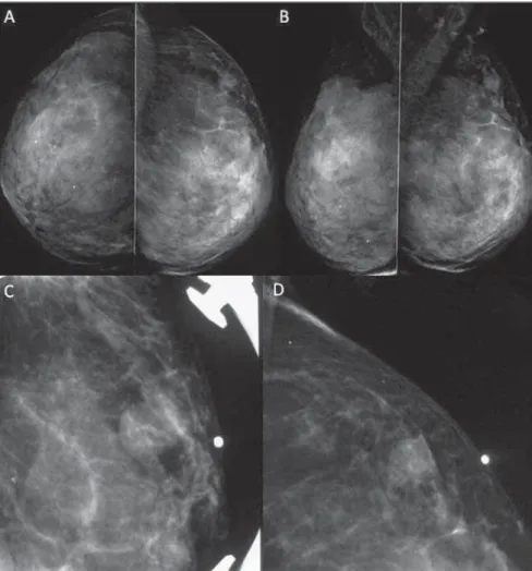
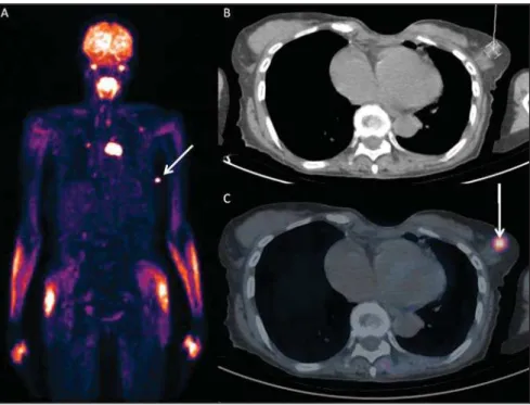
provement of the flogistic signs, but with-out regression of the breast mass. Then, the patient was submitted to imaging studies. Mammography (Figure 1) revealed the presence of a circumscribed mass isodense to fibroglandular tissue in the superolateral quadrant of the left breast, measuring 14 × 10 mm. Breast ultrasonography (Figure 2) demonstrated a complex cyst with solid components and thickened septa located in the superolateral quadrant of the left breast, peripherically to the papilla, measuring 13 × 8 mm, in an area of palpation at physi-cal examination. Positron emission com-puted tomography (PET/CT) (Figure 3) demonstrated anomalous concentration of fluorodeoxyglicose-18F (18F-FDG) in the
left breast mass, with maximum standard-ized uptake value (SUV) of 4.90, and also in other extramammary sites.
After fine needle aspiration biopsy of the breast mass indicating the presence of neoplastic cells, the investigation was pur-sued with US-guided percutaneous core needle biopsy whose microscopy results were compatible with invasive carcinoma. Immunohistochemical analysis (positive cytocheratin 7; positive CA 125; negative mammoglobulin; negative BRST-2; posi-tive WT1) led to the diagnosis of metasta-sis from ovarian carcinoma. Then, the pa-to the breasts(3). In fact, according to a study
developed with 4.051 patients with malig-nant breast lesions, only 0.07% of them presented metastasis to breast originating from ovarian tumors(1,4).
The authors report the case of a patient with ovarian carcinoma who progressed with breast metastasis after about eight years from the initial diagnosis, and present a literature review on the subject with em-phasis on imaging findings.
CASE REPORT
A female, white, 64-year-old, G1P1 pa-tient who had menarche at 12 years and menopause at 50 years of age, former user of oral contraceptive drugs (during 15 years) and hormone replacement therapy (during 10 years), former smoker with fam-ily history of breast cancer (maternal grand-mother and cousins) was submitted to sur-gery and chemotherapy for ovarian serous papilliferous adenocarcinoma. After the initial therapy, the patient presented locoregional and distant metastases.
Eight years after the diagnosis, the pa-tient presented a palpable mass in her left breast in association with flogistic signs on the skin. Initially, the patient was treated with antibiotic therapy, presenting
im-INTRODUCTION
Primary ovarian carcinoma rarely origi-nates breast metastasis and there are few cases reported in the literature(1). Only
about 2% of malignant neoplastic breast lesions are of secondary origin and, from a clinical and radiological point of view, they may mimic both primary benign and malignant tumors(2).
124
Moreira BL et al. Breast metastasis from ovarian carcinoma
Radiol Bras. 2012 Mar/Abr;45(2):123–125 Figure 1. Mammography demonstrating a circumscribed mass isodense to fibroglandular tissue located
in the superolateral quadrant of the left breast, measuring 14 × 10 mm. A: Craniocaudal view. B: Mediolateral oblique view. C,D: Absolute lateral view (C) and localized compression of the region of inter-est on the left breast (D) (metal marker on the skin indicating a palpable mass).
Figure 2. Breast ultrasonography demonstrating a complex cyst with solid components and thickened septa located in the superolateral quadrant of the left breast, peripherically to the papilla, measuring 13 × 8 mm, in a region of palpation at physical examination.
tient progressed with palliative radio-therapy and chemoradio-therapy, dying about two years after the diagnosis of breast me-tastasis.
DISCUSSION
In the presence of breast lesion in ova-rian carcinoma patients, the differentiation between benign and malignant lesions is re-quired. In case of a malignant lesion, it is necessary to know whether the lesion is primary or secondary since the treatment and prognosis differ according to the nature or the lesion(1–3,5).
Frequently, imaging studies demon-strate metastatic lesions to the breast as dense, circumscribed and noncalcified masses. However, as a function of the pres-ence of psamomatous bodies associated with some ovarian tumors, microcalcifi-cations may be seen in cases of ovarian metastasis to the breast(1). But such a
dif-ferentiation may be difficult, considering that metastases to the breast generally do not present specific imaging features(6).
Anyway, like in the present case, the tumor origin must be investigated by means of anatomopathological analysis and taking advantage of the usefulness of immunohis-tochemical analysis in the differentiation of such lesions(7).
recur-125
Moreira BL et al. Breast metastasis from ovarian carcinoma
Radiol Bras. 2012 Mar/Abr;45(2):123–125
rence –, and change the treatment to be adopted. The positive predictive value of this method is 89–98% for cases os ovarian cancer recurrence, with higher sensitivity for detecting extrapelvic lesions(8).
How-ever, in cases of breast lesions, PET/CT
does not exclude the necessity of biopsy, since this imaging method does not allow the differentiation between primary and secondary malignant lesions.
Once the origin of a breast lesion is con-firmed as metastasis from ovarian cancer,
the disease must be considered as systemic, and palliative treatment must be adopted, avoiding unnecessary surgical proce-dures(1–3,5,6). According to some studies in
the literature, the survival time of patients with breast metastasis from ovarian carci-noma is most of times less than one year(1).
REFERENCES
1. Klein RL, Brown AR, Gomez-Castro CM, et al. Ovarian cancer metastatic to the breast presenting as inflammatory breast cancer: a case report and literature review. J Cancer. 2010;1:27–31. 2. Garza-Guajardo R, Mendez-Olvera N,
Flores-Gutierrez JP, et al. Fine needle aspiration biopsy diagnosis of metastatic neoplasms of the breast. A three-case report. Cytojournal. 2005;2:17. 3. Akçay MN. Metastatic disease in the breast. Breast.
2002;11:526–8.
4. Micha JP, Goldstein BH, Epstein HD, et al. Ova-rian cancer metastatic to the breast. Gynecol Oncol. 2006;102:386–90.
5. Özsaran AA, Dikmen Y, Terek MC, et al. Bilateral metastatic carcinoma of the breast from primary ovarian cancer. Arch Gynecol Obstet. 2000;264: 166–7.
6. Bartella L, Kaye J, Perry NM, et al. Metastases to the breast revisited: radiological-histopathological correlation. Clin Radiol. 2003;58:524–31. 7. Lee AH. The histological diagnosis of metastases
to the breast from extramammary malignancies. J Clin Pathol. 2007;60:1333–41.
8. Prakash P, Cronin CG, Blake MA. Role of PET/CT in ovarian cancer. AJR Am J Roentgenol. 2010; 194:W464–70.
Figure 3. PET/CT image revealing anomalous uptake of 18F-FDG on a mass in left breast (arrow) with