Letters to the Editor
Radiol Bras. 2016 Mar/Abr;49(2):126–132
130
Remote cerebellar hemorrhage and intracranial hypotension syndrome following pituitary surgery
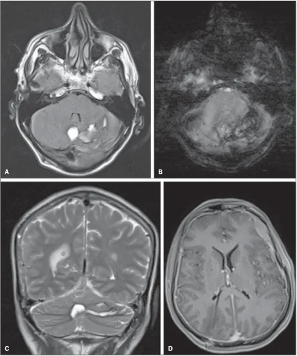
In the postoperative period, the patient evolved to significant worsening of the headache, and a magnetic resonance imaging scan was requested in order to evaluate the condition. The mag-netic resonance imaging showed cerebellar hemorrhage affect-ing the vermis and the left cerebellar hemisphere (Figure 1A), with standard distribution of blood among the cerebellar folia (Figures 1B and 1C), indicative of remote cerebellar hemorrhage. There were also signs suggestive of a loss of cerebrospinal fluid (intracranial hypotension syndrome), such signs including a de-crease in the size of the ventricles, dural thickening, diffuse dural enhancement, and hematic left frontal subdural fluid collection (Figure 1D).
Dear Editor,
A 62-year-old male presented with a several-month history of headache and alteration in his visual field. The diagnostic evalu-ation revealed a suprasellar mass that was causing hydrocephalus by extrinsic compression. We opted for ventricular shunt place-ment and subsequent surgical excision of the mass. The surgical site was accessed through left frontal craniotomy. Histological analysis of the resected mass revealed that it was a craniopharyn-gioma.
Figure 1. MRI scans of the cranium. A: FLAIR sequence in the axial plane, showing foci of hyperintense signals in the vermis and the left cerebellar hemisphere, which is consistent with cerebellar hemorrhage. B: SWI sequence in the axial plane, showing marked hypointensity indicative of hemoglobin degradation products (e.g., hemosiderin), with the zebra sign (pattern of distribution among the cerebellar folia). C: T2-weighted TSE sequence in the coronal plane, confirming cerebellar hemor-rhage, in the different phases of hemoglobin degradation, with alternating areas of hyperintense and hypointense signals (the zebra sign). D: T1-weighted SE sequence in the axial plane after intravenous injection of paramagnetic contrast, showing the terminus of the ventricular shunt catheter at the trigone of the right lateral ventricle. Note the small size of the lateral ventricles, together with the marked dural thickening and significant dural impregnation, as well as the hematic left frontal subdural fluid collection. This combination of findings suggests a loss of cerebrospinal fluid (intracranial hypotension syndrome).
A B
Letters to the Editor
Radiol Bras. 2016 Mar/Abr;49(2):126–132
131
Ultrasound guided injection of botulinum toxin into the salivary glands of children with neurological disorders
Dear Editor,
Here, we report the case of a 2-year-old male patient with corpus callosum atrophy who was under investigation for genetic syndrome. The patient had a gastrostomy and a permanent tra-cheostomy. He had sialorrhea (drooling) that had not responded to clinical treatment with sublingual atropine and had been hos-pitalized for pneumonia on multiple occasions. He was referred for ultrasound-guided injection of botulinum toxin—recom-mended for therapeutic use since 1822(1–7)—into the parotid and submandibular glands.
Ultrasound studies of the parotid and submandibular glands, all conducted by the same physician (with 15 years of experience in ultrasound), revealed that the glands were normal in appear-ance. Prior to, 30 days after, and 60 days after injection of the botulinum toxin, the glands were measured and their volumes were calculated. Ultrasound guidance allowed the best site for
in-jection of the botulinum toxin to be identified, which prevented the toxin affecting structures adjacent to the salivary glands, such as the muscles involved in swallowing and vascular structures (Fig-ure 1).
In follow-up visits, the mother reported that there was a sig-nificant decrease in the number of pads used for cleaning drool and a 50% reduction in the number of tracheal aspirations, with-out any complaints suggesting that the botulinum toxin had pro-voked an inflammatory process. The patient had no episodes of bronchopneumonia during the two-months observation period. The ultrasound studies of the parotid and submandibular glands showed no parenchymal changes subsequent to injection of the botulinum toxin.
The use of ultrasound to guide botulinum toxin injections is important in pediatric patients, especially because the small size of the salivary glands makes them difficult to palpate in such pa-tients. In neurologically impaired children, the use of the ultra-sound guidance is even more relevant, because they can present with increased muscle tone and often have a tracheostomy in an
Luiz de Abreu Junior1, Henrique T. Martucci2, Paulo Tarso Reck de Mendonça3, Gustavo Garcia Marques1, Célia Rodrigues1
1. Universidade São Camilo, São Paulo, SP, Brazil. 2. Clínica São Camilo, Sinop, MT, Brazil. 3. Instituto Neurocirúrgico de Sinop, Sinop, MT, Brazil. Mailing address: Dr. Luiz de Abreu Junior. Rua Baturité, 200, ap. 32B, Aclimação. São Paulo, SP, Brazil, 01530-030. E-mail: [email protected].
Remote cerebellar hemorrhage has been defined as bleed-ing within the cerebellar parenchyma, a rare complication that can occur after neurosurgical intervention. The entity was first described in the 1970s, by Yasargil et al.(1). The reported inci-dence of remote cerebellar hemorrhage after supratentorial in-terventions ranges from 0.08% to 0.6%(2). However, it has been reported to occur after various other surgical procedures involving the cranium or spinal cord(2–7).
Several hypotheses have been suggested to explain the ap-pearance of bleeding in the cerebellum away from the primary (supratentorial or spinal) surgical site. One such hypothesis is that resection of a supratentorial lesion creates a pressure gradient, resulting in suction on the cerebellar veins, particularly in the upper portion of the vermis(8). However, there is another hypothesis that might explain the two findings in the case reported here. That hypothesis is based on the supposition that opening the cisterns or the ventricular system promotes intracranial hypotension, trig-gering the process that culminates in the distension and rupture of cerebellar veins, resulting in cerebellar hemorrhage (9).
Various neurosurgical procedures have been associated with the occurrence of remote cerebellar hemorrhage, including the clipping of aneurysms (ruptured or otherwise), tumor resection, drainage of parenchymal or extra-axial hematomas, and spinal sur-gery(2–7,9). In imaging examinations, remote cerebellar hemor-rhage has a characteristic presentation, with a tendency for the blood to be distributed among the cerebellar folia with a curvilin-ear configuration. This aspect results in the pattern known as the zebra sign(8).
The symptoms of intracranial hypotension syndrome include headache that is orthostatic in presentation, tending to improve in the recumbent position. In imaging studies of the brains of patients with intracranial hypotension(10), findings include dural thickening and diffuse dural enhancement; engorgement and dilatation of venous structures; subdural fluid collections; down-ward displacement of the midbrain; and herniation of the cerebellar tonsils.
The case presented here demonstrates a chain of events that could have collectively resulted in the two central nervous system complications observed. The supratentorial surgical manipulation and the placement of the ventricular shunt could have caused intracranial hypotension, resulting in the traction, distension, and
consequent rupture of cerebellar veins, as well as hemorrhage in the cerebellar parenchyma.
Radiologist knowledge of these entities is relevant, because their proper, early characterization can promote interventions aimed at their correction and at alleviating the associated symp-toms.
REFERENCES
1. Yasargil MG, Yonekawa Y. Results of microsurgical extra-intracranial arterial bypass in the treatment of cerebral ischemia. Neurosurgery. 1977;1:22–4.
2. Bokhari R, Baeesa S. Remote cerebellar hemorrhage due to ventriculo-peritoneal shunt in an infant: a case report. J Med Case Rep. 2012; 6:222.
3. Honegger J, Zentner J, Spreer J, et al. Cerebellar hemorrhage arising postoperatively as a complication of supratentorial surgery: a retrospec-tive study. J Neurosurg. 2002;96:248–54.
4. Smith R, Kebriaei M, Gard A, et al. Remote cerebellar hemorrhage following supratentorial cerebrovascular surgery. J Clin Neurosci. 2014; 21:673–6.
5. Suzuki M, Kobayashi T, Miyakoshi N, et al. Remote cerebellar hemor-rhage following thoracic spinal surgery of an intradural extramedullary tumor: a case report. J Med Case Rep. 2015;9:68.
6. Biasi PR, Mallmann AB, Crusius PS, et al. Hemorragia cerebelar re-mota como complicação de cirurgia de coluna vertebral. Relato de dois casos e revisão da literatura. J Bras Neurocir. 2011;22:66–71. 7. Paola L, Troiano AR, Germiniani FMB, et al. Cerebellar hemorrhage
as a complication of temporal lobectomy for refractory medial tempo-ral epilepsy: report of three cases. Arq Neuropsiquiatr. 2004;62:519– 22.
8. Amini A, Osborn AG, McCall TD, et al. Remote cerebellar hemorrhage. AJNR Am J Neuroradiol. 2006;27:387–90.
9. Chalela JA, Monroe T, Kelley M, et al. Cerebellar hemorrhage caused by remote neurological surgery. Neurocrit Care. 2006;5:30–4. 10. Savoiardo M, Minati L, Farina L, et al. Spontaneous intracranial
hy-potension with deep brain swelling. Brain. 2007;130(Pt 7):1884–93.