Parasite density and impaired biochemical/hematological status are
associated with severe clinical aspects of canine visceral leishmaniasis
Alexandre B. Reis
a,b,*, Olindo A. Martins-Filho
c, Andre´a Teixeira-Carvalho
a,
Maria G. Carvalho
d, Wilson Mayrink
e, Joa˜o C. Franc¸a-Silva
e,
Rodolfo C. Giunchetti
a,b, Odair Genaro
e, Rodrigo Correˆa-Oliveira
baLaborato´rio de Parasitologia e Histopatologia, Nu´cleo de Pesquisas em Cieˆncias Biolo´gicas/NUPEB, Instituto de Cieˆncias Exatas e Biolo´gicas, Universidade Federal de Ouro Preto, Rua Costa Sena, 21 Centro, 35400 000 Ouro Preto, Minas Gerais, Brazil
bLaborato´rio de Imunologia Celular e Molecular, Centro de Pesquisas Rene´ Rachou, Fundac¸a˜o Oswaldo Cruz, Av. Augusto de Lima, 1715 – Barro Preto, Belo Horizonte, Minas Gerais, Brazil
cLaborato´rio de Doenc¸a de Chagas, Centro de Pesquisas Rene´ Rachou, Fundac¸a˜o Oswaldo Cruz, Belo Horizonte, Minas Gerais, Brazil dLaborato´rio de Hematologia Clı´nica, Faculdade de Farma´cia, Universidade Federal de Minas Gerais, Belo Horizonte, Minas Gerais, Brazil
eLaborato´rio de Leishmanioses, Departamento de Parasitologia, Instituto de Cieˆncias Biolo´gicas, Universidade Federal de Minas Gerais, Belo Horizonte, Minas Gerais, Brazil
Accepted 14 September 2005
Abstract
We have performed a detailed investigation in 40 dogs naturally infected withLeishmania infantum(syn. chagasi), subdivided into three groups: asymptomatic (AD = 12), oligosymptomatic (OD = 12) and symptomatic (SD = 16), based on their clinical features. Twenty non-infected dogs (CD) were included as control group. Serological analysis, performed by IFAT and ELISA, demonstrated higher antibodies titers in SD in comparison to the AD. A positive correlation was found between parasite density in the spleen and skin smears as well as the bone marrow parasitism with clinical status of the infection. We observed that the progression of the disease from asymptomatic to symptomatic clinical form was accompanied by intense parasitism in the bone marrow. It is likely that this led to the impaired biochemical/hematological status observed. Finally, we believe that the follow-up of these parameters could be a relevant approach to be used as markers during therapeutic and vaccine evaluations.
Ó2005 Elsevier Ltd. All rights reserved.
Keywords: Canine visceral leishmaniasis;Leishmania infantum(syn. chagasi); Parasite density; Biochemical/hematological status
1. Introduction
Visceral leishmaniasis (VL) is one of the most relevant and emergent diseases worldwide, reaching 98% of mortal-ity in non-treated human cases (Tesh, 1995). Beside its broad epidemiological spectrum, VL is an important zoo-nosis widely spread in tropical and subtropical areas of the globe. In the last 16 years, 37,294 new human cases of VL were reported in Brazil (Vieira and Coelho, 1998).
From the epidemiological standing point, the canine vis-ceral leishmaniasis (CVL) is considered to be more impor-tant than the human disease, due to its higher prevalence and the fact that both asymptomatic and symptomatic dogs are equally infectious to the vectors (Molina et al., 1994). Many asymptomatic animals, in endemic areas, have been detected with parasites in the skin (Marzochi et al., 1985), and from 1980 to 1997, a total of 414,168 sero-positive dogs were identified in Brazil (Vieira and Coelho, 1998).
The CVL may evolve from asymptomatic cases to a sys-temic disease, which mostly culminates in death. From the
0034-5288/$ - see front matter Ó2005 Elsevier Ltd. All rights reserved. doi:10.1016/j.rvsc.2005.09.011
* Corresponding author. Tel.: +55 31 3349 7778; fax: +55 31 3295 3115. E-mail address:alexreis@cpqrr.fiocruz.br(A.B. Reis).
localized cutaneous infection, the parasite can be dissemi-nated via lymphatic or blood vessels, infecting macro-phages of the bone marrow, lymph node, liver and spleen, as well as kidneys and gastrointestinal tract. Initial clinical signs are hypertrophy of lymph node, dermatitis and periorbital and nasal dermatitis that can disseminate. The bristles become opaque and fall, together with onycho-griphosis and edema of the paws. Other signs such as fever, apathy, diarrhea, intestinal hemorrhage, weight loss, hepa-tosplenomegaly, hyperkeratosis, cutaneous ulceration, par-ticularly on the nose, ears, tails, and keratoconjunctivitis are frequent, although not necessarily present in all animals (Genaro et al., 1988; Dias et al., 1999). The CVL presump-tive diagnosis is generally performed by serological tests, such as indirect immunofluorescence assay test (IFAT) and enzyme linked immunosorbent assay (ELISA), con-junction with clinical and epidemiological records. The major problem regarding clinical diagnosis is the fact that CVL signs are very similar to those observed in other infec-tious diseases. The chronic aspect of the disease and its long incubation period may generate a delay or failure in clinical diagnosis (Cardoso and Cabral, 1998). Despite its high sensitivity, serological tests present a broad range of cross-reactions with other protozoan (Costa et al., 1991; Grimaldi and Tesh, 1993). However, the parasitological diagnosis is generally low in sensitivity when parasite den-sity is low.
There are several biochemical and hematological altera-tions in naturally or experimentally infected dogs. The find-ings include normocytic/normochromic anemia, and increase of total serum proteins levels. It seems that bio-chemical alterations are linked to a polyclonal humoral immune response, which leads to raised protein levels in serum (Marzochi et al., 1985). This can be observed by an increase of the total serum proteins with hyperglobulin-emia and hypoalbuminhyperglobulin-emia, with decreased albumin/glob-ulin ratio (Cardoso and Cabral, 1998; Strauss-Ayali and Baneth, 2001). Considering the re-emergent aspect of vis-ceral leishmaniasis, in several parts of the world, the epide-miological importance of dogs, we assessed both the clinical and laboratorial aspects from canine visceral leish-maniasis contributing to monitor the clinical status of dogs during infection.
2. Materials and methods
2.1. Animals
Sixty mixed breed adult dogs of both genders aging from 2- to 6-years old were selected. They were maintained in the kennel of the Institute of Biological Sciences of Federal University of Minas Gerais or provided by the Center of Zoonosis (Zoonosis Center – Belo Horizonte City Council) of Minas Gerais state, Brazil. The clinical pre-selection was carried out in the latter location. The animals were kept in quarantine with drinking water and a balanced feed (KinusÒ
– BRASWEY – AS) given ad libitum.
The dogs inserted in this study were strayed or domi-ciled mongrel dogs, selected based on their serological results on IFAT, used as a ‘‘gold standard’’ immunological test for diagnosis of CVL. Animals presenting IFAT titers P1:40 were considered positive and included into the infected groups. Animals with IFAT negative at 1:40 were considered non-infected and included as a control group. Leishmania infected dogs did not receive any treatment for CVL and were euthanatized to proceed the organs col-lections, including skin, spleen, liver and lymph nodes. As chemotherapeutical practices for CVL is not officially allowed in Brazil, all infected dogs must be submitted to euthanasia.
Infection withLeishmania infantumwas confirmed in all IFAT positive dogs by at least one additional serological and/or parasitologial approach used.
2.2. Blood sample collection
After quarantine, blood samples were collected in 20 c.c. disposable sterile syringes, preferentially from jugular and/ or cephalic veins. Five milliliter-samples were transferred to tubes with EDTA (in the proportion of 1 mg/ml) for the hemogram and 5 ml were transferred at a tube with no anticoagulant. The serum samples were stored in ali-quots at 20°C, until use in the serological and
biochem-ical tests.
2.3. Clinical and parasitological evaluations
The dogs were clinically classified, according to pres-ence/absence of infection signs: asymptomatic (AD, n= 12), with no suggestive signs of the disease; oligosymp-tomatic (OD, n= 12), with maximum three clinical signs including opaque bristles and/or localized alopecia and/ or moderate loss of weight; symptomatic (SD,n= 16), with characteristic clinical signs of visceral leishmaniasis, such as opaque bristles, severe loss of weight, onychogriphosis, cutaneous lesions, apathy and keratoconjunctivitis; non-infected dogs (CD), animals for negative serological and parasitological exams for Leishmania, considered here as control animals.
The assessment of parasitological parameters was per-formed by the isolation of the parasite in bone marrow cul-tures, obtained by aspiration of the bone marrow, in NNN/LIT medium. The cultures were maintained in incu-bators at 23°C and examined three times every 10 days.
Bone marrow aspiration was undertaken from the inferior region of the sternum or from the iliac crest, under sedation (25 mg/kg live weight of sodium tiopental – Thionembu-talÒ
par-asite density evaluation was performed, in bone marrow, skin and spleen smears and the results expressed as ‘‘ Leish-man Donovan Units’’ (LDU index), according to Stauber (1955), which correspond to the number of Leishmania amastigote by 1000 nucleated cells. This study was approved by the Ethical Committee for the use of Experi-mental Animals of the Universidade Federal de Minas Ger-ais, Brazil (CETEA).
2.4. Serological assessment
For the IgG assessment, IFAT, ELISA-extract and ELISA-rK39 were performed. For IFAT,Leishmania ama-zonensispromastigote (MHOM/BR/1960/BH6) were used. These parasites were maintained in logarithmic growth in LIT medium, as described by Camargo and Rebonato, 1969. For IFAT, specific anti-canine IgG antibodies FITC-conjugated diluted 1:200, as recommended by the manufacturer (Biomanguinhos, FIOCRUZ, RJ, Brazil). Animals with antibody titration higher than 1:40 were con-sidered to be positive. The results are expressed as antibody titers.
For ELISA-extract tests, soluble antigens fromL. infan-tum(syn. chagasi) promastigote (MHOM/BR/1972/BH46) were used. The parasites were cultured for 7 days in LIT medium (Mancianti et al., 1995), washed three times by centrifugation at 2000 rpm in phosphate buffer solution (PBS) pH 7.2, for 10 min, followed by three ultra-sound cycles of 1 min at 40 W on ice bath (Sonifier Cell Disrup-torÒ – Branson Sonic Power Co., EUA). The sonicated
material was centrifuged at 18,500 rpm for 1 h and 30 min at 4°C. The supernatant was transferred to dialysis
tubes and dialyzed through PBS for 36 h, and submitted to four PBS changes every 6 h. Finally, the remaining material was filtered in disposable sterile filters of 22lm under
asep-tic conditions; one aliquot was taken for protein dosage by the method ofLowry et al. (1951), and adjusted to the con-centration of 1000lg/ml and stored in small aliquots at
70°C prior to use.
The recombinant antigen rK39, specific for Leishmania of thedonovani complex (Badaro´ et al., 1997) was used.
For ELISA tests, including both ELISA-extract and ELISA-rK39, an anti-canine IgG peroxidase-conjugated was used at 1:8000 dilution (Bethil Laboratories, INC – Montgomery Texas, EUA). Sera samples were tested at the dilution of 1:80 and the optical density obtained with an automated ELISA reader (Bio-Rad – 2550, EUA) at 492 nm.
Results from ELISA-extract test were considered positive when optical density wasP140. Data from ELISA r-K39 were classified considering the optical density P90 as the edge between the negative and positive results.
2.5. Biochemical and hematological evaluation
Serum proteins were determined by the Biuret reagent and the readings performed in a spectrophotometer in
510 nm (CELM E-225D). The electrophoretic profile of the serum proteins was carried out using cellulose acetate strips according to the manufacturer instructions (CELO-GELÒ
). The electrophoretic readings were performed in a spectrophotometer and analyzed with the software CS-93101 PC.
Hemogram was performed by conventional technique of counting erythrocytes and leukocytes (Dace and Lewis, 1984) in an automatic cell counter – CELM CC 510.
2.6. Statistical analysis
Statistical analysis was performed using the Minitab 9.2 software package. In the parametric data, one-way analysis of variance (ANOVA) was used for the comparative study between groups. StudentÕst-test was used for determining the differences between the groupsÕ averages. In the non-parametric data, Kruskal–Wallis test was used for the com-parative study between groups, followed by DunnÕs test. Spearman rank correlation was calculated to compare results of the different methods. The differences were con-sidered significant when the probabilities of equality,p val-ues, were60.05.
3. Results
3.1. Serological parameters
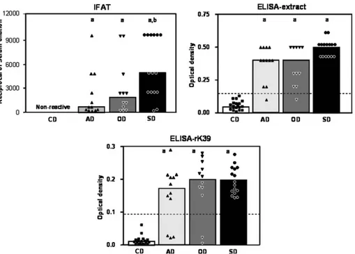
Total IgG reactivity results in infected animal sera as evaluated by IFAT, ELISA-extract and ELISA rk-39 are shown inFig. 1. Statistical analysis confirmed that unin-fected controls selected by presenting negative IFAT, also differ from the infected animals when tested by ELISA-extract and ELISA-rK39 serology. The mean ELISA reac-tivity of all CVL carrier groups was higher than that observed for the control group (p< 0.001).
No significant difference was observed ELISA-extract and ELISA rk-39 in total IgG reactivity among the CVL groups. On the other hand, IFAT reactivity revealed that SD displayed a higher serological titer in comparison to AD.
Despite the methodology applied to evaluate the immu-nological status of infected dogs, a positive correlation was observed between serological reactivity and the clinical sta-tus (IFAT r= 0.4207, p= 0.0069; ELISA-extract r= 0.8041,p< 0.0001; ELISA sK39r= 0.7573,p< 0.001).
3.2. Parasitological parameters
group, whereas higher sensitivity of skin impression smears was identified for OD in comparison to the other tissues.
Further analysis of parasite load, reported as ‘‘Leishman Donovan Units’’ demonstrated that skin, bone marrow and spleen LDU indexes in AD was lower when compared to SD group. No significant differences in LDU values, including those from the skin, bone marrow and spleen, were observed among the OD and all others infected groups (Fig. 2). When we compared LDU values in the dif-ferent tissues analyzed we observed that all infected ani-mals display higher skin LDU indexes in comparison to bone marrow. Moreover, our data demonstrated a positive correlation among bone marrow LDU and spleen LDU (r= 0.6188;p= 0.0319) in AD dogs, while a positive corre-lation was observed between skin LDU and spleen LDU (r= 0.9746;p= 0.0001) in the OD dogs. Only for SD dogs we observed a positive correlation between skin and bone marrow LDU values (r= 0.7994;p= 0.0002).
Correlation analysis of parasite density and serological findings demonstrated that AD showed a positive correla-tion between skin, bone marrow and spleen LDU and IFAT (skin r= 0.8304; p= 0.0008, bone marrow r= 0.8271;p= 0.0009, spleenr= 0.5782;p= 0.0489).
3.3. Biochemical and hematological parameters
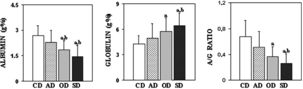
The evaluation of biochemical parameters, related to protein alterations, showed no significant difference in total protein levels among all groups. However, a significant decrease (p< 0.001) in albumin concentrations, in the oli-gosymptomatic and symptomatic dogs, was observed, when compared to control and asymptomatic animals (Fig. 3). Dogs of the oligosymptomatic group presented a significant increase in globulin concentration (p< 0.001), when compared to control group, while the symptomatic group presented a significant increase (p< 0.001) in globu-Fig. 1. Reactivity of Total IgG in serum from CVL infected dogs. IFAT results are shown as scattering of individual values and mean serum dilution. ELISA-extract results (cut-off = 140) and ELISA-rK39 (cut-off = 90) are shown as scattering of individual values and mean optical density. The letters ‘‘a’’ and ‘‘b’’ represent statistically significant differences as compared to control (CD) and asymptomatic dogs (AD), respectively. NR = non-reactive.
lin concentration, when compared to control and asymp-tomatic animals. Those dogs from the oligosympasymp-tomatic group showed a significant decrease (p< 0.001) in A/G ratio when compared to the control group, while the symp-tomatic animals presented a significant decrease (p< 0.001) in A/G ratio in comparison to the control and asymptom-atic groups (Fig. 3). However, only SD dogs presenting a positive correlation between both total protein and globu-lin values and IFAT titers (r= 0.6177; p= 0.0108; r= 0.5947;p= 0.0151, respectively).
The assessment of hematological parameters demon-strated severe anemia in the symptomatic animals, with a significant decrease (p< 0.001) in the number of erythro-cytes, hemoglobin and hematocryt, in relation to the con-trol, asymptomatic and oligosymptomatic groups. As for the white blood cells, statistical analysis showed significant difference among the symptomatic dogs and the other groups in the leukocytes series; however, no significant dif-ference in absolute values of granulocytes neutrophils was observed among the groups of dogs under study. Statistical analysis revealed that the symptomatic dogs showed a sig-nificant decrease (p< 0.025) in leukocyte absolute values (leukopenia) when compared to the control group. Data analysis also demonstrated a significant decrease (p< 0.003) in absolute values of the eosinophylic granulo-cytes subpopulation from symptomatic dogs when com-pared to the control animals. The symptomatic group
showed a significant decrease (p< 0.017) in absolute values of lymphocytes when compared to the control and asymp-tomatic groups. Data analysis showed also a significant decrease (p< 0.0017) in absolute values of monocytes for the symptomatic dogs when compared to control animals (Table 1).
4. Discussion
All clinical signs observed for the OD and SD groups have been extensively reported in the literature for both natural (Mancianti et al., 1988; Genaro et al., 1997) and experimentally infected dogs (Pinelli et al., 1994, 1995; Abranches et al., 1991). The high frequency of clinical signs presented by infected dogs allow us to observe that the dis-ease gradually evolves from an apparently normal clinical picture, presented by AD, to an active intermediate state, which includes some clinical manifestation (OD) that may evolve to a classical severe and terminal form of the disease (SD), characterized by a larger number of clinical signs. These findings suggested that the sub-clinical or asymptomatic disease may indicate that a good balance in parasite–host interaction is ongoing (Oliveira et al., 1993).
Hypergammaglobulinemia is a remarkable characteris-tic of CVL (Almeida et al., 2005). Several studies have reported a high antibody production during active CVL, Fig. 3. Evaluation of biochemical parameters of naturally infected and non-infected dogs. The results are shown as average values ± standard deviation. Control dogs (white bars); asymptomatic dogs (stained bars); oligosymptomatic dogs (gray bars) and symptomatic dogs (black bars). The letters ‘‘a’’ and ‘‘b’’ represent differences statistically significant for the control (CD) and asymptomatic dogs (AD), respectively.
Table 1
Evaluation of hematological parameters of naturally infected and non-infected dogs
Hematological parameters Clinical groups
CD AD OD SD
Erythrocytes – million/mm3 6.8 ± 0.7 5.8 ± 0.7 5.4 ± 1.0 4.2 ± 1.2a,b,c
Hemoglobin – g% 15.7 ± 1.8 14.0 ± 2.3 13.5 ± 3.2 9.8 ± 2.8a,b,c
Hematocryt – % 46.4 ± 5.4 40.6 ± 7.9 38.8 ± 8.1 28.6 ± 8.2a,b,c
Leukocytes – 103/mm3 12.8 ± 2.7 14.1 ± 5.0 13.9 ± 3.2 10.2 ± 3.9a
Granulocytes 9.1 ± 3.1 7.8 ± 3.5 9.4 ± 3.4 8.0 ± 3.2
Neutrophils 7.2 ± 3.1 6.7 ± 3.0 8.5 ± 3.2 7.2 ± 3.0
Eosinophils 1.8 ± 0.8 1.1 ± 1.0 0.9 ± 0.4 0.7 ± 0.9a
Lymphocytes 2.7 ± 1.5 5.2 ± 3.3a 3.7 ± 3.3 1.6 ± 1.0a,b
Monocytes 1.1 ± 0.4 1.0 ± 0.5 0.7 ± 0.3 0.5 ± 0.4a
The letters ‘‘a’’, ‘‘b’’ and ‘‘c’’ represent statistically significant differences as compared to control (CD), asymptomatic (AD) and oligosymptomatic dogs (OD), respectively.
with elevated levels of IgM and IgA circulating immuno-complexes (Margarito et al., 1998). Many reports have focused special attention to the search of alternative meth-odologies, as well as the use of specific antigen prepara-tions, aiming to identify a better diagnostic method and prognostic tool for CVL clinical investigation. Some recombinant antigens have been pointed out as relevant candidates for CVL immunodiagnosis (Badaro´ et al., 1997; Soto et al., 1999; Rosa´rio et al., 2005). Herein, the humoral immune response was evaluated by distinct immu-nological approaches, including IFAT, ELISA-extract and ELISA rK-39. Our data demonstrated that IFAT was able to identify as positive, all infected animals, despite their clinical status and have been used in this study as the gold standard to diagnosis CVL and select positive animals. ELISA-extract failed to detect IgG reactivity on two IFAT-seropositive animals, one AD and another OD. ELISA rK-39 was not able to detect IgG reactivity on five animals (three from AD and two from OD group).Genaro et al. (1997), using the recombinant protein rK-39 on a immunochromatographic assay (TRALd) to diagnosis CVL in an endemic area, demonstrated 92.1% of sensitivity and 99.5% of specificity, with significantly higher ability to detect Leishmania-specific antibodies when compared to IFAT. However, it was demonstrated that rK-39 was not able to predict the active infection when testing dog sam-ples displaying antibody titers between 1:40 and 1:320, esti-mated by IFAT (Genaro et al., 1997). Our data are in agreement with these reports since all five seronegative ani-mals detected by ELISA rK-39 displayed serological titles under 1:320 by IFAT. Together, our data demonstrated that ELISA rK-39 was able to confirm seropositivity in 35 out of 40 IFAT-positive animals (87.5% of sensitivity), re-enforcing previous reports of its applicability for CVL diagnosis (Rosa´rio et al., 2005). The profiles of IgG anti-rK-39 presented by naturally infected dogs showed a positive correlation according to the clinical form of the disease. Similar results were also observed with IFAT and ELISA-extract. However, data analysis demonstrated that only IFAT titers was able to discriminate the mean IgG reactivity among infected dogs, with an enhancement of reactivity detected as more severe clinical status of infec-tion was observed, with significant differences detected between AD and SD groups. According to Abranches et al. (1991), as the clinical signs appear, the antibody levels tend to be higher. However,Abranches et al. (1991) have not found a correlation between severity of the disease and total IgG titers. On the other hand, Pinelli et al. (1994)demonstrated that dogs experimentally infected with L. infantum, which also presented clinical signs, showed the highest antibody titers, similar to the results obtained by Genaro et al. (1988) in dogs naturally infected with L. infantum. Our data are in accordance with this later obser-vation, since we found that SD group displayed the highest antibody titers in IFAT. Despite no significant differences on the mean antibody levels detected by ELISA-extract and ELISA rK-39 observed for SD, it was interesting to
notice that a positive correlation between severity of dis-ease and the antibody titers was detected by all methods.
Following infection, at the site of parasite inoculation, in the dog skin, the parasite disseminates carried out by his-tiocytes and Langerhans cells to the lymph node, bone marrow, spleen, liver, kidney, lungs and gastrointestinal tract, been rarely detected on the blood stream (Cardoso and Cabral, 1998). Infected animals presented hypertrophy of the Phagocytic Mononuclear System, leading to spleno-megaly, hepatomegaly and generalized adenopathy ( Lano-tte et al., 1979). During Leishmania infection, tissue parasitism at different sites may differ and therefore is rel-evant for diagnosis purposes the identification of the major compartments presenting higher parasitism levels, in order to improve CVL diagnostic sensitivity. In this context, although the majority of authors consider the parasite search in the host tissues as the most reliable diagnostic method, its sensitivity and relative value have been ques-tionable, considering the lack of uniformity of parasite dis-tribution during natural infection. The parallel parasite search, at different affected tissues of the mammalian host organism, could contribute as a definitive criterion, which enables an investigator to choose the most appropriate bio-logical specimen to be analyzed during diagnosis and prog-nosis practices. According to Cardoso and Cabral (1998), bone marrow biopsies are more sensitive than lymph nodes and therefore are more indicated for diagnosis purposes. Despite Ashford et al. (1995)reporting similar percentage of parasite detection in cultured versus impression bone marrow smear, our findings from AD and SD pointed out that bone marrow culture showed higher sensitivity as compared to bone marrow impression smears. Abran-ches et al. (1991)state that bone marrow examination led to positive results only during advanced stages of CVL. Our data confirmed such proposal as 88% of SD in con-trast to 25% of AD showed positive results on bone mar-row impression smear (Table 2). In general, our data demonstrated that parasitological survey performed on tis-sue impression smears (skin, spleen, liver and lymph node)
Table 2
Positivity of parasitological search on distinct tissues from dogs naturally infected withL. infantum
Tissues/methodology Number of positive animals (%)
Clinical groups
AD OD SD
Bone marrow/culture 6 (50.0) 6 (50.0) 15 (94.0)a,b Bone marrow 3 (25.0)* 8 (67.0)a* 14 (88.0)a
Skin 5 (42.0) 10 (83.0) 15 (94.0)a Spleen 6 (50.0) 8 (67.0) 15 (94.0)a Liver 5 (42.0) 8 (67.0) 14 (88.0)a Lymph node 5 (42.0) 8 (67.0) 15 (94.0)a
The letters ‘‘a’’ and ‘‘b’’ represent statistically significant differences as compared to AD and OD, respectively.
* Represents statistical differences within a given clinical group.
Results are expressed as number of positive dogs per group; in
as well as cultured bone marrow proved to be more efficient to detect Leishmania organisms in AD than direct bone marrow impression smear, on which was possible to detect amastigotes only in 25% of the specimens analyzed (Table 2). These findings re-enforce that tissue parasitism intensity is parallel with the evolution of the clinical manifestations and point out the importance of the skin parasitism as a useful diagnostic tool when managing asymptomatic animals.
A great advance of parasitological methods is the possi-bility that such tests provide to evaluate the infection load through the parasite density (LDU). Thus, one may esti-mate the real parasite density and its contribution to the development of the pathology associated with the infec-tion. Despite the relevance of quantitative parasite analysis for diagnosis and prognostic approaches there are no pre-vious reports regarding the LDU index in natural or exper-imentally infected dogs. Our data demonstrated that parasite density in skin, bone marrow and spleen compart-ments were higher according to the severity of clinical man-ifestations (Fig. 2). Then, AD presented lower tissue parasitism as compared to SD on which LDU would count up to 7246, 1104 and 2564 amastigotes/1000-nucleated cells in skin, bone marrow and spleen sites, respectively. Inter-estingly, it was observed that all infected dogs, even those AD, presented higher skin parasitism as compared to bone marrow. These findings on the skin LDU indexes re-enforce the importance of cutaneous parasitism density to CVL diagnosis as well as the transmissibility of the infec-tion from AD to susceptible species phlebotomine.
It is important to mention that these findings, regarding skin parasite load, described as LDU, is a pioneer report on canine visceral leishmaniasis. Moreover, the results of this current work highlight the importance quantitative investigations regarding the number of amastigote forms at different tissues. In addition, the assessment of parasite density would also contribute as therapeutic tool when measuring leishmanicidal activities of new promising drugs for CVL, regarding the several affected tissues.
Several biochemical and hematological alterations are observed in dogs natural or experimentally infected by L. infantum. A remarkable CVL characteristic is the dyspro-teinemia, with serum protein electrophoresis revealing decreased levels of albumin (A) and increased levels of globulin fraction (G), leading to inversion of A/G ratio (Keenan et al., 1984). Herein, we have confirmed these findings and also demonstrated that SD group presented lower A/G ratio in comparison to AD and CD, whereas OD differ only from CD (Fig. 3C). This inversion may occur due to decrease of albumin levels and increase of globulin, remarkably observed in SD. Further analysis demonstrated a positive correlation among both total pro-tein and IFAT titers in the SD. These data revealed a rela-tionship between the presences of exacerbated clinical forms of CVL, serum protein levels and humoral immune response. The factors that contribute to the enhancement of gammaglobulin levels in CVL are not yet completely
understood, but this increase is probably due to the poly-clonal activation B-lymphocytes triggered byL. infantum antigens as well as the synthesis of non-specific antibodies and the presence of circulating immunocomplexes ( Marzo-chi et al., 1985; Margarito et al., 1998).
Regarding the hematological picture, it is common to observe normocytic/normochromic anemia, whereas, in the white blood cells, there is a leukopenia with moderated or accentuated lymphopenia (Abranches et al., 1991). The results presented in this study showed that during the symptomatic CVL decreased leukocyte counts, due to a drop in monocytes, eosinophils and mainly in lymphocyte population is one of the most relevant hematological find-ing. Leukopenia, associated to the symptomatic CVL, may be due a multifactorial mechanism on which medular dys-function with diminished hematopoiesis, affected by an intense bone marrow parasitism as well as leukocyte recruitment and trapping into several organs are the major events (Alvar et al., 2004). It could also be related to secre-tion of suppressor cytokines triggered byLeishmania infec-tion (Pinelli et al., 1994, 1995).
In conclusion, our study shows that the clinical evolu-tion of CVL in naturally infected dogs promotes clear alterations in serological, parasitological and biochemi-cal–hematological parameters. Since these alterations are directly correlated with CVL clinical status, they would be taken into account when dealing with diagnosis and prognosis features. The investigation of these laboratorial parameters, associated with the clinical aspect of CVL, is extremely important to be considered in routine clinical follow-up.
Acknowledgments
We are deeply in debt to Dr. GENARO, O., for his long support and enthusiasm to research on canine visceral leishmaniasis. He was also one of the most important investigators in the field of epidemiology, vaccines, and drugs against leishmaniasis with the focus on studies with dogs. He was and will always be remembered by his dedi-cation to the progress of science. We also thank the people from Fundac¸a˜o Nacional da Sau´de, Ministe´rio da Sau´de, Distrito Regional de Belo Horizonte, Minas Gerais, for their special dedication to this work. Finally, we grateful the Prof. Giovanni Gazzinelli by critical analysis of this pa-per. This work was supported by the CNPq/BR/Grant: 521124/98-0 and FAPEMIG/BR/Grant: CBS 2222/97.
References
Abranches, P., Silva-Pereira, M.C.D., Conceic¸a˜o-Silva, F., Santos-Gomes, G.M., Jans, J.G., 1991. Canine leishmaniasis: pathological and ecological factors influencing transmission of infection. J. Paras-itol. 77, 561–577.
Alvar, J., Can˜avate, C., Molina, R., Moreno, J., Nieto, J., 2004. Canine Leishmaniasis. Adv. Parasitol. 57, 1–88.
Ashford, D.A., Bozza, M., Freire, M., Miranda, J.C., Sherlock, I., Eula´dio, C., Lopes, U., Fernades, O., Degrave, W., Barker Jr., R.H., Badaro´, R., David, J.R., 1995. Comparison of the polymerase chain reaction and serology for the detection of canine leishmaniasis. Am. Soc. Trop. Med. Hyg. 58 (8), 251–255.
Badaro´, R., Chun, P., Nakatani, M., Burns, J., Skeiky, Y., Houghton, R.L., Arias, J., Monteiro, P., Genaro, O., Reed, S.G., 1997. TRALd – a rapid test specific for serodianosis of visceral leishmaniasis. Act. Parasitol. Tur. 21 (1), 175.
Camargo, M.E., Rebonato, C., 1969. Cross-reactivity in fluorescence tests for Trypanosoma and Leishmania antibodies. A simple inhibition procedure to ensure specific results. Am. J. Trop. Med. Hyg. 18, 500–505. Cardoso, L., Cabral, M., 1998.Leishmania and canine Leishmaniasis.
Rev. Port. C. Vet. XCIII (527), 122–141.
Costa, C.A., Genaro, O., Lana, M., Magalha˜es, P.A., Dias, M., Michalick, M.S., Melo, M.N., Costa, R.T., Magalha˜es-Rocha, M.N., Mayrink, W., 1991. Leishmaniose visceral canina: avaliac¸a˜o da metodologia sorolo´gica utilizada em inque´ritos epidemiolo´gicos. Rev. Soc. Med. Trop. 24, 21–25.
Dace, J., Lewis, S., 1984. Practical Hematology, sixth ed. Churchil Livingstone, London, 453 pp.
Dias, D.V., Da Costa, C.A., Toledo, V.P.C.P., Bambirra, E., Genaro, O., Michalick, M.S.M., Costa, R.T., Mayrink, W., Ore´fice, F., 1999. Leishmaniose visceral canina – Estudo parasitolo´gico e histolo´gico em olhos de ca˜es – Parte I. Rev. Bras. Oftal. 58 (5), 331–337.
Genaro, O., Costa, R.T., Franc¸a-Silva, J.C., Reis, A.B., Silva, J.C., Vieira, E.P., Arias, J.R., Monteiro, P.S., Reed, S.G., Mayrink, W., Costa, C.A., Netto, E.M., Badaro´, R., 1997. Evaluation of an immunochro-matographic assay for the diagnosis of dogs experimentally and naturally infected withLeishmania chagasi, in Brasil. Acta Parasitol. Turc. 21 (1).
Genaro, O., Mayrink, W., Michalick, M.S.M., Dias, M., Da Costa, C.A., Melo, M.N., 1988. Naturally occurring visceral leishmaniasis in dogs: clinical aspects. Mem. Inst. Oswaldo Cruz. 83, 43.
Grimaldi Jr., G., Tesh, R.B., 1993. Leismanioses of the new world: current concepts and implications for future research. Clin. Microbiol. Ver. 6 (3), 230–250.
Keenan, C.M., Hendricks, L.D., Lightner, L., Webster, H.K., Johnson, A.J., 1984. Visceral leishmaniasis in the German Shepherd dog-I. Infection, clinical disease, and clinical pathology. Vet. Pathol. 21, 74–79. Lanotte, G., Rioux, J.A., Perieres, J., Vollhardt, Y., 1979. E´ cologie des leishmanioses dans le sud de la France. 10. Les forms e´volutives de la leishmanioses visce´rale canine. Elaboration dÕunetypologie bio-cli-nique a` finalite´ e´ epidemie´miologique. Ann. Parasitol. 54, 277–295. Lowry, O.H., Rosebrough, N.J., Farr, A.L., Randall, R.J., 1951. Protein
measurement with the folin phenol reagent. J. Biol. Chem. 193, 263–275. Mancianti, F., Gramiccia, M., Gradoni, L., Pieri, S., 1988. Studies on canine leishmaniais control. I. Evolution of infection of different
clinical forms of canine leishmaniasis following antimonial treatment. Trans. R. Soc. Trop. Med. Hyg. 82, 566–567.
Mancianti, F., Falcone, M.L., Giannelli, C., Poli, A., 1995. Comparison between an enzyme-linked immunosorbent assay using a detergent-solubleLeishmania infantumantigen and indirect immunofluorescence for the diagnosis of canine leishmaniasis. Vet. Parasitol. 59, 13–21. Margarito, J.M., Lucena, R., Lopez, R., Molleda, J.M., Martin, E., Ginel,
P.J., 1998. Levels of IgM and IgA circulating immune complexes in dogs with leishmaniasis. Zent. Vet. 45 (5), 263–267.
Marzochi, M.C.A., Coutinho, S.G., Souza, W.J.S., Toledo, L.M., Grimald Jr., Momen, H., Pacheco, R.S., Sabroza, P.C., Souza, M.A., Rangel Jr., F.B., Tramontano, N., 1985. Canine visceral leishmaniasis in Rio de Janeiro, Brazil. Clinical, parasitological, therapeutically and epidemiological findings (1977–1983). Mem. Inst. Oswaldo Cruz. 80, 349–357.
Molina, R., Amela, C., Nieto, J., San-Andres, M., Gonzalez, F., Castillo, J.A., Lucientes, J., Alvar, J., 1994. Infectivity of dogs naturally infected withLeishmania infantumto colonizedPhlebotomus perniciosus. Trans. Roy. Soc. Trop. Med. Hyg. 88, 491–493.
Oliveira, G.G.S., Santoro, F., Sadigursky, M., 1993. The subclinical form of experimental visceral leishmaniasis in dogs. Mem. Inst. Oswaldo Cruz. 88 (2), 243–248.
Pinelli, E., Killick-Kendrick, R., Wagenaar, J., Bernadina, W., Real, G., Ruitenberg, J., 1994. Cellular and humoral immune responses in dogs experimentally and naturally infected with Leishmania infantum. Infect. Immunol. 62 (1), 229–235.
Pinelli, E., Gonzalo, R.M., Boog, C.J.P., Rutten, V.P.M.G., Gebhard, D., Del Real, G., Ruitenberg, E.J., 1995.. Leishmania infantum-specific T cell lines derived from asymptomatic dogs that lyse infected macro-phages in a major histocompatibility complex-restricted manner. Eur. J. Immunol. 25, 1594–1600.
Rosa´rio, E.Y., Genaro, O., Franc¸a-Silva, J.C., da Costa, R.T., Mayrink, W., Reis, A.B., Carneiro, M., 2005. Evaluation of enzyme-linked immunosorbent assay using crude Leishmania and recombinant antigens as a diagnostic marker for canine visceral leishmaniasis. Mem. Inst. Oswaldo Cruz. 100 (2), 197–203.
Soto, M., Requena, J.M., Quijada, L., Alonso, C., 1999. Antigenicity of theLeishmania infantumhistones H2B and H4 during canine viscero-cutaneous leishmaniasis. Clin. Exp. Immunol. 115 (2), 342–349. Stauber, L.A., 1955. Leishmaniasis in the hamster. In: Cole, W.H. (Ed.),
Some Physiological Aspects and Consequence of Parasitism. Rugers University Press, New Brunswick, NJ, pp. 77–90.
Strauss-Ayali, D., Baneth, G., 2001. Canine visceral leishmaniasis. In: Carmichael (Ed.), Recent Advances in Canine Infectious Diseases. IVIS, Ithaca, NY.
Tesh, R.B., 1995. Control of zoonotic visceral leishmaniasis: is it time to change strategies? Am. J. Trop. Med. Hyg. 52 (3), 287–292. Vieira, J.B., Coelho, G.E., 1998. Leishmaniose visceral ou calazar: