Letters to the Editor
Radiol Bras. 2015 Set/Out;48(5):333–340
333
0100-3984 © Colégio Brasileiro de Radiologia e Diagnóstico por Imagem
Letters to the Editor
Pulmonary artery sarcoma mimicking chronic pulmonary thromboembolism
Sarcoma da artéria pulmonar simulando tromboembolismo pulmonar crônico
Dear Editor,
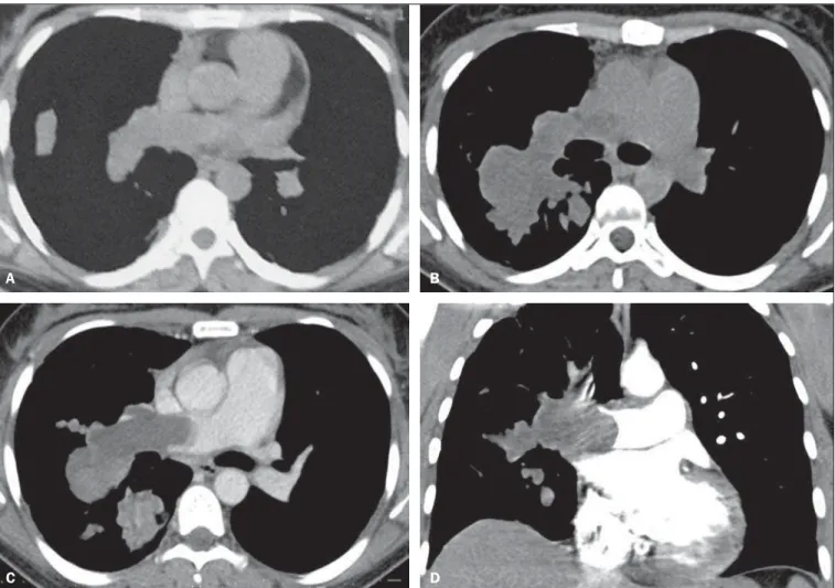
A 35-year-old woman was admitted in our institution with a 2-year history of dyspnea, hemoptysis and chest pain. Chest com-puted tomography (CT) demonstrated filling defects in the right pulmonary artery and some of its branches (Figure 1A). Tran-sthoracic echocardiography showed right heart chambers enlarge-ment and increased pulmonary artery systolic pressure. These test results associated to the patient’s clinical history, allowed for the diagnosis of chronic pulmonary thromboembolism (PTE).
After six months of treatment without clinical improvement, a new contrast enhanced CT revealed a growing intraluminal fill-ing defect and a lobulated mass on the right pulmonary artery and its branches, with areas of contrast enhancement (Figure 1 – B,C,D). In addition to the CT findings, magnetic resonance imaging identified restriction of water diffusion. These imaging findings yielded the diagnosis of pulmonary artery sarcoma (PAS). A significant clinical worsening was observed and the patient died before she could be submitted to a diagnostic/therapeutic surgical procedure.
Vascular lesions of the chest have not been frequently de-scribed in the Brazilian radiological literature(1–5). PAS is a rare
malignant tumor that develops from mesenchymal cells in the intima of the pulmonary artery(6). In general, it affects the central
pulmonary arteries, close to the pulmonary valve(7), resulting in
significant morbidity and high mortality rates(8). There is no
pre-dilection for sex, occurring most commonly in the fifth decade of life(9).
In general, symptoms are nonspecific with dyspnea, cough, hemoptysis, chest pain and weight loss(8), progressing to
pulmo-nary hypertension, right ventricular failure, and possibly chronic
cor pulmonale(9). Clinical and radiological findings are frequently
similar to thromboembolic disease. Due to its rarity and insidious growth pattern, PAS may be diagnosed as chronic PTE, leading to a diagnostic delay and inappropriate therapy such as anticoagu-lation or prolonged thrombolysis(10).
At imaging studies, PAS presents as unilateral, intravascular lobulated masses with heterogeneous contrast enhancement, that may cause vascular distension and local extravascular dissemina-tion(11). Also, the lungs are frequently affected by metastases(7).
According to Yi et al.(12), tomographic findings suggesting the
diagnosis of PAS include low attenuation filling defect of the en-tire luminal diameter of a segment or of the whole extent of the main pulmonary artery, enlargement of the involved arteries and
Figure 1. Axial chest computed tomography (A) demonstrating hypodense mass occupying the lumen of the right pulmonary artery. The luminal diameter is preserved. After seven months, follow-up with contrast-enhanced and non-contrast-enhanced axial (B,C) and coronal (D) computed tomography showed significant enlargement of the intraluminal mass determining dilatation of the affected vessels, with areas of contrast enhancement.
A B
Letters to the Editor
Radiol Bras. 2015 Set/Out;48(5):333–340
334
http://dx.doi.org/10.1590/0100-3984.2015.0046
Central nervous system involvement in sarcoidosis
Envolvimento do sistema nervoso central na sarcoidose
Dear Editor,
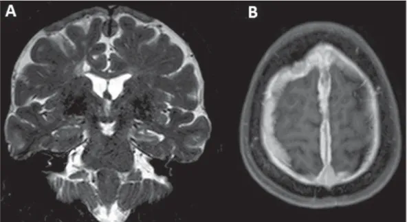
A 51-year-old female patient complained of mild frontotem-poral headache of insidious-onset for two years. One year ago, she had an episode of focal, tonic-clonic seizures (with right lower limb paresthesia) and was prescribed carbamazepine. Cerebrospinal fluid demonstrated increased protein levels and intrathecal im-munoglobulin (IgG) synthesis, suggesting an inflammatory com-ponent. Magnetic resonance imaging was performed (Figure 1). Sarcoidosis is a multisystem disease of unknown etiology characterized by noncaseating granulomatous inflammation(1).
There is a genetic predisposition, with T-lymphocyte receptor
activation by some unknown antigen. The disease affects prefer-entially the respiratory system(1). In the lungs, granulomas are
observed in the interstitial compartment, showing a perilymphatic distribution along the peribronchovascular sheaths, interlobular septa and pleural surface(1).
It is estimated that in about 5% to 15% of cases sarcoidosis affects the central nervous system. Rarely the patient presents with exclusively neurological manifestations like in the present case. Most commonly, neurosarcoidosis is observed in cases of dissemi-nated disease(2).
The clinical manifestations of neurosarcoidosis are pleomor-phic. Cranial nerve compromise, visual alterations, headache, weakness, paresis, paresthesia, psychiatric alterations and signs of meningeal irritation may be observed. Although rare, symp-extraluminal extension of the tumor(6,12). The prognosis is poor,
with mean survival time of approximately one year and a half after symptoms onset(8). Due to pulmonary artery occlusion and acute
symptoms, surgical resection is generally the treatment of choice(8).
In conclusion, the present case reinforces the important role of the imaging methods in the differentiation between pulmo-nary artery intimal sarcoma and chronic PTE. The relevant as-pects for this differentiation, such as contrast enhancement, dis-tention of the affect vessels and extraluminal extension, allow for a correct diagnosis, avoiding delay in the required surgical ap-proach.
REFERENCES
1. Yamanari MGI, Mansur MCD, Kay FU, et al. Bullet embolism of pul-monary artery: a case report. Radiol Bras. 2014;47:128–30. 2. Agnollitto PM, Barreto ARF, Barbieri RFP, et al. Rendu-Osler-Weber
syndrome: what radiologists should know. Literature review and three cases report. Radiol Bras. 2013;46:168–72.
3. Yamada AM, Melo ALKO, Lopes GP, et al. Bilateral breast swelling secondary to superior vena cava obstruction and subclavian vein throm-bosis. Radiol Bras. 2013;46:252–4.
4. Daud DF, Campos MMF, Fleury Neto LAP. Cardiac tamponade in an infant during contrast infusion through central venous catheter for chest computed tomography. Radiol Bras. 2013;46:385–6.
5. Eifer DA, Arsego FV, Torres FS. Unilateral pulmonary veins atresia: evaluation by computed tomography. Radiol Bras. 2013;46:376–8.
6. Chong S, Kim TS, Kim BT, et al. Pulmonary artery sarcoma mimick-ing pulmonary thromboembolism: integrated FDG PET/CT. AJR Am J Roentgenol. 2007;188:1691–3.
7. Grosse C, Grosse A. CT findings in diseases associated with pulmonary hypertension: a current review. Radiographics. 2010;30:1753–77. 8. Wong HH, Gounaris I, McCormack A, et al. Presentation and
manage-ment of pulmonary artery sarcoma. Clin Sarcoma Res. 2015;5:3. 9. Dornas AP, Campos FT, Rezende CJ, et al. Intimal sarcoma of the
pulmonary artery: a differential diagnosis of chronic pulmonary throm-boembolism. J Bras Pneumol. 2009;35:814–8.
10. Cheng HM, Chou ASB, Chiang KH, et al. Serial CT findings of pul-monary artery intimal sarcoma in 4 months: a case report. Chin J Radiol. 2009;34:35–8.
11. Wittram C, Maher MM, Yoo AJ, et al. CT angiography of pulmonary embolism: diagnostic criteria and causes of misdiagnosis. Radiographics. 2004;24:1219–38.
12. Yi CA, Lee KS, Choe YH, et al. Computed tomography in pulmonary artery sarcoma: distinguishing features from pulmonary embolic dis-ease. J Comput Assist Tomogr. 2004;28:34–9.
Marianna Nunes Batista1, Miriam Menna Barreto1,
Renata Fukamati Cavaguti1, Gláucia Zanetti1, Edson
Marchiori1
1. Universidade Federal do Rio de Janeiro (UFRJ), Rio de Janeiro, RJ, Brazil. Mailing Address: Dr. Edson Marchiori. Rua Thomaz Came-ron, 438, Valparaíso. Petrópolis, RJ, Brazil, 25685-120. E-mail: [email protected].
Figure 1. A: Coronal magnetic resonance imaging – T2-weighted sequence demonstrating diffuse pachymeningeal thickening most prominent at the high convexity and extending bilaterally toward the falx, with predominance of hyposignal in association with reduction in volume and hypersignal of the left hippocampus (mesial sclerosis). B: