Frequency of congenital
craniofacial malformations in a
Brazilian Reference Center
Frequência de malformações
congênitas craniofaciais em um
Centro de Referência Brasileiro
Lívia Máris Ribeiro Paranaíba
IRoseli Teixeira de Miranda
IILeila Aparecida Ribeiro
IILetízia Monteiro de Barros
IIHercílio Martelli-Júnior
II, IIII Department of Oral Diagnosis, School of Dentistry, State University of
Campinas. Piracicaba, São Paulo, Brazil.
II Dental School of University of Alfenas, Alfenas, Minas Gerais, Brazil.
III Stomatology Clinic, Dental School, State University of Montes Claros, Montes
Claros, Minas Gerais, Brazil.
Corresponding author: Lívia Máris Ribeiro Paranaíba.Faculdade de Odontologia da Universidade de Alfenas, Rod MG 179, Km 0 - Alfenas, Minas Gerais, Brazil - CEP 37310-000. E-mail: [email protected]
Abstract
Objective: To evaluate the frequency of
cra-niofacial anomalies in patients treated at a Brazilian Reference Center for craniofacial
deformities. Method: Retrospective
epi-demiological study evaluating the clinical records of 1,142 patients: 656 (57.4%) male and 486 (42.6%) female, between 1992 and 2008. Results: Among birth defects, non-syndromic cleft lip and/or palate were the most frequent ones (778 cases; 68.1%), followed by single or multiple congenital anomalies without cleft lip and/or palate (240 cases; 21%), recognized syndromes or sequences (56 cases; 5%), syndromes with orofacial cleft as a component (41 cases; 3.5%), and orofacial clefts in association with systemic malformations (27 cases;
2.4%). Conclusions: Non-syndromic cleft
lip and/or palate was the congenital defect most frequently identiied, although, iso-lated anomalies and syndromes involving craniofacial structures were quite frequent. Furthermore, the need for studies to identify the frequency and risk factors associated with craniofacial anomalies in the Brazilian population is emphasized in order to plan comprehensive strategies and integrated actions for the development of preventive programs and treatment.
Resumo
Objetivo: Avaliar a frequência de anomalias
craniofaciais em pacientes assistidos em um Centro de Referência Brasileiro para
deformidades craniofaciais. Método:
Estu-do retrospectivo epidemiológico avalianEstu-do os prontuários clínicos de 1.142 pacientes, sendo 656 (57,4%) do gênero masculino e 486 (42,6%) do feminino, entre os anos de 1992 e 2008. Resultados: Entre os defeitos congênitos, issura labial e/ou palatina não-sindrômica foi o mais frequente (778 casos; 68,1%), seguido por anomalias congênitas simples ou múltiplas sem fissura labial e/ou palatina (240 casos; 21%), síndromes ou sequências reconhecidas (56 casos; 5%), síndromes com issura orofacial como um componente do quadro sindrômico (41 casos; 3,5%), e issuras orofaciais em associação com malformações sistêmicas
(27 casos; 2,4%). Conclusões: Observou-se
que o defeito congênito identiicado mais frequente foi a issura labial e/ou palatina não-sindrômica, no entanto, anomalias iso-ladas e síndromes envolvendo as estruturas craniofaciais foram bastante encontradas. Além disso, ressalta-se a necessidade de estudos que identiiquem a frequência e os fatores de riscos associados às anomalias craniofaciais na população brasileira, a im de que se planejem estratégias e ações inte-gradas para o desenvolvimento de progra-mas preventivos e de tratamento adequado.
Palavras-chave: Anomalias Congênitas.
Malformações Craniofaciais. Fissura Labial. Fissura palatina. Serviço de informação. Epidemiologia.
Introduction
Congenital anomalies (CA) are changes in structure, function and metabolism present at birth, resulting in physical or mental impairment. They may be simple or multiple, and vary in clinical importance 1,2.
CAs are important causes of mortality and morbidity in childhood and later in life, occurring in approximately 3-5% of new-borns1,3. CAs are currently the second cause
of infant mortality in Brazil, determining 11.2% of these deaths4,5. Of pregnancies
with malformed fetuses more than 20% end in miscarriage; the remaining 80% will be born (alive or dead) with some kind of
congenital anomaly1.
The etiology of most CA remains un-known, although there are a few well esta-blished and avoidable external risk factors6 .
Worldwide surveys have shown that the birth prevalence of CA varies greatly from country to country3,7. It is reported to be as
low as 1.07% in Japan, and as high as 4.3% in Taiwan7. In the USA, the birth prevalence of
CAs is 2-3%, in England 2%, in South Africa 1.49% and, in Brazil 1-3%3,4,8-11. These
va-riations may be explained by ethnic, social, ecological, and economic inluences3,7. In
developing countries like Brazil and others of Latin America, childbearing-age women are exposed to potential risk factors like infectious agents and poverty diseases, environmental chemical compounds, un-healthy working conditions during preg-nancy, use of medication, and maternal metabolic diseases. More than that, these risk factors interact with low schooling and low socioeconomic status in the population and scarce resources in the public health care system targeting the prevention and treatment of congenital anomalies4,12
In general, the most common CAs are nervous system anomalies (especially neural tube defects, such as spina biida, anencephaly, and encephalocele), cleft lip and/or palate, musculoskeletal system anomalies (such as polydactyly, syndactyly and congenital clubfoot), and cardiovas-cular anomalies4,5,9,13,14
urogenital anomalies are also included as the most frequent2.
Among CA, craniofacial anomalies are a large and complex group including skull and/or facial feature contour alterations. Among them are cleft lip and/or palate, craniosynostosis, holoprosencephaly, oto-mandibular defects, neural tube defects that affect the cephalic pole, and multisystem syndromes as Apert, Crouzon syndrome, among others15.
Undoubtedly, cleft lip and/or palate (CL/P) are the most common examples of CA and may occur in up to one in every 600 newborns, which means the birth of a patient every 2.5 minutes in the world15,16.
CL/P etiology is complex with both en-vironmental and genetic factors playing important roles, and the intensive effort of current research has not revealed a single major risk factor for human clefting16. It has
long been known that CL/P can be associa-ted with other systemic defects, although the reported prevalence and the type of associated alterations vary among different studies, ranging from 3% to 63.4%16-19. Also,
reporting of cleft patients in the immediate postnatal period can underestimate the true frequency of associated congenital anoma-lies because many are still not diagnosed at birth or in the neonatal period20.
Clefting has been proposed as part of a complex malformation associated with other anomalies. The identiication of spe-ciic associations with CL/P is important for improving the deinition of the classi-fication, and the genetics, epidemiology and morphology of this malformation17,20.
A combination of these approaches may be useful for public health, treatment, and preventive strategies17.
Although most patients with craniofacial anomalies have normal life expectancy, these anomalies can lead to signiicant ab-normalities in speech, hearing, appearance, and cognition, leading to long-term adverse health events and social integration prob-lems for individuals affected15.
In general, population-based studies on congenital malformations are rare in
Brazil and are represented by studies from hospital sources, such as the ECLAMC net-work (Latin American Collaborative Study on Congenital Malformations)4. In order to
better acknowledge, understand and treat the patients seen at our Service, this study aimed to evaluate the frequency of congeni-tal craniofacial malformations in the Center for Rehabilitation of Craniofacial Anomalies of the University of Alfenas, which is a refe-rence Center for the State of Minas Gerais.
Methods
The medical records of 1,142 patients aged 1 month to 61 years (average age, 19.1 yrs; standard deviation, 14.9) treated at the Center for Rehabilitation of Craniofacial Anomalies of the state of Minas Gerais, Brazil, between 1992 and 2008, were exami-ned retrospectively. All incomplete medical records were excluded, such as incomplete clinical information and/or inconclusive diagnoses. This sample of patients was derived from individuals living in the state of Minas Gerais, which is mostly formed by an admixed population of Africans and Europeans (most from Portugal, Spain and Italy), with a very small percentage of indi-genous Brazilians. The Brazilian population is comprised by an intense admixture of Europeans, Africans and native Indians, making the differentiation between ethnic groups dificult21. This reference service of
the Brazilian Health Department compri-ses a multidisciplinary team of health care specialists, including plastic and dental surgeons, dentists, psychologists, pedia-tricians, genetic counseling professionals, nutritionists, and a speech therapist. The initial physical examination was performed by a pediatrician, followed by the multidis-ciplinary team.
Information obtained from medical records was analyzed according to the type of congenital craniofacial anomalies as follows (adapted from Vallino-Napoli
et al., 2004 and Jaruratanasirikul et al., 2008)22,23 : (1) non-syndromic cleft lip and/
absence of any other structural or cognitive abnormality, (2) syndromic cleft lip and/or palate (SCL/P), an oral cleft in association with other phenotypes characterizing a syndrome, (3) recognized syndromes or sequences in the absence of cleft lip and/ or palate (RSS), (4) single or multiple con-genital anomalies without cleft lip and/or palate (SMCA) – such as congenital tumors and malformations of ears, eyes, and the maxillo-mandibular complex, and (5) cleft lip and/or palate and associated malfor-mations (CL/PAM) without deinition of a syndrome.
Oral clefts were classiied in 4 groups with the incisive foramen as reference24 - (1)
isolated cleft lip (CL): complete or incom-plete, uni or bilateral pre-foramen cleft, (2) cleft lip and palate (CLP): uni or bilateral trans-foramen cleft, (3) isolated cleft palate (CP): complete or incomplete post-foramen cleft, and (4) rare orofacial cleft (ROC). Des-criptive statistical analysis was performed by using the SPSS program, version 17.0 (Chicago, EUA). The study was approved by the Ethical Research Committee of the University. There is no conlict of interest.
Results
The majority of patients affected by CA were male (n=656, 57.4%) with a male-to-female ratio of 1.35. The frequency of con-genital craniofacial anomalies was 68.1% (n=778) of NSCL/P, 21% (n=240) of SMCA,
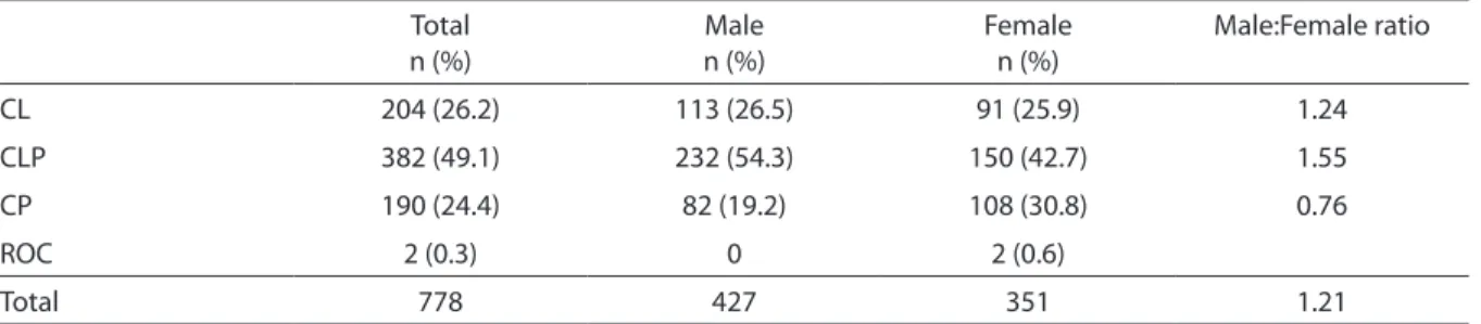
5% (n=56) of RSS, 3.7% (n=41) of SCL/P and, 2.4% (n=27) of CL/PAM. Among NSCL/P patients, 204 (26.2%) had CL, 382 (49.1%) CL/P, 190 (24.4%) CP and, 2 presented ROC (0.3%) (Table 1). CL and CL/P were more common in males than in females (1.24:1 and 1.55:1 respectively), whereas CP was more frequent in females (1.26:1). When all cases were considered together, the male:female ratio was 1.21 (Table 1).
Eye anomalies represented the most common alteration in the SMCA group, cor-responding to 145 out of 240 cases (60.4%), followed by ear anomalies, maxillo-mandi-bular defects, multiple system defects, limb/ extremity anomalies, facial hemangioma, nervous system anomalies, and tongue mal-formations (Table 2). Patients were included as having multiple system defects when they demonstrated more than one organ/system affected by CAs. In this group, we identiied 4 patients with an association of limb/extre-mity CA and malformations in the face and respiratory system, 3 subjects with facial and respiratory anomalies, 2 cases of limb/ extremity deformities in association with cardiac and facial malformations, 2 patients demonstrating limb/extremity and facial anomalies associated with nervous system alterations, 2 patients with an association of malformations in the face and nervous sys-tem, and 2 patients with congenital disor-ders in both cardiac and nervous systems. Tongue malformations were represented by ankyloglossia, issured tongue, and aglossia.
Table 1 - Distribution of speciic types of cleft according to patient sex among 778 patients with non-syndromic cleft lip/ palate (NSCL/P) seen in a Brazilian reference center, 1992-2008.
Tabela 1 - Distribuição dos tipos especíicos de issuras de acordo com o gênero entre os 778 pacientes com issura labial e/ou palatina não-sindrômica vista em um centro de referência Brasileiro, 1992-2008.
Total n (%)
Male n (%)
Female n (%)
Male:Female ratio
CL 204 (26.2) 113 (26.5) 91 (25.9) 1.24
CLP 382 (49.1) 232 (54.3) 150 (42.7) 1.55
CP 190 (24.4) 82 (19.2) 108 (30.8) 0.76
ROC 2 (0.3) 0 2 (0.6)
Total 778 427 351 1.21
CL: isolated cleft lip; CLP: cleft lip and palate; CP: isolated cleft palate; ROC: rare orofacial cleft.
A total of 56 RSS were identiied. The most frequent ones were Goldenhar syn-drome (6 cases, 10.7%), Treacher Collins syndrome (5 cases, 9%), Pierre Robin sequence (4 cases, 7.1%), and Moebius syndrome (4 cases, 7.1%). Other relatively common syndromes were also identiied
such as Crouzon syndrome, Down syn-drome, and Apert syndrome. Moreover, 16 (28.6%) patients had non-diagnosed syndromes (Table 3).
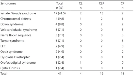
Forty-one patients had SCL/P, and the most frequent one was van der Woude syndrome (17 cases, 41.5%) (Table 4). Of Table 2 - Frequency of single or multiple congenital malformations without cleft lip and/or palate (SMCA) identiied in this study.
Tabela 2 - Frequência das malformações congênitas únicas ou múltiplas sem issura labial e/ou palatina identiicadas neste estudo.
Single or Multiple Anomalies n (%)
Eye anomalies 145 (60.4)
Ear anomalies 26 (10.8)
Maxillo-mandibular defects 23 (9.6)
Multiple system defects 15 (6.3)
Limb/extremity anomalies 11 (4.6)
Facial hemangioma 10 (4.2)
Nervous system anomalies 6 (2.4)
Tongue malformations 4 (1.7)
Total 240
Table 3 - Frequency of syndromes and sequences without cleft lip and/or palate (RSS) identiied in this study.
Tabela 3 - Frequência das síndromes e sequências sem issura labial e/ou palatina identiicadas neste estudo.
Syndromes and sequence n (%)
Goldenhar syndrome 6 (10.7)
Treacher Collins syndrome 5 (9.0)
Pierre Robin sequence 4 (7.1)
Moebius syndrome 4 (7.1)
Crouzon syndrome 3 (5.3)
Down syndrome 3 (5.3)
Frontonasal syndrome 3 (5.3)
Apert syndrome 2 (3.6)
Sturge Weber syndrome 2 (3.6)
Hereditary ectodermal dysplasia 2 (3.6)
Orofaciodigital syndrome 2 (3.6)
West syndrome 1 (1.8)
Sotos syndrome 1 (1.8)
Rubstein-Taybi syndrome 1 (1.8)
Ellis van Creveld syndrome 1 (1.8)
Non-diagnosed syndromes 16 (28.6)
the remaining SCL/P group, patients were diagnosed with chromosomal defects, in-cluding 46XX, t(8;11)(p.10,p10) and 46XY, inv(18)(p.11.1p11.32, Down syndrome, velocardiofacial syndrome, Pierre Robin sequence, Turner syndrome, ectrodactyly, ectodermal dysplasia and cleft lip/palate (EEC) syndrome, Optiz syndrome, among others. Table 4 shows the syndromes and the type of cleft associated with them.
Of the 27 reported cases with CL/PAM, 9 (33.3%) had tongue anomalies and CL/P (Table 5), and the association of CP with ankyloglossia was the most common (6 cases, 66.7%). Other malformations iden-tiied were nervous system anomalies plus CL/P, extremity/limb alterations (2 patients with CL and 2 with CLP), ear and growth anomalies associated with CLP, and 2 eye malformations plus CLP (Table 5).
Table 4 - Distribution of clefts in the syndromes (SCL/P) observed in this study. Tabela 4 - Distribuição das issuras em síndromes observadas neste estudo.
Syndromes Total
n (%)
CL n
CLP n
CP n
van der Woude syndrome 17 (41.5) 2 13 2
Chromosomal defects 4 (9.8) 1 2 1
Down syndrome 4 (9.8) 0 2 2
Velocardiofacial syndrome 3 (7.1) 0 0 3
Pierre Robin sequence 3 (7.1) 0 0 3
Turner syndrome 3 (7.1) 0 0 3
EEC 2 (4.9) 0 2 0
Optiz syndrome 2 (4.9) 0 0 2
Dysplasia Diastrophic 1 (2.4) 0 0 1
Orofaciodigital syndrome 1 (2.4) 1 0 0
Cystic Fibrosis 1 (2.4) 0 0 1
Total 41 4 19 18
CL: isolated cleft lip; CLP: cleft lip and palate; CP: isolated cleft palate; EEC: Ectrodactyly, ectodermal dysplasia and cleft lip/ palate syndrome.
CL: issura labial isolada; CLP: issura lábio palatina; CP: issura palatina isolada; EEC: Ectrodatilia, displasia ectodermal e síndrome de issura labial/palatina.
Table 5 - Frequency of malformations identiied in this study in association with oral clefts (CL/ PAM).
Tabela 5 - Frequência das malformações identiicadas neste estudo em associação com issuras orofaciais.
Anomaly Total
n (%)
CL n
CL/P n
CP n
Tongue malformations 9 (33.3) 1 2 6
Nervous system 6 (22.2) 0 4 2
Extremity/limb alterations 4 (14.9) 1 1 2
Ear anomalies 3 (11.1) 0 3 0
Growth anomalies 3 (11.1) 0 3 0
Eye anomalies 2 (7.4) 0 2 0
Total 27 2 (7.4%) 15 (55.6%) 10 (37%)
CL: isolated cleft lip; CLP: cleft lip and palate; CP: isolated cleft palate.
Discussion
The most common congenital cranio-facial anomaly identiied in this study was NSCL/P with 778 cases (68.1%). NSCL/P (OMIM 119530) is one of the most common congenital malformations observed in hu-mans, and represents the most common craniofacial anomaly, with an incidence of approximately 1 in 600 live births world-wide16,25. The birth incidence may vary
according to ethnic factors, geographic origin, and socioeconomic level, with Asian and Native American populations having the highest rate and African ances-tries having the lowest one16. In Brazil, the
prevalence of NSCL/P at birth is 1.46/1000 live births26. The management of NSCL/P
is complex, and families find difficulties and lack of specific guidance on how to take care of children with clefts. The most common problems of NSCL/P children are breast feeding dificulties, repeated middle ear infections, speech disarticulations, ma-locclusion, and staged surgical repair. Con-sequently, NSCL/P patients require the care of a variety of healthcare professionals (i.e., plastic surgeon, dentist, speech pathologist, otolaryngologist, geneticist, and psycholo-gist) working as a team in the planning and delivery of treatment. There is no standard treatment strategy, timing of intervention, or surgical repair technique, but the gold standard goal is aimed at achieving intelli-gible speech, proper dentition, and normal facial appearance 27,28.
Of the 778 NSCL/P patients, 54.9% were males with a male/female ratio of 1.21:1, which is similar to previous reports26,29-31.
The most common form of cleft observed was CLP, corresponding to 49% of medical records, followed by CL (26.2%), CP (24.4%), and ROC (0.3%). CP and ROC were more frequent in females, while CL and CLP were more observed in males. In general, our results were compatible with previous investigations16,23,26,30,32-34. The gender ratio
has been reported in the literature to vary among the types of oral cleft34. Males are
more likely than females to have a CL with
or without CP, whereas females are at greater risk for isolated CP22. In our study, there was
a difference in gender ratio for all types of oral clefts. Females were more frequent than males for isolated CP and ROC, but without statistical signiicance.
Oral cleft associated with other CAs or in a syndromic pattern was found in 6.1% of patients, which is within the 4.3% to 63.4% range reported by other studies23,35,36.
The most frequent syndrome was van der Woude syndrome, which is also similar to previous reports29,37. Tongue malformations,
nervous system alterations, and extremity/ limb anomalies were the most common disorders in the CL/PAM group, which is in disagreement with the literature. In gene-ral, malformations of the extremities and/ or skeletal system and nervous system are
the most common33,36-38. CLP was the most
prevalent orofacial cleft among infants with additional malformations, which is similar to other studies23,33,36.
Evidence suggests that the presence of structural and cognitive brain anomalies in CL/P are important because the brain and the face are intimately related in normal and pathological conditions with a close relationship between the development of the face, the craniofacial skeleton, and the brain. Moreover, among patients with clefting disorders, some level of cognitive dysfunction is frequent and has been rela-ted to brain pathology (i.e., schizophrenia, bipolar diseases and mental retardation)39,40.
Calzolari et al. (2006) suggested that clini-cal trials should be undertaken to evaluate whether neuroimaging studies in patients with apparently isolated CL/P and cognitive dysfunction should become part of clinical practice.
clefts), musculoskeletal disorders, uroge-nital and cardiovascular anomalies5,7,9,14. In
part, our results are similar to these studies; although, eye, ear, face, and neck anomalies were the most common, which is probably associated with the intrinsic characteristic of the service with specialists in craniofacial deformities. In Brazil, the most frequent site and/or system of malformations are the central nervous system (e.i., spina biida, hydrocephalus, and encephalocele), face (CL/P), osteomuscular system (e.i., gastros-chisis and omphalocele), extremities/limbs (e.i., congenital clubfeet, polydactyly), and urogenital system 2,4,9. Brazilian studies show
some risk factors that contribute to CA in the population, such as deicient folic acid supplementation, maternal diseases (parti-cularly diabetes mellitus), maternal age, his-tory of abortion, maternal low schooling2,4,9.
It should be emphasized that the pre-vention of a significant proportion of malformations is possible, especially in the central nervous system4,9,41. The use of folic
acid supplementation during the period surrounding conception signiicantly redu-ces the incidence of neural tube defects41. In
Brazil, fortiication of wheat and corn lour with folic acid has been mandatory since 20044. Furthermore, the control of maternal
diabetes, and possibly vitamin supplemen-tation may also reduce the occurrence of malformations resulting from uncontrolled diabetes4.
The main syndromes and sequences identified were Goldenhar syndrome, Treacher Collins syndrome, Pierre Robin sequence, and Moebius syndrome. Accor-ding to several studies, the most common syndromes observed in newborn infants with birth defects are Trisomy of the 13, 18 and 213,8,14. However, even with a different
grouping of the syndromes identiied in our classiication, the results were different from most studies3,8,23. Likewise, these syndromes
identiied in our service represent mainly disorders involving craniofacial structures
that are the main focus of the center. The knowledge and the identification of all these CAs in our population allow us to lead a comprehensive strategy and integrated actions for the best possible treatment and prevention through community education, population screening, genetic counseling, and the availability of early diagnosis.
Unfortunately, it was not possible to assess the risk factors (previous abortions, maternal and/or paternal age, parity, edu-cation, family history, and outhers1,2,42)
associated with congenital malformations identiied at the Reference Center becau-se of lack of access to medical records in hospitals. Research of possible risk factors related to congenital malformations are es-sential to determine which prevention and management policies should be planned and enforced. It would be interesting for the country to implement a national registry of craniofacial anomalies comprising detailed information on mothers during pregnancy, family history of affected patients, health status of newborns, re-registration, and a new assessment at a later age, among others.
Conclusion
The most common CA identiied was NSCL/P; however, isolated anomalies and syndromes were also recognized and treated in this Brazilian Reference Center. This study allowed us to acknowledge the universe of patients who come to our service, providing insight to improve the planning and treat-ment strategy for CA patients. Furthermore, it reinforces the importance of a multidis-ciplinary team for the treatment of patients affected by CAs.
Acknowledgements: LMR Paranaíba is
References
1. Jones KL. Dysmorphology. In: Berman RE, Kliegman
RM, Jenson HB. Nelson Textbook of Pediatrics. 17th ed.
Philadelphia: WB Saunders; 2004. p. 616-23.
2. Castro MLS, Cunha CJ, Moreira PB, Fernández RR, Garcias GL, Martino-Röth MG. Frequency of multiple neonatal malformations in Pelotas, Rio Grande do Sul,
Brazil, and associated socio-demographic factors. Cad
Saúde Pública 2006; 22(5): 1009-15.
3. Canield MA, Honein MA, Yuskiv N, Xing J, Mai CT,
Collins JS et al. National estimates and
race/ethnic-speciic variation of selected birth defects in the United
States, 1999-2001. Birth Defects Res A Clin Mol Teratol
2006; 76(11): 747-56.
4. Costa CMS, Gama SGN, Leal MC. Congenital
malformations in Rio de Janeiro, Brazil: prevalence and
associated factors. Cad Saúde Pública 2006; 22(11):
2423-31.
5. Victora CG, Barros FC. Infant mortality due to perinatal causes in Brazil: trends, regional patterns and possible
interventions. Sao Paulo Med J 2001; 119: 33-42.
6. Varela MM, Nohr EA, Llopis-González A, Andersen AM, Olsen J. Socio-occupational status and congenital
anomalies. Eur J Public Health 2009; 19(2): 161-7.
7. Temtamy SA, Abdel Meguid N, Mazen I, Ismail SR, Ramzy MI. A genetic epidemiological study of
malformations at birth in Egypt. East Mediterr Health J
1998; 4: 252-9.
8. Dolk H. EUROCAT: 25 years of European surveillance of
congenital anomalies. Arch Dis Child Fetal Neonatal Ed
2005; 90(5): F355-8.
9. Amorin MMR, Vilela PC, Santos ARVD, Lima AMV,
Melo EFP, Bernardes HF, et al. Impact of congenital
malformations on perinatal and neonatal mortality
in an university maternity hospital in Recife. Rev Bras
Saúde Matern Infant 2006; 6 (S1): S19-S25.
10. Melo LL, Carvalho MR, Wojciechowski M, Bianchim MM. Natimortalidade e malformaçöes congênitas em natimortos: estudo de frequência, fatores de risco e padräo de defeitos congênitos em uma populaçäo de
Porto Alegre. Rev AMRIGS 1989; 33: 10-4.
11. Nóbrega FJ. Antropometria, patologias e malformações congênitas do recém-nascido brasileiro e estudos de
associação com algumas variáveis maternas. J Pediatr
1985; 59 (S1): 6-140.
12. Schuler-Faccini L, Leite JCL, Sanserino MTV, Peres RM.
Avaliação de teratógenos na população brasileira. Ciênc
Saúde Coletiva 2002; 7: 66-71.
13. Pinto CO, Nascimento LSC. Prevalence study of birth
defects in Vale do Paraíba, São Paulo, Brazil. Rev Paul
Pediatr 2007; 25(3): 233-9.
14. Tomatir AG, Demirhan H, Sorkun HC, Köksal A, Ozerdem F, Cilengir N. Major congenital anomalies: a
ive-year retrospective regional study in Turkey. Genet
Mol Res 2009; 13(1): 19-27.
15. Monlleó IL, Gil-da-Silva-Lopes VL. Craniofacial anomalies: description and evaluation of treatment
under the Brazilian Uniied Health System. Cad Saúde
Pública 2006; 22(5): 913-22.
16. Mossey PA, Little J. Epidemiology of oral clefts: an
international perspective. In: DF, ed. Cleft Lip and
Palate: From Origin to Treatment. New York: Oxford University Press; 2002. p. 127-58.
17. Calzolari E, Pierini A, Astoli G, Bianchi F, Neville AJ, Rivieri F. Associated anomalies in multi-malformed infants with cleft lip and palate: An epidemiologic study
of nearly 6 million births in 23 EUROCAT registries. Am J
Med Genet A 2007;143(6): 528-37.
18. Fraser FC. The genetics of cleft lip and cleft palate. Am J
Hum Genet 1970; 22: 336-352.
19. Shprintzen RJ, Siegel-Sadewitz VL, Amato J, Golberg RB. Anomalies associated with cleft lip, cleft palate, or both.
Am J Med Genet 1985; 20: 585-95.
20. van der Veen FJ, van Hagen JM, Berkhof J, Don Griot JP. Regional underreporting of associated congenital
anomalies in cleft patients in the Netherlands. Cleft
Palate Craniofac J 2006; 43(6): 710-4.
21. Vieira AR, Karras JC, Orioli IM, Castilla EE, Murray JC. Genetic origins in a South American clefting population.
Clin Genet 2002; 62: 458-63.
22. Vallino-Napoli LD, Riley MM, Halliday JL. An
epidemiologic study of isolated cleft lip, palate, or both
in Victoria, Australia from 1983 to 2000. Cleft Palate
Craniofac J 2004; 41: 185-94.
23. Jaruratanasirikul S, Chichareon V, Pattanapreechawong N, Sangsupavanich P. Cleft lip and/or palate: 10 years experience at a pediatric cleft center in Southern
Thailand. Cleft Palate Craniofac J 2008; 45(6): 597-602.
24. Spina V, Psillakis JM, Lapa FS, Ferreira MC. Classiicação
das issuras lábiopalatinas. Rev Hosp Clin Fac Med S
Paulo 1972; 27: 5-6.
25. Vieira AR. Unraveling human cleft lip and palate
research. J Dent Res 2008; 87: 119-25.
26. Martelli-Júnior H, Orsi-Júnior J, Chaves MR, Barros LM, Bonan PRF, Freitas JAS. Estudo epidemiológico das issuras labiais e palatais em Alfenas, Minas Gerais, de
1986 a 1998. Rev Fac Odontol Univ São Paulo 2006; 13(1):
31-5.
27. Strauss RP, Broder H. Interdisciplinary team care of cleft
lip and palate: social and psychosocial aspects. Clin
28. Paranaíba LMR, Almeida H, Barros LM, Chaves MR,
Martelli DRB, Orsi-Júnior J, et al. Surgical rehabilitation
of cleft lip-palate in Minas Gerais state, Brazil. RBOtorrin
2009; 75(6): 839-43.
29. Hagberg C, Larson O, Milerad J. Incidence of cleft lip
and palate and risks of additional malformations. Cleft
Palate Craniofac J 1997; 35(1): 40-5.
30. Forrester MB, Merz RD. Descriptive epidemiology of oral clefts in a multiethnic population, Hawaii, 1986–2000.
Cleft Palate Craniofac J 2004; 41: 622–8.
31. Martelli-Junior H, Porto LCVP, Barbosa DRB, Bonan PRF, Freitas AB, Coletta RD. Prevalence of nonsyndromic oral clefts in a reference hospital in Minas Gerais State,
between 2000-2005. Braz Oral Res 2007; 21(4): 314-7.
32. Loffredo LCM, Souza JMP, Freitas FAS, Mossey PA.
Oral clefts and vitamin supplementation. Cleft Palate
Craniofac J 2001; 38(1): 76-83.
33. Shaw GM, Carmichael SL, Yang W, Harris JA, Lammer EJ. Congenital malformations in births with orofacial clefts
among 3.6 million California births. Am J Med Genet
2004; 125: 205-6.
34. Cooper ME, Ratay JS, Marazita ML. Asian oral-facial cleft
birth prevalence. Cleft Palate Craniofac J 2006; 43:
580-89.
35. Wyszynski DF, Sárközi A, Czeizel AE. Oral clefts with
associated anomalies: Methodological issues. Cleft
Palate Craniofac J 2006; 43: 1-6.
36. Stoll C, Alembik Y, Dott B, Roth MP. Associated
malformations in cases with oral clefts. Cleft Palate
Craniofac J 2000; 37: 41-7.
37. Milerad J, Larson O, Hagberg C, Ideberg M. Associated malformations in infants with cleft lip and palate: A
prospective, population based study. J Pediatr 1997;
100(2): 180-6.
38. Sandrini FAL, Robinson WM, Paskulin G, Lima MC. Family study in orofacial clefts associated anomalies patients in the Serviço de Defeitos de Face of Pontifícia
Universidade Católica of Rio Grande do Sul. Rev Cir
Traumatol 2006; 6(2): 57-68.
39. Nopoulos P, Berg S, Canady J, Richman L, Van Denmark D, Andreasen NC. Structural brain abnormalities in
adult males with clefts of the lip and/or palate. Genet
Med 2002a; 4: 1-9.
40. Nopoulos P, Berg S, Van Denmark D, Richman L, Canady J, Andreasen NC. Cognitive dysfunction in adult males with non-syndromic clefts of the lip and/or palate.
Neuropsychologia 2002b; 40: 2178-84.
41. Wolff T, Witkop CT, Miller T, Syed SB, U.S. Preventive Services Task Force. Folic acid supplementation for the prevention of neural tube defects: an update of the
evidence for the U.S. Preventive Services Task Force. Ann
Intern Med 2009; 150: 632-9.
42. Vrijheid M, Dolk H, Stone D, Abramshy L, Alberman E, Scott JES. Socioeconomic inequalities in risk of
congenital anomaly. Arch Dis Child 2000; 82: 349-52.