ORIGIN
AL RESER
CH
Cardiorespiratory adjustments during the
accentuation of respiratory sinus arrhythmia:
influence from time of maneuver on minute volume,
fraction of expired CO
2, and heart rate variability
Ajustes cardiorrespiratórios durante a manobra de acentuação da arritmia sinusal
respiratória: inluência do tempo da manobra sobre o volume minuto, fração expirada de CO₂
e variabilidade da frequência cardíaca
Ajustes cardiorrespiratorios durante la maniobra de acentuación de la arritmia sinusal
respiratoria: inluencia del tiempo de maniobra sobre el volumen minuto, fracción espirada de
CO
2y variabilidad de la frecuencia cardiaca
Alexandre Fenley1,2, Leonardo da Costa Silva1,2, Hugo Valverde Reis1,2, Luciana Malosá Sampaio3, Audrey Borghi-Silva4, Michel Silva Reis1,2
Mailing address: Michel Silva Reis – Departamento de Fisioterapia, Faculdade de Medicina, Universidade Federal do Rio de Janeiro – Rua Prof. Rodolpho Paulo Rocco, s/n, Hospital Universitário Clementino Fraga Filho, 8º andar, ala E, sala 3 (8E-03), Ilha do Fundão – Rio de Janeiro (RJ), Brazil – CEP: 21941-913 – Phone: (55 21) 2562-2223 – E-mail: [email protected] – Financing source: Fundação Carlos Chagas de Apoio à Pesquisa do Estado do Rio de Janeiro (FAPERJ, process: E-26/110.878/2013) and Conselho Nacional de Desenvolvimento Cientíico e Tecnológico (CNPq, process: 487375/2012-2) – Conlict of interest: Nothing to declare – Presentation: May 2015 – Accepted for publication: Mar. 2016 – Approved by the Ethics Committee of Hospital Universitário Clementino Fraga Filho da Universidade Federal do Rio de Janeiro nº 970.098/2015.
Study developed by the Grupo de Pesquisa em Avaliação e Reabilitação Cardiorrespiratória (GECARE) – Departamento de Fisioterapia e Programa de Pós-Graduação em Educação Física e Medicina (Cardiologia) of the Universidade Federal do Rio de Janeiro (UFRJ) – Rio de Janeiro (RJ), Brazil.
1Group of Research on Cardiorespiratory Evaluation and Rehabilitation (GECARE), department of Physiotherapy, School of Medicine,
Federal University of Rio de Janeiro – Rio de Janeiro (RJ), Brazil.
2Graduate Program in Physical Education and Medicine (Cardiology), center of Health Science, Federal University of Rio de Janeiro –
Rio de Janeiro (RJ), Brazil.
3Graduate Program in Rehabilitation Sciences, University Nove de Julho – São Paulo (SP), Brazil.
4Laboratory of Cardiopulmonary Physiotherapy, Department of Physiotherapy, Federal University of São Carlos – São Carlos (SP),
Brazil.
ABSTRACT | Heart rate (HR) luctuate during the respiratory cycle. This phenomenon is known as respiratory sinus arrhythmia. The deep breathing test is to keep a paced breathing in six breathing per minute and I:E relationship 1:1. The purpose of this study is to access minute volume, expired fraction of carbon dioxide (EFCO2) and autonomic control of heart rate during deep breathing test longer than 90 seconds. Sixteen young healthy male (18 – 25 years old) were assessed. The subjects were instructed to perform inspirations and expirations with duration of 10 seconds per cycle, I:E = 1:1, and consequently respiratory rate of 6 cycles per minute, for about four minutes with one minute after and before, totaling six minutes. HR was recorded beat-to-beat using a cardio frequencimeter; MV and EFCO2 was measured and recorded using a mobile ergoespirometer. To analyse statistics diferences, ANOVA one way (Tuckey hoc) and Kruskall Wallis (Dunn post-hoc) were used (p<0.05). When deep breathing test in course, EFCO2, MV and time domain heart rate variability shows no statistics diference over time. To perform deep
breathing test in young healthy male, longer than 90 seconds, can be safety, without risks of hypocapnia and no interference from EFCO2 changes in time domain heart rate variability analysis of M-RSA.
Keywords | Heart Rate; Respiratory Sinus Arrhythmia;
Men; Healthy Volunteers.
de 10 segundos por ciclo, TI:TE de 1:1 e consequente frequência respiratória de seis incursões por minuto, durante quatro minutos. Durante a avaliação foi coletada a frequência cardíaca (FC) batimento a batimento por meio de um cardiofrequencímetro, o volume minuto (VM) e a FeCO2 através de um ergoespirômetro. Para análise estatística empregou-se ANOVA one-way (com post-hoc de Tukey) ou teste de Kruskal-Wallis (com post-post-hoc de Dunn) quando conveniente (p<0,05). Durante a M-ASR, a FeCO2, o VM e os índices do domínio do tempo da variabilidade da frequência cardíaca (VFC) não sofreram alterações signiicativas ao longo do tempo. A realização da M-ASR em jovens saudáveis, por mais de 90 segundos, pode ser executada com segurança, sem o risco de hipocapnia e sem a interferência das alterações de FeCO2 nos índices do domínio do tempo da análise de VFC da M-ASR.
Descritores | Frequência Cardíaca; Arritmia Sinusal Respiratória; Homens; Voluntários Saudáveis.
RESUMEN | La frecuencia cardíaca sufre oscilaciones durante el ciclo respiratorio, fenómeno conocido como arritmia sinusal respiratoria. La maniobra para acentuación de la arritmia sinusal respiratoria (M-ASR) consiste en mantener ventilación educada con frecuencia respiratoria de seis ciclos por minuto con relación al tiempo inspiración/espiración (TI:TE) de 1:1. En este estudio
se propone a evaluar la conducta del volumen minuto, de la fracción espirada de CO2 (FeCO₂ iniere sobre el PaCO2) y el control autonómico de la frecuencia cardíaca durante la M-ASR con duración mayor de 90s. Se evaluaron 16 varones jóvenes sanos (de 18 a 25 años de edad). Se les orientaron para que realizasen inspiraciones y espiraciones pausadas de 10 segundos de duración por ciclo, TI:TE de 1:1, y consecuente frecuencia respiratoria de seis incursiones por minuto, durante cuatro minutos. Durante la evaluación se recolectaron la frecuencia cardíaca (FC), latido a latido a través de un monitor de frecuencia cardíaca, el volumen minuto (VM) y la FeCO2 mediante un ergoespirómetro. Para el análisis estadístico se empleó ANOVA one-way (con post-hoc de Tukey) o test de Kruskal-Wallis (con post-hoc de Dunn) cuando necesario (p<0,05). Durante la M-ASR, la FeCO2, el VM y los índices del dominio del tiempo para la variabilidad de la frecuencia cardíaca (VFC) no presentaron alteraciones signiicativas al largo del tiempo. Puede ejecutarse con seguridad la realización de la M-ASR en jóvenes sanos por más de 90 segundos, sin riesgo de hipocapnia y sin interferencia de las alteraciones de la FeCO2 en los índices del dominio del tiempo para analizar la VFC de la M-ASR.
Palabras clave | Frecuencia Cardíaca; Arritmia Sinusal Respiratoria; Hombres; Voluntarios Sanos.
INTRODUCTION
Heart rate variability (HRV) is a simple, quick, and inexpensive means to assess the integrity and adjustments of the autonomic nervous system (ANS). It is able to provide information for the interpretation of the sympathovagal balance by means of analyses performed in
the time domain, spectral analysis, and nonlinear analysis1,2.
Some speciic protocols enable measuring separately the inluence from the sympathetic and parasympathetic
ANS on the autonomic modulation of heart rate (HR)3-5.
Accentuation of respiratory sinus arrhythmia (RSA) has been used to assess parasympathetic modulation on autonomic control of HR in patients with chronic
cardiorespiratory and metabolic disorders6-8. In addition,
this maneuver has also been applied as a therapeutic
strategy in patients with systemic hypertension3, chronic
obstructive pulmonary disease4, diabetes mellitus5, and
chronic heart failure6 to improve sympathovagal balance.
he RSA-M consists in maintaining ventilation with a respiratory rate of ive to six cycles per minute with an
inspiration/expiration ratio of 1:17.
During respiratory cycles, HR sufers variations during
the inspiratory and expiratory phases8. his
phenome-non is known as respiratory sinus arrhythmia (RSA)9-11.
During the inspiratory phase, there is inhibition of the parasympathetic ANS and consequent increase in HR, while, during the expiratory phase, there is recovery of the
parasympathetic ANS and decrease in HR9. herefore, the
application of RSA-M for evaluation of vagal integrity and as therapeutic intervention has been widely studied. On the other hand, the literature is incipient regarding the implications in the parasympathetic autonomic modula-tion on minute volume (MV) and on the partial pressure
of carbon dioxide (PaCO2) during RSA-M.
In this context, our study aims to evaluate the behavior of
the minute volume, of the fraction of expired CO2 (FeCO₂
- which allows inferring about PaCO2), and the autonomic
control of heart rate during the RSA-M. Our hypothesis is
that there will be no alteration in MV and FeCO2, because
even if there is an increase in tidal volume during RSA-M, respiratory rate will remain reduced, with little variation in
MV with no alterations in FeCO2 and, consequently, in the
METHODOLOGY
Subjects
Observational and transversal study in which 16 healthy young males were selected. As inclusion criteria for this study, volunteers should be aged 18-25 years, and be male. We excluded individuals who were smokers, users of illegal drugs and medication, with known cardiopulmonary, musculoskeletal, neurological, autoimmune and/or metabolic disease. his research was approved by the Research Ethics Committee of the University Hospital Clementino Fraga Filho, Federal University of Rio de Janeiro, under written opinion No. 970,098/2015. All participants signed a free and informed consent form.
Experimental protocol
Volunteers received a form with guidelines for preparation in the previous day and in the day of the evaluations. hey were instructed with respect to no ingestion of stimulant drinks (cofee, soda, energy drinks, and teas), no vigorous physical activities, and to have proper night’s sleep. Research was conducted in the Laboratory of the Group of Research on Cardiopulmonary Evaluation and Rehabilitation (GECARE) in heated room with temperature ranging from 22-24°C in the period from 9 a.m. to 3 p.m. Initially, volunteers were introduced to the experimental environment and to the researchers involved. Before starting the tests, volunteers were evaluated and examined to ensure the guidelines given had been followed strictly. Vital signs (HR and BP) were checked before, during, and after each test.
For the conduct of RSA-M7, the volunteers were
instructed, by verbal and tactile command (with abdominal stimulation), to inspire by nose and expire by mouth, deeply and slowly, varying lung volume from total lung capacity to residual volume. Each cycle was performed in 10 seconds (ive seconds for the inspiratory phase and ive seconds for the expiratory phase), in which maximum RSA was expected. he protocol had total time of 6 minutes and was conducted as follows: 1) 1 minute at rest and spontaneous ventilation; 2) 4 minutes during the RSA-M; and 3) 1 minute at rest and spontaneous ventilation. he protocol was conducted twice considering the efect of learning and selected the second procedure.
During RSA-M, instant HR was collected, beat
to beat, using a heart monitor (Polar® RS800CX).
he heart monitor has a sampling frequency of 1000 Hz, ixed with an elastic belt in the lower third of the breastbone and with simultaneous transmission to the watch where they were stored. Subsequently, through USB interface, data were transported and stored in a notebook (Intel Core i3-2330M) to be analyzed in the
Polar® Precision Performance software. Numerical data
of the R-R intervals were extracted and exported to
Microsoft Excel®, in which irstly we deleted artifacts
and ectopic heartbeats. hen, data were exported to
the Kubios HRV® software, and segments from each
minute of RSA-M were analyzed in the time domain, through the mean HR, mean iR-R, standard deviation of the normal iR-R (SDNN), which is the square root of the variance, and the root mean square of successive
diferences between adjacent iR-R (RMSSD)1,12.
Concomitantly, MV and FeCO2 were taken through
the ergospirometry system VO2000 (Medigraphics). For
that, the low-low pneumotachometer was ixed through a neoprene face mask with size chosen according to the anthropometric characteristics of each individual. he
numerical values for MV and FeCO2 were exported to
Microsoft Excel® and then, for each minute of maneuver,
we calculated the mean values of these variables during the corresponding minute.
Statistical analysis
Sample calculation was carried out based on the outcome variable SDNN of a pilot study in our laboratory. hus, for a power of 80%, with efect size of 5 and alpha of 5%, we determined the need for 12 individuals (GPower 3.0.1.0 for Windows). In the statistical analysis, the data were submitted to the tests of normality (Shapiro-Wilk test) and homogeneity (Levene test). Subsequently, we employed the one-way ANOVA (with post-hoc Tukey) for variables with normal distribution and the Kruskal-Wallis test (with post-hoc Dunn) for variables without normal distribution. All measures are expressed as mean±SD. he signiicance level was 5% (p<0.05). he analyses were performed with the SIGMA PLOT
software for Windows version 11.0, copyright© 2008
Systat Software, Inc.
RESULTS
volunteers studied. he sample of individuals has normal demographic (age) and anthropometric distribution.
Table 1. Characteristics of the population
Volunteers (n=16)
Age (years) 22.4±1.26
Height (m) 1.77±0.07
Body mass (kg) 76.2±9.6
BMI (kg/m2) 24.4±3.07
Values in mean±SD.BMI: body mass index
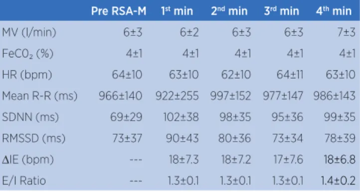
Table 2 presents the mean values (±SD) for VE,
FeCO2, HR, and the time domain indices for HRV
of the individuals evaluated. here was no diference for these variables, independent of the time taken to perform the maneuver.
Table 2. Ventilatory variables and heart rate variability obtained during the RSA-M
Pre RSA-M 1st min 2nd min 3rd min 4th min
MV (l/min) 6±3 6±2 6±3 6±3 7±3
FeC0₂ (%) 4±1 4±1 4±1 4±1 4±1
HR (bpm) 64±10 63±10 62±10 64±11 63±10
Mean R-R (ms) 966±140 922±255 997±152 977±147 986±143
SDNN (ms) 69±29 102±38 98±35 95±36 99±35
RMSSD (ms) 73±37 90±43 80±36 73±34 78±39
∆IE (bpm) --- 18±7.3 18±7.2 17±7.6 18±6.8
E/I Ratio --- 1.3±0.1 1.3±0.1 1.3±0.1 1.4±0.2
Values in mean±SD;
MV: Minute volume; FeCO2: fraction of expired CO2; HR: heart rate; SDNN: standard deviation
of the iR-R; RMSSD: root mean square of successive diferences between adjacent iR-R; ∆IE: inspiration-expiration delta in bpm; E/I ratio: expiration/inspiration ratio. One-way ANOVA with p>0.05
DISCUSSION
he main results of this study show that, during the
RSA-M, the FeCO2, the MV, and the time domain
indices for HRV sufered no signiicant alterations over time. Compared to the minute before the maneuver, we observed no signiicant statistical diference in the
values for FeCO2 and MV, as well as in the time domain
indices for HRV.
he RSA-M allows evaluating the parasympathetic
modulation on the autonomic control of HR2, in
addition to being applied as a therapeutic strategy in
patients with systemic arterial hypertension3, chronic
obstructive pulmonary disease4, diabetes mellitus5, and
chronic heart failure6. In this study, we evaluated the
parasympathetic modulation on the autonomic control
of HR in healthy, young persons and the behavior of
FeCO2 and MV at the beginning, during, and at the
end of the respiratory sinus arrhythmia maneuver. herefore, one of the major issues of our study concerns the uncertainties regarding the inluence from the time of the RSA-M on the response of HRV. his is because the literature is inconsistent as whether the responses of the ventilatory variables determined by lung volume variation, in protocols in which the respiratory frequency is controlled, can generate efects on the cardiovascular adjustments. hese responses may be determined by the central command and peripheral
aferent impulses (such as chemoreceptors)13,14 along
the conduct of the RSA-M. In this sense,
Guillén-Mandujan et al.15, who evaluated the inluence from
diferent respiratory frequencies and lung volumes on the RSA-M, showed that both conditions are able to determine cardiovascular adjustments independently.
Interestingly, Shields2 suggested that one of the factors
that can decrease HRV during the RSA-M performed with duration greater than 90 seconds is the possibility of inducing hypocapnia. However, in our study, the RSA-M was conducted for 240 seconds and, regardless of the time taken, the time domain indices for HRV, the
MV, and the FeCO2 remained constant from the
pre-maneuver period until the fourth minute of pre-maneuver. hese results can be attributed to the fact that our educated breathing protocol for the conduct of RSA-M has a low RF with high expiratory time, which results
in constant FeCO2. A condition that was conirmed
in the study of Lopes et al. (2011)16, which evaluated
healthy individuals in six diferent educated breathing patterns: two with diferent inspiratory/expiratory time ratio (TI:TE) (of 1:1 and of 1:2) for each ixed RF of 6, 12, and 20 incursions per minute. For each ixed RF, a target TV was determined. As a result, we observed that the efects of TI:TE on RSA are dependent on RF and these efects are more pronounced in the lower RFs and with higher TEs. Considering that in our study TE was 5 seconds, it may have resulted in the maintenance of
constant FeCO2.
In 2004, Cooper et al.17 evaluated 12 normal,
in hypocapnia, the amplitude of RSA decreased signiicantly (40±5 ms). In our study, the maintenance of RSA-M for more than 90 seconds did not alter the
FeCO2, a fact that probably contributed to the absence
of alteration in the variables for HRV in the time domain during the RSA-M.
Studies on RSA-M are specially important for cardiorespiratory physiotherapy. Considering that the RSA-M can be applied to evaluate the vagal modulation and as therapeutic strategy of the sympathovagal balance, understanding its mechanisms will ensure a more secure and appropriate handling, thus preventing inadvertent use.
As limitations of this study, the measurement
of FeCO2 during the RSA-M was performed
non-invasively, with analysis of gas expired; therefore, our results should be limited to non-invasive analyses
of PaCO2. In this sense, an invasive direct analysis
of PaCO2 could conirm our results in future trials.
Furthermore, it would be important to collect with systems that allow for the measurement of tidal volume during the RSA-M. Finally, our results apply to healthy individuals. In the future, other studies with patients with cardiorespiratory disorders should be encouraged.
CONCLUSION
he performance of RSA-M in healthy, young individuals, for more than 90 seconds, did not alter
the FeCO2 and the time domain indices for HRV
during RSA-M. Our study brings initial information for the safe conduct of spontaneous ventilation in a controlled way whether for RSA-M in the evaluation of parasympathetic modulation or for its therapeutic application. Finally, future studies with patients with cardiorespiratory disorders are feasible for a greater understanding of the mechanisms of the RSA-M.
ACKNOWLEDGMENTS
he authors would like to thank the Carlos Chagas Foundation for Research Support of the State of Rio de Janeiro (FAPERJ- process: E-26/110.878/2013) and the National Council for Scientiic and Technological Development (CNPq – process: 487375/2012-2) for the inancial support. Additionally, we thank the
colleagues of the Research Group on Cardiopulmonary Evaluation and Rehabilitation (GECARE) of the Department of Physiotherapy, Federal University of Rio de Janeiro.
REFERENCES
1. Heart rate variability. Standards of measurement, physiological interpretation, and clinical use. Task Force of the European Society of Cardiology and the North American Society of Pacing and Electrophysiology. Eur Heart J. 1996;17(3):354-81.
2. Shields RW. Heart rate variability with deep breathing as a clinical test of cardiovagal function. Cleve Clin J Med. 2009;76(Suppl 2):S37-40.
3. Joseph CN, Porta C, Casucci G, Casiraghi N, Mafeis M, Rossi M, et al. Slow breathing improves arterial barorelex sensitivity and decreases blood pressure in essential hypertension. Hypertension. 2005;46(4):714-8.
4. Reis MS, Arena R, Deus AP, Simões RP, Catai AM, Borghi-Silva A. Deep breathing heart rate variability is associated with respiratory muscle weakness in patients with chronic obstructive pulmonary disease. Clinics (Sao Paulo). 2010;65(4):369-75.
5. Rosengård-Bärlund M, Bernardi L, Sandelin A, Forsblom C, Groop PH, Group FS. Barorelex sensitivity and its response to deep breathing predict increase in blood pressure in type 1 diabetes in a 5-year follow-up. Diabetes Care. 2011;34(11):2424-30.
6. Reis MS, Deus AP, Simões RP, Aniceto IA, Catai AM, Borghi-Silva A. Autonomic control of heart rate in patients with chronic cardiorespiratory disease and in healthy participants at rest and during a respiratory sinus arrhythmia maneuver. Rev Bras Fisioter. 2010;14(2):106-13.
7. Hayano J, Mukai S, Sakakibara M, Okada A, Takata K, Fujinami T. Efects of respiratory interval on vagal modulation of heart rate. Am J Physiol. 1994;267(1 Pt 2):H33-40.
8. Moreira GL, Ramos EMC, Vanderlei LCM, Ramos D, Manzano BM, Fosco LC. Efeito da técnica de oscilação oral de alta frequência aplicada em diferentes pressões expiratórias sobre a função autonômica do coração e os parâmetros cardiorrespiratórios. Fisioter Pesq. 2009;16(2):113-9.
9. Grossman P, Wilhelm FH, Spoerle M. Respiratory sinus arrhythmia, cardiac vagal control and daily activity. Am J Physiol Heart Circ Physiol. 2004;287(2):728-34.
10. Hirsch JA, Bishop B. Respiratory sinus arrhythmia in humans: how breathing pattern modulates heart rate. Am J Physiol. 1981;241(4):H620-9.
11. Reis MS, Arena R, Archiza B, de Toledo CF, Catai AM, Borghi-Silva A. Deep breathing heart rate variability is associated with inspiratory muscle weakness in chronic heart failure. Physiother Res Int. 2014;19(1):16-24.
13. Williamson JW. The relevance of central command for the neural cardiovascular control of exercise. Exp Physiol. 2010;95(11):1043-8.
14. Mitchell JH. Neural control of the circulation during exercise: insights from the 1970-1971 Oxford studies. Exp Physiol. 2012;97(1):14-9.
15. Guillén-Mandujano A, Carrasco-Sosa S. Additive efect of simultaneously varying respiratory frequency and tidal volume on respiratory sinus arrhythmia. Auton Neurosci. 2014;186:69-76.
16. Lopes TC, Beda A, Granja-Filho PC, Jandre FC, Giannella-Neto A. Cardio-respiratory interactions and relocation of heartbeats within the respiratory cycle during spontaneous and paced breathing. Physiol Meas. 2011;32(9):1389-401.