Instituto de Investigaciones Biotecnologicas, Instituto Tecnologico de Chascomus (IIB-INTECH, UNSAM-CONICET) Av. General Paz 5445, INTI, edificio 24, 1650 San Martin, Provincia de Buenos Aires, Argentina
Manuscript received on August 13, 2007; accepted for publication on August 17, 2007; presented byLUCIAMENDONÇAPREVIATO
ABSTRACT
Trypanosoma cruziis highly sensitive to oxidative stress caused by reactive oxygen species. Trypanothione, the parasite’s major protection against oxidative stress, is kept reduced by trypanothione reductase, using NADPH; the major source of the reduced coenzyme seems to be the pentose phosphate pathway. Its seven enzymes are present in the four major stages in the parasite’s biological cycle; we have cloned and expressed them inEscherichia coli as active proteins. Glucose 6-phosphate dehydrogenase, which controls glucose flux through the pathway by its response to the NADP/NADPH ratio, is encoded by a number of genes per haploid genome, and is induced up to 4fold by hydrogen peroxide in metacyclic trypomastigotes. The genes encoding phosphogluconolactonase, 6-phosphogluconate dehydrogenase, transaldolase and transketolase are present in the CL Brener clone as a single copy per haploid genome. 6-phosphogluconate dehydrogenase is very unstable, but was stabilized introducing two salt bridges by site-directed mutagenesis. Ribose-5-phosphate isomerase belongs to Type B; genes encoding Type A enzymes, present in mammals, are absent. Ribulose-5-phosphate epimerase is encoded by two genes. The enzymes of the pathway have a major cytosolic component, although several of them have a secondary glycosomal localization, and also minor localizations in other organelles.
Key words:Trypanosoma cruzi, Chagas disease, pentose phosphate pathway, oxidative stress, NADPH generation.
INTRODUCTION
Trypanosoma cruzi, a flagellated protozoan parasite, is the causative agent of the American trypanosomiasis, Chagas disease. The parasite has a complex life cy-cle, involving two forms present in the gut of the in-sect vector, the replicative epimastigote and the infec-tive metacyclic trypomastigote, and two forms present in the infected mammal, the intracellular amastigote and the bloodstream trypomastigote, released from infected cells into the blood. The infection is endemic in Latin
*Present address: ICP, Université Catholique de Louvain, Brussels, Belgium.
Correspondence to: Juan J. Cazzulo
America, where its prevalence is estimated at 16-18 mil-lion cases, with about 120 milmil-lion people at risk. No vaccines have been developed so far, and the low ef-fectiveness of the chemotherapeutic agents available (at present, only benznidazole), together with their unde-sirable side effects, makes treatment of Chagas disease difficult. There is, therefore, an urgent need for the iden-tification of novel drug targets to improve the treatment of this disease (Barrett et al. 2003).
which have different substrate specificities and subcellu-lar localization. Wilkinson et al. demonstrated the pres-ence of two glutathione peroxidases, TcGPXI (Wilkin-son et al. 2002a) and TcGPXII (Wilkin(Wilkin-son et al. 2002b), localized in different cell compartments; the former in the cytosol and glycosomes, the latter in the endoplasmic reticulum. They also detected two trypanothione depen-dent members of the peroxiredoxine family TcMPX and TcCPX, (Wilkinson et al. 2000), and an ascorbate depen-dent hemoperoxidase TcAPX (Wilkinson et al. 2002c). These enzymes belong to an intricate network converg-ing to reduced trypanothione T[SH]2, which is
main-tained in its reduced form by trypanothione reductase, with the utilization of NADPH. Therefore, it is abso-lutely essential for the parasite to have reliable pathways for the maintenance of a suitable pool of this reduced coenzyme. Several enzyme reactions may be responsi-ble for this supply of reducing power: the NADP-linked glutamate dehydrogenase (Juan et al. 1978, Barderi et al. 1998), the NADP-linked malic enzyme (Cannata et al. 1979), and the two dehydrogenases of the Pentose Phosphate Pathway (PPP).
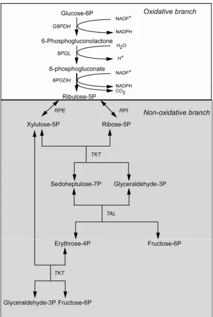
In most organisms glucose is metabolized through two major pathways: the glycolytic, or Embden-Meyer-hof, pathway, and the PPP. The latter one (Fig. 1), also starting from glucose 6-phosphate (G6P), is involved in the production of the ribose 5-phosphate (R5P) re-quired for nucleotide synthesis, and of reducing power in the form of NADPH. The pathway can be separated in two branches, namely an oxidative branch, involv-ing glucose phosphate dehydrogenase (G6PDH), 6-phosphogluconolactonase (6-PGL) and 6-phosphoglu-conate dehydrogenase (6PGDH), and a non-oxidative, or sugar interconversion, branch, involving ribose 5-phos-phate isomerase (RPI), ribulose 5-phos5-phos-phate epimerase (RPE), transaldolase (TAL) and transketolase (TKT) (Fig. 1). The PPP has also been known as the pentose phosphate cycle, since, when functioning as a whole, the fructose 6-phosphate (F6P) and glyceraldehyde 3-phosphate (Gly3P) formed can be converted back into glucose 6-phosphate, entering the oxidative branch again. However, the PPP needs not to act as a cycle, and the different enzymatic reactions will be operative according to the cell needs (Stryer 1999).
Differing from the glycolytic pathway, which has
been thoroughly studied in Trypanosomatids over the last three decades, the PPP has received much less at-tention up to the last decade. In the case ofT. cruzi, early
studies showed that both dehydrogenases were present in epimastigotes (Raw 1959), and one of them (G6PDH) was partially purified and some of its properties were determined (Funayama et al. 1977). Moreover, studies with labeled glucose suggested that the PPP was func-tional in some strains of the parasite (Mancilla and Náquira 1964). However, until recently most of the en-zymes of the pathway had not been detected, and nothing was known about their properties and subcellular local-ization. Over the last few years, we have studied in detail this subject. This review will describe the present situ-ation in T. cruzi, comparing, whenever possible, with
the findings made inTrypanosoma bruceiand Leishma-niaspp.
FUNCTIONALITY OF THE PENTOSE PHOSPHATE PATHWAY INTrypanosoma cruzi
The early studies of Mancilla and Náquira (1964) sug-gested that the PPP was functional in two strains of T. cruzi. Recently we performed similar experiments with
intact epimastigotes of the CL Brener clone, using glu-cose labeled with14C in C
1or C6. An increased
produc-tion of14CO
2from14C1-glucose over that from14C6
-glucose indicates that the PPP is functional (Katz and Wood 1963). From our experiments we could conclude that in normal conditions, 10% of the glucose metabo-lized goes through the PPP (Maugeri and Cazzulo 2004). Similar assays were performed in two strains ofL. mexi-canashowing that 5.5-5.8% of the glucose consumed is
metabolized by the PPP (Maugeri et al. 2003). Atamna et al. (1994) showed that the addition of a permeant scavenger of NADPH, such as methylene blue, increases glucose flux through the PPP by rising the NADP/ NADPH ratio, and thus releasing the inhibition of the first enzyme of the pathway, G6PDH, by the reduced coenzyme. This was indeed the case inT. cruzi, since in the presence of 0.2 mM methylene blue the glucose flux through the PPP doubled, from 10 to 20% of the total glu-cose utilization (Maugeri and Cazzulo 2004). A similar situation was observed for promastigotes of both strains ofL. mexicanatested (Maugeri et al. 2003). These
Fig. 1 – Scheme of the pentose phosphate pathway. The enzymes involved are glu-cose phosphate dehydrogenase (G6PDH), phosphogluconolactonase (PGL), 6-phosphogluconate dehydrogenase (6PGDH), ribose 5-phosphate isomerase (RPI), ribu-lose 5-phosphate epimerase (RPE), transaldolase (TAL) and transketolase (TKT).
at least of its oxidative branch, in the insect stages of both organisms, and suggest that G6PDH activity is also regulated by the NADP/NADPH ratio. InT. cruzi the production of ribose 5-phosphate from glucose through the action of the PPP has not been studied but in pro-mastigotes from L. mexicana about 11% of the taken
glucose was incorporated into nucleic acids (Maugeri et al. 2003).
All seven enzymes of the PPP are present in the four major stages of the biological cycle ofT. cruzi, epi-mastigotes, metacyclic trypoepi-mastigotes, amastigotes and bloodstream-like trypomastigotes (Maugeri and Cazzulo 2004). With the only exception of RPE, the other en-zymes presented their highest activities in metacyclic trypomastigotes. InT. brucei, on the other hand, all the
trypomastigotes, but some of them could not be detected in the bloodstream form of the parasite (Cronin et al. 1989). In this organism, three of the seven enzymes of the conventional PPP (G6PDH, TAL, RPE) do not have any glycosomal targeting signals although the whole pathway is expected to be present inside this organelle. In epimastigotes from T. cruziall the enzymes of the PPP seem to present multiple subcellular localizations (Maugeri and Cazzulo 2004); however, the bulk of the enzyme activities were detected in the cytosolic tions, both in digitonin extraction and in subcellular frac-tionation experiments. The only exception was the RPE, that seems to be localized in a highly digitonin-access-ible subcellular compartment, as described below. The three trypanosomatid genomes code for a sedoheptulose-1,7 bisphosphatase (SBPase), an enzyme typical of the Calvin cycle of photosynthetic organisms that can be found only in the chloroplast of green algae and plants. However, the presence of a functional Calvin cycle in kinetoplastids may be excluded. In the three organisms, the protein sequences presented a peroxisomal targeting signal-1 (PTS1)in their C-terminal suggesting a
possi-ble glycosomal localization. Since the Calvin cycle and the PPP are processes which are similarly organized and share a number of enzymes, it has been postulated that in trypanosomatids the SBPase could be involved in a modified PPP (Hannaert et al. 2003). The possible par-ticipation of this enzyme in the PPP ofT. cruzihas not
been evaluated until present.
THE ENZYMES OF THE OXIDATIVE BRANCH
GLUCOSE6-PHOSPHATE DEHYDROGENASE
InT. cruziCL Brener clone, G6PDH is encoded by sev-eral genes located in three of the parasite chromosomes (Igoillo-Esteve and Cazzulo 2006). We identified two pseudo-genes and several others that could be clustered in three groups. Those genes were 98% identical in their coding region, but differed considerably in the se-quences upstream and downstream of their ORFs. Most of the amino acid changes predicted were conservative, and none involved residues important for catalysis. Se-quence comparisons showed that theseT. cruzigenes had
69% and 64% identity, respectively, with theirT. brucei
and L. major counterparts (Fig. 2). The T. cruzi and
T. brucei G6PDH genes presented two candidate start
codons, 111 bp apart, while inL. major a unique ATG
codon, equivalent to the first one observed in T. cruzi
andT. brucei, was present. If the T. cruzi G6PDH is
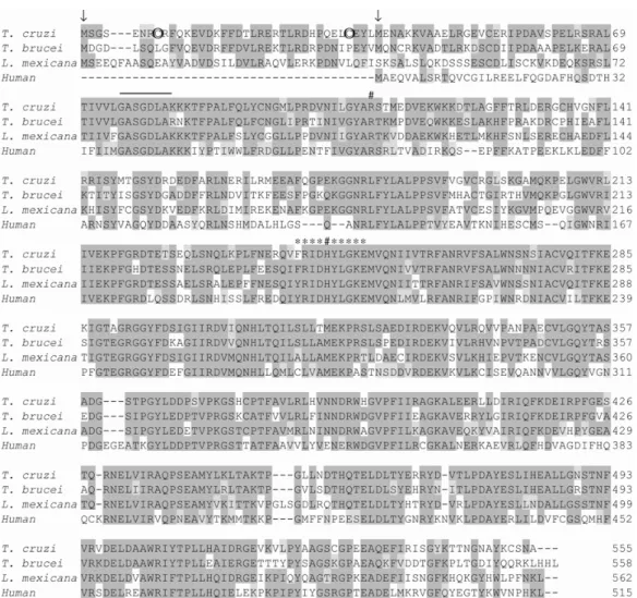
translated from the first start codon, a polypeptide chain of 518 amino acid residues is obtained in which the 37 first residues constitute an N-terminal extension com-pared to the human enzyme. We expressed theT. cruzi
G6PDH in both, its long and short forms (Igoillo-Esteve and Cazzulo 2006) and compared both recombinant en-zymes with the native one. On the other hand, the T. bruceiG6PDH was only expressed in its short form, start-ing from the second Met residue (Duffieux et al. 2000). Due to the low levels of expression of the native G6PDH in epimastigotes fromT. cruzi,added to the instability
of the enzyme, we were unable to purify it to homo-geneity by standard procedures. A particular feature of the native enzyme was the loss of enzymatic activity in the presence of reducing agents, likeβ-mercaptoethanol (β-ME) or dithiothreitol (DTT). When the effects ofβ -ME, as well as those of reduced glutathione (GSH) and DTT were tested on both purified recombinant enzyme forms, we found that the long G6PDH was inhibited by the three reagents, the more prominent effect being ob-tained when the enzyme was incubated in the presence of DTT (90% inhibition after 15 min incubation with the reducing agent, at 25 mM). Similar but less striking ef-fects were observed while incubating the enzyme with the same concentration of GSH orβ˜ME (30% and 50%
inhibition respectively, after 15 min incubation). Con-versely, the short enzyme was activated (up to 80% by 15 mM GSH after the same incubation time). The in-hibition of the long G6PDH by reducing agents corre-lates with the presence of two additional Cys residues in the N-terminal extension of the protein, also observed in redox-regulated G6PDHs from plant chloroplasts and cyanobacteria (Wenderoth et al. 1997). The similar ex-tension predicted from theL. majorgene does not
con-tain these Cys residues and in good agreement, assays performed with the native enzyme showed that it was not inhibited by DTT (Igoillo-Esteve and Cazzulo 2006). These results suggested that theT.cruziG6PDH, at
Fig. 2 – Alignment of the deduced amino acid sequences of different glucose 6-phosphate dehydrogenases. The sequences corresponding to theT. cruziG6PDH (DQ408239) with the highest identity to itsT. bruceicounterpart, theT. bruceiG6PDH (CAC07816),L. mexicanaG6PDH (AAO37825) and human G6PDH (NP_000393) were aligned using the Clustal program. The first and second methionine in theT. cruziandT.
bruceisequences are marked with arrows. The two cysteine residues belonging to theT. cruziN-terminal extension are circled. The cofactor binding site is overlined, and the sequence corresponding to the G6PDH signature, that is part of the substrate binding site, is marked with asterisks. Key residues involved in binding the substrate and cofactor are marked with #.
By Western blot analysis performed using an anti-serum developed against the N-terminal polypeptide of the long G6PDH, absent in the short form of the en-zyme, we could demonstrate that the former is expressed in the four main life stages ofT. cruziCL Brener clone. In good agreement, the apparent Km for the substrate, G6P, was similar for the long recombinant enzyme and the native one.
Inhibition kinetics by NADPH performed using the purified short and longT. cruzi G6PDHs showed that
molecules like H2O2 or dehydroascorbate are present,
the enzyme is oxidized attaining its more active form. This process would constitute a fast response to increase the NADPH level to counteract the oxidative stress.
The relevance of the G6PDH in the defense mech-anisms ofT. cruziagainst oxidative stress was demon-strated by incubating metacyclic trypomastigotes in the presence of different concentrations of H2O2. These
ex-periments showed that after 6 hr incubation in the pres-ence of the oxidizing agent at 70µM, there was a 46-fold increase in G6PDH specific activity, together with an important increase in the protein levels, determined by Western blots. On the other hand, the same experi-ment performed in epimastigotes, revealed a deleterious effect on the G6PGH since a time-dependent decrease in its specific activity and protein amount was observed (Igoillo-Esteve and Cazzulo 2006).
These experiments showed that in metacyclic try-pomastigotes, a form of the parasite naturally exposed to ROS, the G6PDH is strongly induced by oxidative stress. Considering thatT. cruziis more sensitive to ROS
than mammalian cells, the G6PDH can be considered a suitable target for chemotherapy. Further studies will be required to develop suitable inhibitors, which might become lead compounds for the design of new drugs.
Preliminary studies on the subcellular localization of the G6PDH by immunofluorescence suggest that, in addition to its main cytosolic localization, the enzyme would be also present in glycosomes in amastigotes, trypomastigotes and metacyclic trypomastigotes of T. cruzidespite the apparent lack of a PTS1or a PTS2
sig-nal. These experiments, together with G6PDH-GFP fu-sion analysis, also suggested that the enzyme is probably associated to the Golgi complex in epimastigotes and metacyclic trypomastigotes (Igoillo-Esteve 2005). This localization is not completely unusual since it has been previously suggested for the G6PDH of rabbit intestine cells (Ishibashi et al. 1999). Nevertheless, its biolog-ical relevance is not very clear until now; we suggest that inT. cruzi a G6PDH fraction associated with the
Golgi complex would be indirectly protecting the lipid peroxidation in the organelle membranes via the micro-somal Tc-GPXII (Wilkinson et al. 2002b). The G6PDH from procyclic and bloodstream trypomastigotes from
T. bruceihas a dual subcellular localization, cytosolic
and glycosomal (Heise and Opperdoes 1999), despite the apparent lack of a PTS signal (Duffieux et al. 2000).
6-PHOSPHOGLUCONOLACTONASE
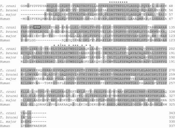
Studies still in progress show that the T. cruzi 6-PGL
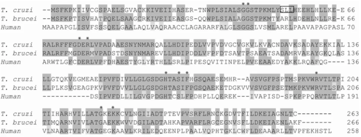
gene is present in the CL Brener clone as a single copy per haploid genome. A comparison of its sequence with that of the enzyme fromT. brucei(Fig. 3) indicates that they are 62% identical, and both predict a possible PTS1
glycosomal targeting signal, placed at the C-terminus in the case of T. brucei and internal in the T. cruzi 6-PGL (P. Beluardi and J.J. Cazzulo, unpublished results, Duffieux et al. 2000). TheT. cruzi6-PGL behaves as a monomeric enzyme, with a molecular mass of 29 kDa. 6-PGL has been recently crystallized from T. brucei,
and its structure determined by X-ray diffraction. Al-though the crystals suggest that the enzyme might be a dimer, the authors concluded, by other evidence, that it is most probably a monomer (Delarue et al. 2007). Both trypanosomatid enzymes have been expressed as soluble active recombinant proteins inE. coli, but theT. brucei enzyme was not characterized, due to the
diffi-culties in the enzyme assay. In fact, the substrate, 6-phosphogluconolactone, must be generated in situ by
the action of G6PDH on G6P, and undergoes sponta-neous hydrolysis, which must be subtracted to get the actual initial reaction velocity. Despite these limitations, we were able to obtain an approximate Km value of 50µM, close to one of the values reported for the rat liver enzyme (80µM, Schofield and Sols 1976). TheT. cruzi6-PGL seems to be distributed between the cytosol
and the glycosome, as reported for theT. bruceienzyme.
6-PHOSPHOGLUCONATE DEHYDROGENASE
The 6PGDH fromT. cruzi is encoded by a single copy gene per haploid genome in the CL Brener clone. The protein has 78.6% identity with itsT. bruceicounterpart (Igoillo-Esteve and Cazzulo 2004). Both, the native and the recombinant 6PGDHs fromT. cruziproved to be un-stable, and the native enzyme could only be partially purified from parasite extracts. Such instability has not been reported for theT. bruceienzyme. Structural
anal-ysis of theT. brucei6PGDH dimer suggested that 5 salt
Fig. 3 – Alignment of the deduced amino acid sequences of different 6-phosphogluconolactonases. The accession numbers for the sequences aligned using the Clustal program are the following:T. cruziEU077554,T. bruceiAJ249255, Human AJ243972. Boxes shaded in dark grey show correspond to identical residues. The asterisks show the residues strictly conserved in the 6PGL family. Previous studies (Delarue et al. 2007) suggest that R77, F170, and R198 in theT. cruzisequence could constitute the 6PGL signature. The grey box shows a putative internal PTS in the
T. cruzisequence.
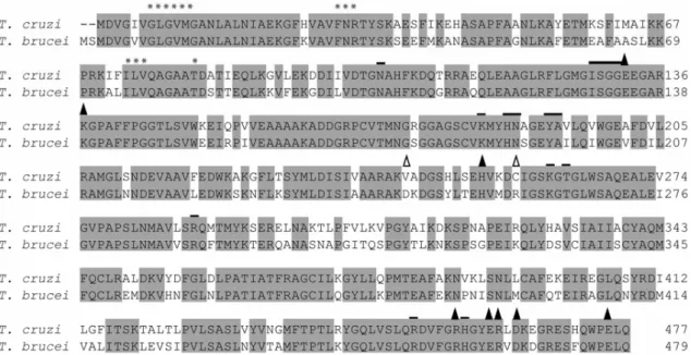
A comparison of theT. cruziandT. brucei6PGDH
se-quences (Fig. 4) showed that in the former two residues which are involved in the formation of two out of the five mentioned salt bridges are lacking. Considering that this might be the reason for the instability of the enzyme fromT. cruzi, we performed site-directed mutagenesis,
leading to a double mutant protein which, according to computational modeling, would have the five bridges. This mutant proved to be considerably more stable than the wild type enzyme and non mutated recombinant 6PGDH, therefore allowing its kinetic study. By gel fil-tration analysis we could determine that the double mu-tant enzyme remained as a dimer under conditions which led to very significant dissociation of the non mutated one (Igoillo-Esteve and Cazzulo 2004).
Hanau et al. (1996) performed detailed kinetic analysis of theT. brucei6PGDH; the enzyme presented unusually low Km values, 3.5µM for the substrate and 1.5µM for the coenzyme. In contrast we found that for theT. cruzienzyme, the Km for 6-phosphogluconate
was similar to that of the human 6PGDH, but the Km for NADP (5.9µM), although 4-fold higher than that for theT. bruceienzyme, was still 5-fold lower than the one
reported for the human one (30µM) (Igoillo-Esteve and Cazzulo 2004).
It is noteworthy that deletion of 6PGDH is lethal
for all cells studied, since accumulation of 6-phospho-gluconate inhibits phosphoglucose isomerase, thus blocking glycolysis. Therefore, specific inhibitors for the parasite’s 6PGDH could become good lead com-pounds for the development of new drugs. Dardonville and co-workers performed inhibition studies on the
T. brucei6PGDH utilizing several substrate and
cofac-tor analogues (Dardonville et al. 2003), as well as high-energy intermediates and transition-state analogues (Dar-donville et al. 2004). A few of these compounds were tested on the purified recombinant double-mutantT. cruzi
6PGDH and the best result, Ki 0.16µM, was obtained
for 4-phospho-D-erythronate, an analogue of the enediol-reaction intermediate (M. Igoillo-Esteve and J.J. Caz-zulo, unpublished results). This compound also showed a selective inhibition of 6PGDH fromT. bruceiover the sheep liver enzyme, (Ki (T. brucei) 0.13 µM and Ki
(sheep) 10.7µM) (Pasti et al. 2003). Additionally Dar-donville et al. (2003) have reported that some inhibitors developed for theT. bruceienzyme were toxic toT. cruzi
amastigotes at concentrations lower than 10µM. These results indicate that it would be possible to extrapolate toT. cruzithe utilization of some of the compounds
de-signed to inhibit theT. brucei6PGDH that nowadays are
Fig. 4 – Alignment of the deduced amino acid sequences of different 6-phosphogluconate dehydrogenases. The 6-PGDHs fromT. cruzi(AY300924) andT. brucei(P31072) were aligned using the Clustal program. The asterisks indicate the residues comprising the coenzyme binding site, and those involved in substrate binding are overlined. The triangles indicate the residues involved in the formation of salt bridges in theT. brucei enzyme; empty triangles indicate those absent in theT. cruzienzyme.
The subcellular localization of theT. cruzi6PGDH
was analyzed by immunofluorescence. These experi-ments revealed that in addition to the main cytosolic localization, a small fraction of the enzyme would be present inside the glycosome, at least in epimastigotes and cell culture-trypomastigotes (Igoillo-Esteve 2005). This localization is in agreement with a possible inter-nal PTS1– like sequence (SHL) localized in a predicted
exposed loop of the protein (Fig. 4).
THE ENZYMES OF THE NON-OXIDATIVE, OR SUGAR INTERCONVERSION, BRANCH
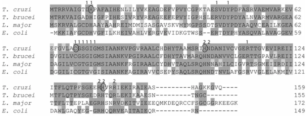
RIBOSE5-PHOSPHATE ISOMERASE
The haploid genome of the CL Brener clone ofT. cruzi
contains one gene coding for a Type B RPI. However, genes encoding Type A RPIs, most frequent in eukary-otes, seem to be absent. So far, RPI B eukaryotic genes have been detected only inGiardia lamblia,Entamoeba histolytica, some fungi and the insectAnopheles gam-biae. The deduced amino acid sequence of theT. cruzi
RPI B does not predict a PTS1or PTS2glycosomal
tar-geting signal (Fig. 5). The RPI fromT. cruziis another
example of an enzyme which is absent in upper
eukary-otic genomes (Stern et al. 2007), since Type A and B RPIs are totally unrelated. A recombinant enzyme was expressed as an active dimeric protein, able to catalyze the reversible isomerization of R5P to Ru5P, with Km values of 4 mM and 1.4 mM, respectively. We were able to study the reaction mechanism of this Type B RPI using several approaches, including site-directed mutagenesis. The competitive inhibition of the enzyme by 4-phospho D-erythronohydroxamic acid showed that this RPI acts
via a mechanism involving the formation of a 1,2-cis enediol. The reaction in the direction R5P to Ru5P, but not the reverse reaction, must involve the opening of the ribose furanose ring. Using site-directed mutagenesis, based on the modeling of the enzyme fromT. cruziusing theE. coli RPI B structure, we were able to show that His102, but not His138, was essential for ring opening.
Moreover, we were able to confirm the essential role of Cys69 in catalysis. These results not only gave
Fig. 5 – Alignment of the deduced amino acid sequences of different ribose 5-phosphate isomerases. The TcRPIB (gi: 110984574) is aligned with those ofT. brucei(gi:70834348),L. major(gi:68127548) andE. coli(gi: 85676843 W3110). Mutagenizated aminoacid residues are circled. Residues that lie within the RPI active-site pocket are annotated with 1 for one subunit and with 2 for the second.
RIBULOSE5-PHOSPHATE EPIMERASE
RPE catalyses the interconversion of two phosphory-lated pentoses, R5P and xylulose 5-phosphate (X5P). The genome of the CL Brener clone ofT. cruzicontains
two genes encoding RPEs. One of them predicts a PTS1
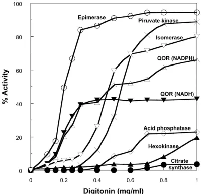
glycosomal targeting signal (SHL) at the C terminus (Fig. 6). Digitonin extraction studies showed that RPE activity is extracted from the epimastigotes by very low concentrations of the detergent (about 0.3 mg/ml), lower than those required to extract the pyruvate kinase used as cytosolic marker (Maugeri and Cazzulo 2004). Other enzymes, like some quinone oxidoreductases (QOR), have a similar extraction pattern (J.J.B. Cannata and D. Maugeri, unpublished results, Fig. 7), suggesting the possibility of the existence of a new, highly digitonin-accessible, subcellular compartment. It is noteworthy that, in cellular fractionation experiments, almost 50% of the RPE activity behaves as particulate, with latency. Both RPE forms have been cloned and expressed as ac-tive proteins in E. coli, and are being characterized at present, in order to define their subcellular localization and possible functions (A.L. Stern and J. J. Cazzulo, un-published results).
TRANSALDOLASE
Transaldolase (D-sedoheptulose-7-phosphate: D-glyce-raldehyde - 3 - phosphate - dihydroxyacetone transferase, EC 2.2.1.2, TAL), transfers a dihydroxyacetone unit from
F6P to erythrose 4-phosphate (E4P), leading to the syn-thesis of sedoheptulose 7-phosphate and Gly3P. A Schiff base involving a Lys residue present in the active site and dihydroxyacetone is essential in the reaction mechanism. Different lines of evidence suggest that TAL has an es-sential regulatory role in the non-oxidative branch of the PPP, at least in higher eukaryotes (Reitzer et al. 1980, Banki et al. 1996, Heinrich et al. 1976).
We have cloned the gene encoding TAL from the CL Brener clone ofT. cruzi, which is present as a
sin-gle copy per haploid genome (Fig. 8), and expressed the recombinant protein inE. colias an active enzyme,
with apparent Km values of 1 mM for F6P and 0.02 mM for E4P. Preliminary results suggest the presence of four TAL isoforms in epimastigotes. At this moment we are working in the purification of the native isoforms, in an attempt to elucidate their role in the parasite metabolism (A.L. Stern and J.J. Cazzulo, unpublished results).
TRANSKETOLASE
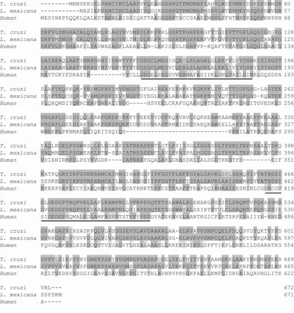
The genome of the CL Brener clone contains a gene en-coding TKT, which seems to be present as a single copy per haploid genome. The gene, which predicts 67% identity with the recently characterized TKT from L. mexicana(Veitch et al. 2004, Fig. 9), was cloned and
the protein expressed as a fusion with a polyHis tag in the N-terminus, inE. coliBL21 Codon Plus. The
Fig. 6 – Alignment of the deduced amino acid sequences of different ribulose 5-phosphate epimerases. The two epimerases fromT. cruzi(TcRPE1, EU075265 and TcRPE2, EU075366) are aligned with those ofT. brucei(XP_823426),L. major(CAJ06401) and human (NP_954699). Tc Rpe2 presents a PTS1(SHL) in the C-terminus. The asterisks indicate the residues conserved in the RPE signature.
0 20 40 60 80 100
0 0.2 0.4 0.6 0.8 1
%
A
c
ti
v
ity
Digitonin (mg/ml) Epimerase
Isomerase Piruvate kinase
QOR (NADPH)
QOR (NADH)
Acid phosphatase
Hexokinase
Citrate synthase
Fig. 8 – Alignment of the deduced amino acid sequences of different transaldolases. TheT. cruziTAL (EU075264) is aligned with those of
T. brucei(XP_847390 ),L. major(CAJ03645) and human (NP_006746). A putative PTS1(SRL) is boxed. The asterisks indicate the residues
conserved in the TAL signature.
a dimer (146 kDa) acting on R5P, E4P and X5P with ap-parent Km values of 1.34 mM, 0.1 mM and 0.07 mM, respectively, at the optimum pH value of 8.0. The en-zyme is expressed in the four main stages ofT. cruzi,
with an apparent subunit molecular mass of 75 kDa, as determined by Western blot (P. Beluardi and J.J. Caz-zulo, unpublished results). TheL. mexicana TKT has been recently characterized, and its 3-D structure has been determined (Veitch et al. 2004). It has been demon-strated that the enzyme, that bears a C-terminal PTS1, has
a dual localization, cytosolic and glycosomal, in Leish-maniapromastigotes. Possibly theT. cruziTKT shares the same location since it also presents a PTS1signal in
its C-terminal region (Fig. 9) but this point still needs to be confirmed.
CONCLUSIONS
The PPP has been shown to be functional inT. cruzi, and
the seven enzymes of the pathway have been cloned,
ex-pressed and are being fully characterized. The biochem-ical evidence obtained so far suggest that the oxidative branch of the PPP is essential for the protection of the parasite against oxidative stress. In addition, one of the enzymes of the non-oxidative branch is of prokaryotic type, and has no counterpart in the higher eukaryotic genomes sequenced until now.
Apart from its main cytosolic localization, the PPP is expected to be present, at least partially, inside the glycosomes, in order to produce the NADPH and R5P required for other enzymatic pathways present inside the organelle. R5P will be converted into 5-phosphoribosyl-1-pyrophosphate that will serve in purine and pyrimi-dine biosyntheses, processes that occurred inside the gly-cosomes. On the other hand, NADPH would be involved in the defense against reactive oxygen species (ROS) since, as mentioned above, Tc-GPXI is located inT. cruzi
Fig. 9 – Alignment of the deduced amino acid sequences of different transketolases. The accession numbers for the sequences aligned using the Clustal program are the following:T. cruziEU077555,L. mexicanaAj427448, human P29401. Boxes shaded in dark grey show correspond to identical residues. The conserved TKT and ThDP boxes, which are involved in cofactor and substrate union, are underlined.
the cytosol but also in the glycosomes of T. cruzi and
T. brucei. These defense systems are, via trypanothione reductase, ultimately dependent on NADPH produced through the PPP (Hannaert et al. 2003). Although we have demonstrated that at least the oxidative branch of the pathway might be present inside the organelle in some developmental stages ofT. cruzi, further experiments are
required in order to confirm or discard the glycosomal localization of the whole PPP in this parasite.
Finally, more studies, involving inhibitor kinetics
and knock-out experiments, will be required to validate the PPP as a suitable target for the development of new drugs for the treatment of Chagas Disease.
ACKNOWLEDGMENTS
fel-sentes nos quatro principais estágios do ciclo biológico do para-sita; nós clonamos e expressamos as enzimas emEscherichia colicomo proteínas ativas. Glucose 6-fosfato desidrogenase, que controla o fluxo da glucose da via em resposta à relação NADP/NADPH, é codificada por um número de genes por genoma haplóide e é induzida até 46-vezes por peróxido de hidrogênio em trypomastigotas metacíclicos. Os genes que codificam 6-fosfogluconolactonase, 6-fosfogluconato desidro-genase, transaldolase e transcetolase estão presentes no clone CL Brener como cópia única por genoma haplóide. 6-fosfo-gluconato desidrogenase é muito instável, mas foi estabilizada introduzindo duas pontes salinas por mutagênese sítio-dirigida. A Ribose-5-fosfato isomerase pertence ao Tipo B; genes que codificam enzimas Tipo A, presentes em mamíferos estão au-sentes. A Ribulose-5-fosfato epimerase é codificada por dois genes. As enzimas da via têm um componente citosólico prin-cipal, embora várias delas tenham uma localização glicosomal secundária e também, localizações em menor número em ou-tras organelas.
Palavras-chave: Trypanosoma cruzi, doença de Chagas, via das pentoses fosfato, estresse oxidativo, produção de NADPH.
REFERENCES
ATAMNAH, PASCARMONAGANDGUINSBURG H. 1994. Hexosemonophosphate shunt activity in intact Plasmo-dium falciparum– infected erythrocytes and in free para-sites. Mol Biochem Parasitol 67: 78–89.
BANKIK, HUTTERE, COLOMBOE, GONCHOROFFNJAND PERLA. 1996. Glutathione levels and sensitivity to apop-tosis are regulated by changes in transaldolase expression. J Biol Chem 271: 32994–33001.
BARDERI P, CAMPETELLA O, FRASCH ACC, SANTOMÉ JA, HELLMANU, PETTERSSON UANDCAZZULOJJ. 1998. The NADP-linked glutamate dehydrogenase from
play differential activities in procyclic and bloodstream forms ofTrypanosoma brucei. FEBS Lett 244: 26–30. CSÉKEC, BALOGHAANDFARKASGL. 1981. Redox
mod-ulation of glucose-6-P dehydrogenase inAnacystis nidu-lansand its “uncoupling” by phage infection. FEBS Lett 126: 85–88.
DARDONVILLE C, RINALDIE, HANAUS, BARRETT MP, BRUNR ANDGILBERT IH. 2003. Synthesis and bio-logical evaluation of substrate-based inhibitors of 6-phos-phogluconate dehydrogenase as potential drugs against African trypanosomiasis. Bioorg Med Chem 11: 3205– 3214.
DARDONVILLE C, RINALDI E, BARRETT MP, BRUNR, GILBERTIHANDHANAUS. 2004. Selective inhibition ofTrypanosoma brucei6-phosphogluconate dehydroge-nase by high-energy intermediate and transition-state ana-logues. J Med Chem 47: 3427–3437.
DELARUEM, DUCLERT-SAVATIERN, MICLETE, HAOUZ A, GIGANTID, OUAZZANIJ, LOPEZP, NILGESMAND STOVENV. 2007. Three dimensional structure and impli-cations for the catalytic mechanism of 6-phosphogluco-nolactonase fromTrypanosoma brucei. J Mol Biol 366: 868–881.
DOCAMPOR. 1990. Sensitivity of parasites to free radical damage by anti parasitic drugs. Chem Biol Interact 73: 1–27.
DUFFIEUXF, VANROYJ, MICHELSPAMANDOPPERDOES FR. 2000. Molecular characterization of the first two en-zymes of the pentose phosphate pathway ofTrypanosoma brucei. J Biol Chem 275: 27559–27565.
FUNAYAMA S, FUNAYAMA S, ITO IY AND VEIGA LA. 1977. Trypanosoma cruzi: kinetic properties of glucose-6-phosphate dehydrogenase. Exp Parasitol 43: 376–381. HANNAERT V, BRINGAUD F, OPPERDOES FR AND
HANAUS, RIPPAM, BERTELLIM, DALLOCCHIO F AND BARRETT MP. 1996. 6-Phosphogluconate dehydroge-nase fromTrypanosoma brucei. Kinetic analysis and in-hibition by trypanocidal drugs. Eur J Biochem 240: 592– 599.
HEINRICHPC, MORRISHPANDWEBERG. 1976. Behav-ior of transaldolase (EC 2.2.1.2) and transketolase (EC 2.2.1.1) activities in normal, neoplastic, differentiating, and regenerating liver. Cancer Res 36: 3189–3197. HEISENANDOPPERDOESFR. 1999. Purification,
localiza-tion and characterizalocaliza-tion of glucose-6-phosphate dehydro-genase ofTrypanosoma brucei. Mol Biochem Parasitol 99: 21–32.
IGOILLO-ESTEVE M. 2005. Clonado, expresión caracteri-zación y localicaracteri-zación subcelular de las dos dehidrogenasas de la vía de las pentosas fosfato enTrypanosoma cruzi (Glucosa 6-fosfato dehidrogenasa y 6-fosfogluconato de-hidrogenasa). Tesis Doctoral, Universidad Nacional de General San Martin, Argentina.
IGOILLO-ESTEVEMANDCAZZULOJJ. 2004. The 6-phos-phogluconate dehydrogenase from Trypanosoma cruzi: the absence of two inter-subunit salt bridges as a reason for enzyme instability. Mol Biochem Parasitol 133: 197–207. IGOILLO-ESTEVEMANDCAZZULOJJ. 2006. The glucose-6-phosphate dehydrogenase fromTrypanosoma cruzi: its role in the defense of the parasite against oxidative stress. Mol Biochem Parasitol 149: 170–181.
ISHIBASHI T, TAKIZAWA T, IWASAKI H, SAITO T, MAT-SUBARA S, NAKAZAWA E ANDKANAZAWA K. 1999. Glucose-6-phosphate dehydrogenase cytochemistry us-ing a copper ferrocyanide method and its application to rapidly frozen cells. Histochem Cell Biol 112: 221–232. JUANSM, SEGURAELANDCAZZULOJJ. 1978.
Purifica-tion and some properties of the NADP-linked glutamate dehydrogenase fromTrypanosoma cruzi. Int J Biochem 9: 395–400.
KATZJ ANDWOODHG. 1963. The use of C14O2 yields
from glucose-1- and -6-C14for the evaluation of the path-ways of glucose metabolism. J Biol Chem 238: 517–523. MANCILLARANDNÁQUIRA C. 1964. Comparative meta-bolism of C14-glucose in two strains of Trypanosoma cruzi. J Protozool 11: 509–513.
MAUGERIDANDCAZZULOJJ. 2004. The pentose phosphate pathway in Trypanosoma cruzi. FEMS Microbiol Lett 234: 117–123.
MAUGERIDA, CAZZULOJJ, BURCHMORERJS, BARRETT MP ANDOGBUNUDE POJ. 2003. Pentose phosphate
metabolism inLeishmania mexicana. Mol Biochem Par-asitol 130: 117–125.
PASTI C, RINALDI E, CERVELLATI C, DALLOCCHIO F, HARDRÉ R, SALMON L AND HANAU S. 2003 Sugar derivatives as new 6-phosphogluconate dehydrogenase in-hibitors selective for the parasite Trypanosoma brucei. Bioorg Med Chem 11: 1207–1214.
PHILLIPSC, DOHNALEKJ, GOVERS, BARRETTMPAND ADAMS MJ. 1998. A 2.8 Å resolution structure of 6-phosphogluconate dehydrogenase from the Protozoan parasiteTrypanosoma brucei: comparison with the sheep enzyme accounts for differences in activity with coenzyme and substrate analogues. J Mol Biol 282: 667–681. RAWI. 1959. Some aspects of carbohydrate metabolism of
cultural forms ofTrypanosoma cruzi. Rev Inst Med Trop São Paulo 1: 192–194.
REITZERLJ, WICEBMANDKENNELLD. 1980. The pen-tose cycle. Control and essential function in HeLa cell nucleic acid synthesis. J Biol Chem 255: 5616–5626. SCHOFIELDPJ ANDSOLSA. 1976. Rat liver
6-phospho-gluconolactonase: a low Km enzyme. Biochem Biophys Res Commun 71: 1313–1318.
STERN AL, BURGOS E, SALMON L AND CAZZULO JJ. 2007. Ribose 5-phosphate type B from Trypanosoma cruzi: kinetic properties and site-directed mutagenesis reveal information about the reaction mechanism. Bio-chem J 401: 279–285.
STRYERL. 1999. Biochemistry. 4th ed., W.H. Freeman and Co., New York, p. 559–565.
VEITCH NJ, MAUGERIDA, CAZZULO JJ, LINDQVIST Y ANDBARRETTMP. 2004. Transketolase from Leishma-nia mexicanahas a dual subcellular localisation. Biochem J 382: 759–767.
WENDEROTHI, SCHEIBERAND VONSCHAEWENA. 1997. Identification of the cysteine residues involved in redox modification of plant plastidic glucose-6-phosphate dehy-drogenase. J Cell Biol 272: 26985–26990.
WILKINSON SR, TEMPERTON NJ, MONDRAGONA AND KELLY JM. 2000. Distinct mitochondrial and cytoso-lic enzymes mediate trypanothione dependent peroxide metabolism inTrypanosoma cruzi. J Biol Chem 275: 8220–8225.