Luciana Fonseca MERIGUE(a)
Ana Cláudia de Castro Ferreira CONTI(b)
Paula Vanessa Pedron OLTRAMARI-NAVARRO(a)
Ricardo de Lima NAVARRO(c)
Marcio Rodrigues de ALMEIdA(a)
(a)Universidade do Norte do Paraná – UNOPAR, Faculdade de Odontologia, Department of Orthodontic, Londrina, PR, Brazil.
(b) Universidade do Sagrado Coração - USC, Faculdade de Odontologia, Department of Orthodontics, Bauru, SP, Brazil.
(c)Universidade Estadual de Maringá - UEM, Department of Dentistry, Area of Oral and Maxillofacial Surgery, Maringá, PR, Brazil.
Tomographic evaluation of the
temporomandibular joint in
malocclusion subjects: condylar
morphology and position
Abstract: The aim of this study was to investigate condyle concentricity and morphology, and their association with Class I and II malocclusions (Angle). The sample consisted of 49 individuals of both genders, between 11 and 35 years old, divided into two groups, G1: 26 patients with Class I malocclusion, and G2: 23 patients with Class II malocclusion, selected for orthodontic treatment. Evaluation of the condyle morphology and position was performed by the same previously calibrated examiner using cone-beam computed tomography (CBCT) images of the subjects. The CBCT scans were analyzed by means of a 3D program (Dolphin 11.5, Dolphin Imaging & Management Solutions, Chatsworth, CA, USA), with a 25% level of sensitivity. The images obtained from the coronal slices were
employed for the condyle morphology analysis, which classiied the condyle form as rounded, as lat or convex, and as triangular or angled. The sagittal
slices were used to classify further the condyles as concentric and displaced anteriorly or posteriorly. A clinical examination was also performed, including TMJ and muscle palpation. The kappa test was used to evaluate investigator calibration; the Chi-square and paired t-tests were used for analysis. The convex and anteriorly positioned condyles were found most frequently, regardless of the type of malocclusion. No association was observed between the groups regarding condylar characteristics.
Keywords: Mandibular Condyle; Temporomandibular Joint; Malocclusion.
Introduction
The temporomandibular joint (TMJ) is one of the most complex joints in the body and its harmonious functioning is very important to maintain a normal masticatory system.The morphologic alterations and
the asymmetrical position of the TMJ structures may be inluenced by
different factors, such as dental absence, abrasion, premature contacts, parafunction, unilateral crossbite and dentoskeletal asymmetries.1
The morphology of the TMJ varies among individuals, and one of the
factors that could inluence its shape concerns the differences in functional
loads imposed on it. This is based on the intimate relationship between form
and function, and justiies the assumed differences in condyle and mandibular
fossa morphology among subjects with different types of malocclusion.2
However, the inluence of the occlusion is not completely understood.1,2,3
declaration of Interests: The authors certify that they have no commercial or associative interest that represents a conflict of interest in connection with the manuscript.
Corresponding Author:
Ana Cláudia de Castro Ferreira Conti E-mail: [email protected]
dOI: 10.1590/1807-3107BOR-2016.vol30.0017
Submitted: May 28, 2015
Although there are studies showing how joint characteristics relate to facial morphology,2,4 data are sparse and most studies focus mainly on the position of the condyle in the mandibular fossa, without mentioning its morphology.2 Conversely, several studies have evaluated condylar concentricity on tomographic scans, by using both symptomatic and/or asymptomatic samples,5,6,7,8 normal occlusion,9 or different modalities of malocclusion.1,10,11,12,13 Despite the numerous studies, the condylar position in the population remains a controversial topic. Further investigation is required to understand the high prevalence of posteriorly positioned condyles in subjects with symptoms of temporomandibular joint disorder (TMD),14 considering the wide variation in condyle positioning observed in the population. Additionally, there are few data on how the anatomical architecture of the TMJ may predict a normal function or dysfunction, or even the progression of symptoms.15
In orthodontics, the condyle position may be of interest for two main reasons, its relation either to TMJ dysfunction or to different mandibular corpus positions, which could affect orthodontic diagnosis and treatment.16
Another important issue during patient treatment planning concerns changing the nonconcentric position of the condyles or leaving them unchanged, especially when the treatment involves orthodontic/prosthetic and surgical approaches that could potentially lead to changes in the condyle position. Prognosis-related issues in cases of changing the condyle position due to orthodontic and surgical procedures, and in cases of condyle fracture, still remain unsolved.17
Another issue of discussion has been the best method of evaluating the morphology and positioning of the condyles. Some authors5 have demonstrated the precision of computed tomographic (CT) images to evaluate the joint spaces, as compared with transcranial x-rays. Moreover, cone-beam computed tomographic (CBCT) scans are considered the most appropriate images to evaluate the anatomic structures of patients for best diagnosis and treatment planning.18,19
Considering that a malocclusion is a factor that
could inluence TMJ variation, the purpose of this
study was to evaluate condylar morphology and
concentricity in patients with Angle Class I and II malocclusion, using CBCT scans.
Methodology
This study was approved by the Research Ethics Committee of Universidade do Norte do Paraná - UNOPAR, protocol number Pt/0088/11. During the screening process, patients and parents/guardians were fully informed of both the objectives of the study and all the clinical procedures, and could participate if they so wished. All participants or parents/guardians signed an informed consent form.
The study sample comprised 49 patients of both genders, aged 11-35 years (mean age, 16.40 years),
selected from the patient iles of UNOPAR. The sample
was divided into 2 groups: G1, 26 Class I subjects; and G2, 23 Class II, division 1 subjects, with a bilateral distal molar relationship equal to or greater than one-half cusp width.
Complete permanent dentition or a maximum of two missing teeth in different quadrants, excluding the third molars, was deemed as inclusion criteria. The patients had no history of previous orthodontic or
TMD treatment. The malocclusion classiication was
based on plaster models according to Angle criteria. Evaluation of the condylar morphology and position was performed by the same previously calibrated examiner, using CBCT images of the subjects. The images were obtained with i-CAT
tomography (Imaging Sciences International, Hatield,
USA). The scanning protocol was 120kV, 36.9 mA,
13 x 23 cm ield of view, and 0.4-mm voxel, with patients
in a natural head position. The images generated were exported to the DolphinTM 11.5 program (Dolphin Imaging & Management Solutions, Chatsworth, USA) in Digital Imaging Communication in Medicine (DICOM) format.
Initially, the head orientation images were standardized. Observed from a front view, the horizontal plane was aligned with the orbits. The skull was repositioned according to the Frankfort horizontal plane. After this procedure, a sagittal reconstruction of the TMJ was obtained (Figure 1), and the central point of the condyle was marked to reconstruct the images of the sagittal and coronal
morphology was performed on a coronal slice, based on that proposed by Kinzinger et al.,10 which deines
condylar forms as rounded (A), lat or convex (B),
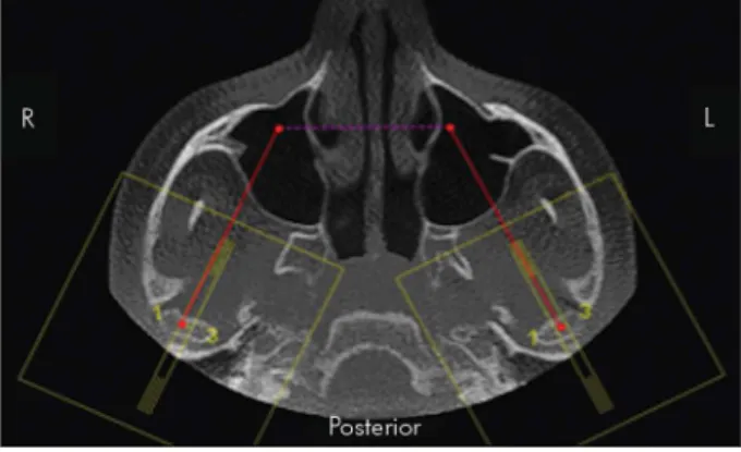
and triangular or angled (C), as shown in Figure 3. Condylar concentricity was measured on the most centered sagittal slice, based on the formula proposed by Pullinger and Hollender,20 as shown below.
P - A
x 100 (%) P + A
The narrowest anterior (A) and posterior (P) articular spaces were calculated as shown in Figure 4. The concentric condyle in the articular fossa was indicated by a zero result, whereas a negative value indicated a posterior location and a positive value indicated an anterior location.
Clinical examinations were performed by a previously calibrated examiner. The presence of joint pain was detected during TMJ palpation performed bilaterally in the TMJ lateral and posterior aspects.
The masticatory muscle examination comprised the palpation of the masseter and temporalis muscle.
Statistical Analysis
Statistical analysis was performed using GraphPad Prism 5.0 (GraphPad Software, San Diego, USA), BioStat 5.0 (Instituto Mamirauá, Tefé, Brazil) and G Power 3.0 (UCLA Institute for Digital Research and Education,
Los Angeles, USA). A conidence interval of 95% and a signiicance level of 5% (p < 0.05) were adopted
for all the tests. After performing the Shapiro-Wilk normality test, the quantitative data were described by the mean and standard deviation of the parameters and by the absolute (n) and relative frequency (%) of the qualitative data.
In order to avoid interexaminer error, a single investigator performed the measurements and the kappa test was used to evaluate investigator calibration in determining condyle morphology and concentricity.
Figure 1. Sagittal view of the TMJ to perform the axial cuts.
Figure 2. Axial view of the central condyle point to reproduce the sagittal and coronal TMJ slices.
Figure 3. Coronal cuts for classification of condylar morphology: (A) Round, (B) convex (C) Angulated (Kinzinger et al.10).
The Chi-square test was applied for comparison of morphology and condylar concentricity between the groups, and the paired t-test, for comparison between the anterior and posterior joint spaces between right and left side for the two groups.
Results
A pilot study for examiner calibration was conducted in order to evaluate the condylar morphology and
concentricity. The kappa coeficient was 0.68 for condyle
concentricity and 0.84 for condylar morphology.
There was no statistically signiicant intergroup
difference in age (t = -0.11; p = 0.90). The age range of G1 was 11–35.6 years (mean, 16.25 ± 5.6 years) and that of G2 was 10.9-31.5 years (mean, 16.4 ± 6.0 years). Both groups showed a similar gender distribution (Chi-square = 0.64; p = 0.61). G1 comprised 8 male (30.77%) and 18 female (69.23%) patients, and G2, 10 male (41.67%) and 14 female (58.33%) patients. Thus, it was assumed that the groups were matched for gender and age.
C on s ide r i ng t h e condyl a r mor pholog y, no association was found regarding the type of malocclusion (Chi-square test with Yates correction = 3.34; p = 0.18) (Table 1).
The measurement of the articular spaces was similar for the right and left sides, and for both the anterior and posterior TMJ in G1. Similar data were observed in G2, except for the posterior articular space,
which showed a statistically signiicant difference in
values (p = 0.007) between the two sides. These data are presented in Table 2. Moreover, no difference was observed regarding condylar concentricity between the two groups (Chi-square test with Yates correction = 4.84; p = 0.08) (Table 3).
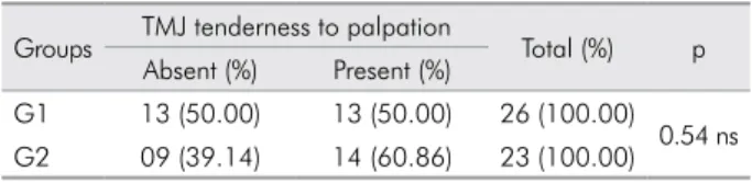
In relation to the clinical examination, no difference was detected between TMJ tenderness and malocclusion (Chi-square test 0.79; p = 0.54, Table 4). Muscle tenderness to palpation was not associated with malocclusion type (Chi-square test = 2.26; p = 0.22, Table 5).
discussion
The understanding of articular characteristics related to malocclusions may have clinical implications that are important for diagnosis and orthodontic treatment plans,19,21 which may change the condyle fossa relationship.
According to the results of this study, the condylar shape found was: G1, convex (57.7%), round (34.61%), and angulated (7.69%), and G2, convex (75%), round (20.83%), and angulated (4.17%),
with no signiicant intergroup differences (Table 1).
These numbers are in accordance with those of Kinzinger et al.,10 in regard to condyle morphology being convex (55%), round (25%), and angulated (20%) in Class II patients, based on coronal magnetic resonance (MR) images. Katzavrias et al.21 found predominantly oval (60.4%) and rounded (29.2%) condyles in sagittal slices. Likewise, Karlo et al.22 found rounded condylar forms in children, also observed in sagittal slices. Solberg et al.23 found
predominantly rounded (66%), followed by lattened
Table 1. Comparison of condylar morphology between groups (Chi-square test with Yates correction,p > 0.05).
Condylar morphology
Total (%) p Convex (%) Angulated (%) Round (%)
Group 1 30 (57.70) 04 (7.69) 18 (34.61) 52 (100.00)
0.18 ns
Group 2 36 (75.00) 02 (4.17) 10 (20.83) 48 (100.00)
ns: non-significant difference.
Figure 4. Sagittal cuts for measurement of the smallest joint spaces (anterior and posterior) to evaluate condyle concentricity: (A) Right condyle, (B) Left condyle.
(17%), and angular (17%) forms, in individuals of the same age, but the authors did not consider malocclusion in their study.
The controversy regarding the results cited may be justified by the difficulty in finding the best imaging method and suitable cuts for evaluating the morphology of the mandibular condyle. It is believed that the basic morphology of the condyle is established early in life, but changes during the individual’s lifetime, according to the functional load.24 Perhaps the most important avenue of investigation would be to determine whether different malocclusions might indeed generate functional overload. Analyzing
the inluence of Class I, II, and III malocclusions on
the mandibular fossa, Burley25 emphasized that the occlusal contacts in patients with malocclusions produce no functional stimulation that may alter the contour of the mandibular fossa.
Condylar concentricity was another parameter evaluated in this study. The anterior condyle position was prevalent in both groups. In G1, 73% of the condyles were anterior, 25% were posterior, and only 2% were concentric, whereas in G2, 52% of the condyles were anterior, 45% were posterior, and 2% were concentric.
The anterior and posterior articular spaces were similar for the right and left joints. G1 presented an average of 1.6 ± 0.5 mm for the anterior articular spaces, and 2.0 ± 0.6 mm for the posterior articular spaces, on both sides (Table 2). Rodrigues et al.12 also observed the anterior condyle displacement in a Class I sample, but with minor differences in the values of anterior (average of 1.3 mm) and posterior (average of 1.7 mm) articular spaces.
Similar data were observed for G2, with 2.0 ± 0.5 mm and 2.1 ± 0.8 mm for the anterior spaces, on the left and right sides, respectively. Regarding the posterior
articular spaces, there was little statistically signiicant
difference between the right (2.1 ± 0.8 mm) and left (2.3 ± 0.8 mm) sides (Table 2). Kikuchi et al.26 found similar values for anterior (mean, 1.8 mm) and posterior (mean, 2.29 mm) articular spaces, in a sample of adolescents.
Anterior condylar displacement was also observed in several other studies, both in samples with Class I12 and Class II7,11,12,27 malocclusions, and in patients with normal occlusion,9 as well as in samples where the type of malocclusion was not considered.6
In contrast, some studies10,21 that also evaluated the condylar concentricity in Class II patients showed posteriorly positioned condyles; the former study10 was based on MR images. Although not statistically different, our results for G2 showed 45.83% of the condyles posteriorly positioned, whereas only 25% of the Class I patients presented this condition (Table 3). On the other hand, another study with Class II patients reported concentric condyles.2
As shown in the literature, a posteriorly aligned condyle observed in Class II division 2 subjects, unlike Class I or Class II division 1 patients, may occur due to distinct muscle characteristics. These subjects may present anteriorly positioned mastication muscles,
which result in signiicant differences with respect to
Table 2. Comparison of anterior and posterior joint space, mean (M), standard deviation (SD) and p value (p) between the two groups, in both joints (paired t-test).
Joint space Anterior
(M ±SD) p
Posterior (M ±SD) p Group 1
Right side 1.65 ± 0.45
0.96 ns 2.04 ± 0.69 0.45 ns Left side 1.64 ± 0.56 1.97 ± 0.67
Group 2
Right side 2.00 ± 0.57
0.47 ns 2.00 ± 0.81 0.007 * Left side 2.10 ± 0.86 2.33 ± 0.83
ns: non-significant difference.
* Statistically significant difference (p < 0.05).
Table 3. Comparison of condylar position between the groups (Chi-square test with Yates correction, p > 0.05).
Condylar position
Total (%) p Anterior (%) Posterior (%) Concentric (%)
Group 1 38 (73.07) 13 (25.00) 01 (1.93) 52 (100.00)
0.08 ns
Group 2 25 (52.09) 22 (45.83) 01 (2.08) 48 (100.00)
the mechanical occlusal forces and their magnitude.4 Therefore, it follows that the muscle overload on the TMJ in Class II division 2 patients differs from that of patients with other dentofacial morphologies.28 Bearing in mind that TMJ morphology depends to some extent on its load, Class II division 2 patients
must have speciic morphological characteristics.2 During palpation procedures, 55.10% of the sample presented at least one TMJ tender site: G1- 50% and G2- 60.86% (Table 4). It is important to highlight that
similar indings have been observed regardless of
the type of malocclusion. This value is smaller than the 22.5% found in a similar Class I and II sample.29 Some differences are expected due to the variation in palpation techniques and pressure; this makes comparisons very unreliable.
Regarding muscle tenderness to palpation, 40.81% of the study sample had at least two tender sites (Table 5). Another study found a lower value (26%) in Class I and II malocclusion patients, but
this could be justiied, because only one tender site
was considered.29
Although the clinical evaluation results showed a high number of nonconcentric condyles, this fact may
not inluence TMJ clinical signs. The few number of
concentric condyles with reduced anterior articular spaces found in our study seems to be a common
inding in different types of malocclusion patients.7,12,21
These indings are relevant because patients with
anteriorly displaced condyles do not require a different orthodontic approach. This clinical implication is important, since it has been reported that a more posterior relative position of the condyle in the mandibular fossa could be one of the reasons for anterior disc displacement, which frequently results in TMJ sounds.30
Conclusions
The convex condyle shape was the most prevalent in this study and Class I and II patients seem to present similar condyle morphology.
Regarding the condyle position, anterior displacement was more prevalent regardless of the type of malocclusion.
Table 5. Association of muscle tenderness to palpation between the two groups (Chi-square test, p > 0.05).
Groups Muscle tenderness to palpation Total (%) p Absent (%) Present (%)
G1 13 (50.00) 13 (50.00) 26 (100.00) 0.22 ns G2 16 (69.57) 07 (30.43) 23 (100.00) ns: non-significant difference.
Table 4. Association of TMJ tenderness to palpation between the two groups (Chi-square test, p > 0.05).
Groups TMJ tenderness to palpation Total (%) p Absent (%) Present (%)
G1 13 (50.00) 13 (50.00) 26 (100.00) 0.54 ns G2 09 (39.14) 14 (60.86) 23 (100.00) ns: non-significant difference.
1. Rodrigues AF, Fraga MR, Vitral RWF. Computed tomography evaluation of the temporomandibular joint in Class II Division 1 and Class III malocclusion patients: condylar symmetry and condyle-fossa relationship. Am J Orthod Dentofacial Orthop. 2009;136(2):199-206. doi:10.1016/j.ajodo.2007.07.033
2. Katsavrias EG. Morphology of the temporomandibular joint in subjects with Class II Division 2 malocclusions. Am J Orthod Dentofacial Orthop. 2006;129(4):470-8. doi:10.1016/j.ajodo.2005.01.018
3. Barrera-Mora JM, Espinar EE, Abalos LC, Llamas CJM, Ballesteros EJ, Solano RE, Rocabado M,et al. The relationship between malocclusion, benign joint hypermobility
syndrome, condylar position and TMD symptoms. J Cranio. 2012;30(2):121-30. doi:10.1179/crn.2012.018
4. Kurusu A, Horiuchi M, Soma K. Relationship between occlusal force and mandibular condyle morphology. Evaluated by limited cone-beam computed tomography. Angle Orthod. 2009;79(6):1063-9. doi:10.2319/120908-620R.1 5. Pullinger AG, Hollender L, Solberg WK, Petersson A. A
tomografic study of mandibular condyle position in an asymptomatic population. J Prosthet Dent. 1985;53(5):706-13. 6. Ikeda K, Kawamura A. Assessment of optimal condylar
position with limited cone-beam computed tomography. Am J Orthod Dentofacial Orthop. 2009;135(4):495-501. doi:10.1016/j.ajodo.2007.05.021
7. Pullinger AG, Solberg WK, Hollender L, Petersson A. Relationship of mandibular condylar positon to dental occlusion factors in an asymptomatic population. Am J Orthod Dentofacial Orthop. 1987;91(3):200-206. doi:10.1016/0889-5406(87)90447-1
8. Bonilla-Aragon H, Tallents RT, Katzberg RW, Yrkanides S, Moss ME. Condyle position as a predictor of temporomandibular joint internal derangement. J Prosthet Dent. 1999;82(2):205-8. doi:10.1016/S0022-3913(99)70157-5
9. Vitral RW, Campos MJS, Rodrigues AF, Fraga MR. Temporomandibular joint and normal occlusion: Is there anything singular about it? A computed tomographic evaluation. Am J Orthod Dentofacial Orthop. 2011;140(1):18-24. doi:10.1016/j.ajodo.2009.07.030
10. Kinzinger G, Kober C, Diedrich P. Topography and morphology of the mandibular condyle during fixed functional orthopedic treatment - a magnetic resonance imaging study. J Orofac Orthop. 2007;68(2):124-47. doi:10.1007/s00056-007-0650-0
11. Krisjane Z, Urtane I, Krumina G, Zepa K. Three-dimensional evaluation of TMJ parameters in Class II and Class III patients. Stomatologija. 2009;11(1):3236.
12. Rodrigues AF, Fraga MR, Vitral RWF. Computed tomography evaluation of the temporomandibular joint in Class I malocclusion patients: condylar symmetry and condyle-fossa relationship. Am J Orthod Dentofacial Orthop. 2009;136(2):192-8. doi:10.1016/j.ajodo.2007.07.032.
13. Vitral RW, Telles CS. Computed tomography evaluation of temporomandibular joint alterations in Class II Division 1 subdivision patients: condylar symmetry. Am J Orthod Dentofacial Orthop. 2002;121(4):369-75. doi:10.1067/mod.2002.121664
14. Pullinger AG, Solberg WK, Hollender L, Guichet D. Tomographic analysis of mandibular condyle position in diagnostic subgroups of temporomandibular disorders. J Prosthet Dent. 1986;55(6):723-9.
15. Okeson JP. Critical commentary 1: Evaluation of the research diagnostic criteria for temporomandibular disorders for the recognition of an anterior disc displacement with reduction. J Orofac Pain. 2009;23(4):312-5; author reply 323-4.
16. Williamson EH, Evans DL, Barton WA, Williams BH. The effect of bite plane use on terminal hinge axis location. Angle Orthod. 1977;47(1):25-33.
17. Pullinger AG, Seligman DA, John MT, Harkins S. Mu lt i fac tor ia l model i ng of temporom a nd ibu la r anatomic and orthopedic relationships in normal versus undifferentiated disk displacement joints. J Prosthet Dent. 2002 Mar;87(3):289-97. doi:10.1067/mpr.2002.121741.
18. Mah JK, Huang JC, Choo H. Practical applications of cone-beam computed tomography in orthodontics. J Am Dent Assoc. 2010;141 Suppl 3:7S-13S.
19. Hodges RJ, Atchison KA, White SC. Impact of cone-beam computed tomography on orthodontic diagnosis and treatment planning. Am J Orthod Dentofacial Orthop. 2013;143(5):665-74. doi:10.1016/j.ajodo.2012.12.011
20. Pullinger A, Hollender L. Variation condyle-fossa relationships according to different methods of evaluation in tomograms. Oral Surg Oral Med Oral Pathol. 1986;62(6):719-27.
21. Katsavrias EG, Halazonetis DJ. Condyle and fossa shape in Class II and Class III skeletal patterns: a morphometric tomographic study. Am J Orthod Dentofacial Orthop. 2005;128(3):337-46. doi:10.1016/j.ajodo.2004.05.024
22. Karlo CA, Stolzmann P, Habernig S, Muller L, Saurenmann T, Kellenberger CJ. Size, shape and age-related changes of the mandibular condyle during childhood. Eur Radiol. 2010;20(10):2512-7. doi:10.1007/s00330-010-1828-1
23. S olb erg W K, Ha n sson TL, Nord st rom B. T he temporomandibular joint in young adults at autopsy: a morphologic classification and evaluation. J Oral Reabil. 1985;12(4):303-321.
24. Cimasoni G. Histopathology of the temporomandibular joint following bilateral extractions of molars in the rat. A preliminary report. Oral Surg Oral Med Oral Pathol. 1963;16:613-21. 25. Burley M. An examination of the relation between the
radiographic appearance of the temporomandibular joint and some features of the occlusion. Br Dent J. 1961;110:195-200. 26. Kikuchi K, Takeuchi S, Tanaka E, Shibaguchi T, Tanne K.
Association between condylar position, joint morphology and craniofacial morphology in orthodontic patients without temporomandibular joint disorders. J Oral Rehabil. 2003;30(11):1070-5. doi:10.1046/j.1365-2842.2003.01194.x 27. Uzel A, Özyürek Y, Öztunç H, Condyle position in Class
II Division 1 malocclusion patients: correlation between MPI records and CBCT images. J World Fed Orthod. 2013;2(2):65-70. doi:10.1016/j.ejwf.2013.03.002
28. O´Ryan F, Epker BN. Temporomandibular joint function and morphology observations on the spectra of normalcy. Oral Surg Oral Med Oral Pathol. 1984;58(3):212-219.
29. Conti A, Freitas M, Conti P, Henriques J, Janson G. Relationship between signs and symptoms of temporomandibular disorders and orthodontic treatment: a cross-sectional study. Angle Orthod. 2003;73(4):411-7.