COMPARISON BETWEEN PHARYNGEAL FLAP
SURGERY AND SPHINCTEROPLASTY:
NASOMETRIC AND AERODYNAMIC ANALYSIS
Comparação entre cirurgia do retalho faríngeo e esfincteroplastia:
análise nasométrica e aerodinâmica
Renata Paciello Yamashita(1), Carla Aparecida Curiel(2), Ana Paula Fukushiro(1), Maria Natália Leite de Medeiros(3), Inge Elly Kiemle Trindade(1)
(1) Laboratório de Fisiologia do Hospital de Reabilitação de
Anomalias Craniofaciais da Universidade de São Paulo – HRAC-USP, Bauru, São Paulo, Brasil.
(2) Faculdade de Odontologia de Bauru da Universidade de
São Paulo - FOB-USP, Bauru, São Paulo, Brasil.
(3) Programa de Pós-Graduação em Ciências da Reabilitação
do Hospital de Reabilitação de Anomalias Craniofaciais da Universidade de São Paulo (HRAC-USP), Bauru, São Paulo, Brasil.
Conlict of interest: non-existent
the current air is deviated to the nasal cavity leading to the occurence of symptoms that may impair speech in different ways1-5. VPI’s most
represen-tative symptom is hypernasality, which may persist even after primary correction of the palate. In such cases, secondary surgery is required6-10. Pharyngeal
lap and sphincteroplasty are among the surgical
techniques used for VPI correction. Both proce-dures aim to reduce the space between oro and
nasopharynx, thus reducing symptoms resulting from insuficient velopharyngeal closure5,6,11.
The pharyngeal lap technique consists of the construction of a myomucosal lap uniting the posterior wall of the pharynx to the soft palate,
INTRODUCTION
Velopharyngeal insuficiency (VPI) is deined as
a fault in the velopharyngeal closure, where part of ABSTRACT
Purpose: to compare the effect of pharyngeal lap surgery and sphincteroplasty on hypernasality and
velopharyngeal closure in the velopharyngeal insuficiency management, by means of instrumental
assessment. Methods: thirty patients with repaired cleft palate±lip, submitted to surgical treatment for
velopharyngeal insuficiency (15 pharyngeal lap and 15 sphincteroplasty) were evaluated before and,
at least, 1 year after surgery. Hypernasality was estimated by means of nasalance scores (acoustic correlate of nasality) obtained by nasometry considering a cutoff score of 27%. Velopharyngeal closure was determined by the velopharyngeal area measurement. Nasalance scores were obtained by
nasometry, during the reading of a set of 5 sentences containing exclusively oral sounds, considering
the cutoff value of 27%. Velopharyngeal area was provided by the measurement of velopharyngeal
area by means of pressure-low technique and was classiied as: 0 to 4.9 mm2=adequate; 5 to 19.9
mm2=borderline and ≥20mm2 inadequate. Differences between the two techniques were accepted
as signiicant when p < 0.05. Results: before surgery nasalance mean scores were 43±8.4% and
45±14.2% and velopharyngeal area mean were 51±35.4mm2 and 69±29.2mm2 for the pharyngeal lap
and sphincteroplasty groups, respectively. After surgery, nasalance mean scores were 27±10.1% and
31±14.2% and velopharyngeal area mean were 3.6±5.5mm2 and 24±32.7mm2 for the pharyngeal lap
and sphincteroplasty groups, respectively. The reduction of the nasalance scores and velopharyngeal
area was statistically signiicant in both groups. Conclusion: these results suggest that pharyngeal
lap was shown to be more eficient than sphincteroplasty in the elimination of hypernasality and
adequacy of velopharyngeal closure in the patients studied.
techniques employed for VPI treatment, using different methodologies for the analysis of surgical results. Some employed direct instrumental evaluations, through nasoendoscopy and
video-luoroscopy12,19-22 and others, indirect instrumental
evaluations such as nasometry and pressure-low
technique12,17,21,23-26. Previous studies conducted at
the Laboratory of Physiology17,26 investigated the
effect of the pharyngeal lap surgery on the speech
and breathing of patients with residual VPI, since this is a routine surgery performed at HRAC-USP.
An investigation of the effect of the pharyngeal lap on upper airways revealed that the lap led to the
appearance of permanent respiratory symptoms,
such as, oral breathing, snoring and dificulty in
breathing during sleep in 36% of the patients as a result of the reduction in nasopharyngeal dimen-sions after surgery, evaluated by the
pressure-low technique27. Another study analyzed speech
outcomes obtained before and after pharyngeal lap surgery in 241 individuals, using nasometric and aerodynamic evaluations. The authors veriied that the pharyngeal lap was effective in reducing hyper
-nasality in 68% of the cases in accordance with
nasometry and in improving velopharyngeal closure in 66% of the patients, according to aerodynamic
evaluation (pressure-low technique)17. Recently,
the effect of the pharyngeal lap was compared to
another technique for VPI correction, secondary palatoplasty with intravelar veloplasty. The authors
veriied that, in patients submitted to pharyngeal lap surgery, hypernasality was absent in 70% and velopharyngeal closure was adequate in 80%. In
those submitted to secondary palatoplasty with intravelar veloplasty, hypernasality was absent in
34% and velopharyngeal closure was adequate in 50%. Therefore, the pharyngeal lap was more eficient than intravelar veloplasty for correcting
hypernasality and for adequate velopharyngeal closure26.
Sphincteroplasty surgical results have also been frequently compared to other surgical techniques, in isolation or combined, aimed at establishing the most effective technique for correcting velopharyngeal
insuficiency12,21,28. A large group of researchers compared the speech results obtained before and
after sphincteroplasty in 45 individuals, and the pharyngeal lap in 52 individuals, using perceptual,
nasometric and nasoendoscopy evaluations. The
authors veriied that both surgical techniques were
equally effective in reducing nasalance scores and eliminating hypernasality, which occurred in 76% of the cases submitted to sphincteroplasty
and 81% of the pharyngeal lap cases21. Likewise,
other researchers did not verify signiicant differ -ences between one group of 26 patients submitted constituting a bridge between both, delimiting two
lateral oriices. Flap height and width should be
determined in accordance with the size of velopha-ryngeal gap and the degree of movement of the
pharynx’s lateral walls. These should be evaluated
prior to surgery, enabling the construction of the
lap in accordance with the needs of each case2.
Sphincteroplasty was proposed as a physiological solution for correcting VPI. In this technique, the
myomucosal laps are removed from the posterior pillars and from the lateral walls of pharynx, on
each side. They are then sutured to each other
and inserted in the posterior wall of the pharynx. This creates a single central oriice surrounded
by mucosa and muscle at the level of the velum palatinum. The technique aims to create a “dynamic sphincter” that controls air passage from the oral portion to the nasal portion during speech2,6,12.
The determination of surgical results for VPI correction in general is done by auditory-perceptual assessment of speech associated with instrumental evaluation. For such, use of at least one of the following instrumental methods is recommended:
nasoendoscopy, videoluoroscopy, nasometry or pressure-low technique13. The latter two, which
were used in this study, are considered indirect methods, the results of which lead to verifying the functional status of the velopharyngeal mechanism.
Nasometry and pressure-low technique, by
providing quantitative data, contribute greatly to following up on surgical treatment using pre- and post-surgical comparisons14.
Nasometry is a non-invasive technique that permits an indirect check of speech resonance, that is, hypernasality or hyponasality, by measuring
nasalance, a physics measures that relects the
quantity of acoustic nasal energy during speech
expressed in percentage14. Nasalance is deter-mined, primarily, by velopharyngeal sphincter activity, which is why nasalance deviations are indicative of VPI3,15,16. The pressure-low technique
evaluates velopharyngeal mechanism in its functional aspect, providing objective data about the aerodynamic repercussions of any failure in velopharyngeal function14. It provides quanti-tative data about the velopharyngeal function in a non-invasive manner, making it possible to check
the extension of velopharyngeal closure during
production of the plosive phone [p]17. The literature
demonstrated that areas smaller than 5mm2 are
suggestive of adequate velopharyngeal closure, 5 to 9mm2, of adequate-borderline closure, 10 to
19mm2, of borderline-inadequate closure and, equal
to or greater than 20mm2, of inadequate closure18.
captures the signals from the nasal component of speech. The bottom one captures the signals from
the oral component, which are iltered, digitized and analyzed using speciic software. The exam
is conducted while reading a set of 5 sentences in
Brazilian Portuguese, containing exclusively oral
sounds, to identify hypernasality29. Patients who are
unable to read the text are asked to repeat each sentence to the examiner. As the individual reads the text shown on the computer screen connected
to the system, the signals captured by the micro-phone appear as points on the screen, forming the
coniguration of a curve. Nasalance is calculated
using the numeric ration between nasal acoustic energy and total acoustic energy (sum of nasal and oral acoustic energy), multiplied by 100. A cutoff of 27% is considered the upper limit of normality. That is, values greater than 27% are considered indic-ative of hypernasality14. Figure 1 shows the system
coniguration in a schematic format.
Velopharyngeal area measurement - Pressure-low technique
Determination of the velopharyngeal area during
speech was conducted using the pressure-low technique (modiied anterior rhinomanometry),
using a PERCI-SARS (computer system - version 3.50)30. The principle of the technique is based on
the minimal cross sectional area of a constriction
(or oriice) may be estimated by the simultaneous
measurement of differential pressure between the
two sides of the constriction and the airlow that
crosses through it31.
The velopharyngeal area is determined during the production of the voiceless plosive phone [p],
inserted in the word “rampa”, produced 4 to 6 times
in succession, positioning a catheter inside the oral cavity and another in one of the nostrils. The nasal catheter is kept in position by a nasal obturator that blocks the nostril. Both catheters measure static air pressures transmitted to pressure transducers.
Nasal air low is measured by a plastic tube adapted
to the other nostril, connected to a pneumotacho-graph previously heated and connected to a pressure transducer. The signals from the three transducers
(nasal pressure, oral pressure and nasal low) are sent to the PERCI system for analysis by a speciic
program. The area considered for this analysis represents the average for multiple productions. Based on the equation, the program itself calculates it: A= V/k(2DP/d)1/2, where A=minimum nasal
cross-sectional area of the oriice in cm2; V=nasal low in
cm3/s; k=0.65; DP=oral-nasal pressure in dynes/
cm2; d=density of air (0.001g/cm3). Figure 2 shows
the system coniguration in a schematic format.
to sphincteroplasty and one group of 22 patients
submitted to pharyngeal lap surgery, evaluated using nasometry, nasoendoscopy and videoluo -roscopy. The authors showed a VPI rate of 11.5% after sphincteroplasty and 9% after pharyngeal
lap12. Through perceptual assessment, a study
recently compared the speech results of 20 patients
submitted to isolated sphincteroplasty, 38 submitted to pharyngeal lap and 38 submitted to sphinc -teroplasty combined with the Furlow technique. The
authors veriied a signiicant reduction in hyper -nasality after surgery in the three groups studied. However, they demonstrated that the resonance
results were signiicantly better for the groups with pharyngeal lap and sphincteroplasty combined
with the Furlow technique, compared to the isolated sphincteroplasty group28.
In the current study, the aim was to compare the
effect of the pharyngeal lap and of sphincteroplasty
on speech nasality and on velopharyngeal closure using instrumental evaluations for such.
METHODS
This retrospective study was developed in the Laboratory of Physiology of the Hospital for Rehabilitation of Craniofacial Anomalies of the University of São Paulo, Bauru-SP with the approval of the local ethics committee for human research, number 153/2011.
Casuistics
Thirty patients with residual VPI were evaluated. They were submitted to surgical correction of the VPI at least 12 months ago; 15 submitted to pharyngeal
lap (PF group) and 15 submitted to sphinctero -plasty (SP group). The age of the patients ranged
between 6 and 38 (average ages of 19±16 for the PF group and 18±11 for the SP group); 12 patients
with isolated cleft palate, 13 with unilateral cleft lip
and palate, 4 with bilateral cleft lip and palate and 1
with noncleft VPI.
Procedures
The patients were evaluated before surgery
(PRE) and, at least, 12 months after pharyngeal lap
and sphincteroplasty surgery (POST).
Source: Trindade et al. Diagnóstico instrumental da disfunção velofaríngea. In: Trindade e Silva Filho. Fissuras labiopalatinas: uma abordagem interdisciplinar. São Paulo: Editora Santos; 2007.p.134.
Figure 1 - Representative schematic of instrumentation for measuring nasalance (Nasometer 6200-3 IBM, Kay Elemetrics Corp., Lincoln Park, NJ, USA).
Nasometer
Print PC
Source: Trindade et al. Diagnóstico instrumental da disfunção velofaríngea. In: Trindade e Silva Filho. Fissuras labiopalatinas: uma abordagem interdisciplinar. São Paulo: Editora Santos; 2007.p.137.
Figure 2 - Representative schematic for determining velopharyngeal area (PERCI-SARS System, Microtronics Corp., Chapel Hill, NC, USA).
Pressure transducer
Pressure transducer
Pressure transducer
Ampliier
Ampliier
Ampliier
Pneumota-chograph
RESULTS
Mean nasalance scores obtained in the PRE
condition were 43±8.4% in patients from the PF group and 45±12.4% in patients from the SP
group. In both cases, the values were indicative of hypernasality. In the POST condition, average nasalance decreased to 27±10.1% in the PF group
and 31±14.2% in the SP group. Statistical analysis
revealed that after surgery both groups presented
mean nasalance scores signiicantly lower than
those obtained before surgery. There was no statisti-cally difference between mean nasalance in the two groups in the PRE as well as the POST condition (Table 1). Results showed that after surgery, the PF group began to present an mean nasalance score
indicative of normality (≤27%), whereas the mean
score for the SP group remained indicative of hyper-nasality. Figure 3 illustrates these results.
The values found for the velopharyngeal area are analyzed in accordance with the proposed
velopharyngeal function classiication criteria: 0 to 4.9mm2=adequate velopharyngeal closure; 5
to 9.9mm2=adequate-borderline velopharyngeal
closure; 10.0 to 19.9mm2
=borderline-inade-quate velopharyngeal closure and, 20mm2 or
more=inadequate velopharyngeal closure19.
Data Analysis
Nasalance is expressed in % and velopha -ryngeal area in mm2. The comparison of average
values of nasalance and of the pre- and post-surgical velopharyngeal area for the same post-surgical technique was done using Student’s t test for paired samples and the comparison of these two variables between the surgical techniques was done using
Student’s t test for independent samples. P<0.05 values were accepted as signiicant.
Figure 3: Mean nasalance scores obtained after surgery in patients submitted to pharyngeal lap and
to sphincteroplasty.
Table 1 - Mean scores (±SD) of nasalance obtained before (PRE) and after (POST) surgery in the
group of patients submitted to pharyngeal lap and to sphincteroplasty
NASALANCE (%)
Mean (±SD) PF Group
(n=15)
SP Group (n=15)
PRE 43(±8.4) 45(±12.4)
POST 27(±10.1)# 31(±14.2)* PF-Pharyngeal Flap; SP=Sphincteroplasty; SD=Standard Deviation
#Pre vs post (PF group): statistically signiicant difference-paired T test (p=0.000)
*Pre vs post (SP group): statistically signiicant difference-paired T test (p=0.010)
PF Group vs SP Group (PRE): non-signiicant difference-T test (p=0.550) PF Group vs SP Group (POST): non-signiicant difference-T test (p=0.359)
Me
a
n
N
a
s
a
la
n
c
e
(%
surgery, the mean velopharyngeal area reduced to 3.6±5.5mm2 in the PF group and 24±32.7 mm2 in the
SP group. The statistical analysis showed that the mean velopharyngeal area obtained after surgery
was signiicantly smaller than that obtained before
surgery in the two groups studied and that the post-surgical velopharyngeal area for the PF group was
signiicantly smaller than for the SP group. After
surgery, the PF group also began to present values indicative of adequate closure, on average, whereas the SP group continued with inadequate
velopha-ryngeal closure. Figure 4 illustrates these results.
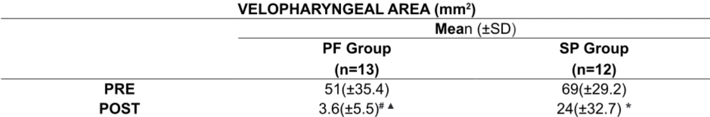
In Table 2, the mean values are shown (±SD) for the velopharyngeal area, obtained in patients from the PF and SP groups, before (PRE) and after (POST) surgery. The patients who presented compensatory articulation in phone [p] were not included in this analysis (2 individuals from the PF group and 3 from the SP group). Before surgery, it was determined that the mean velopharyngeal area
in the PF group was 51±35.4mm2 and in the SP
group, 69±29.2mm2. Both cases are indicative of
inadequate velopharyngeal closure. There was no statistically difference between the velopharyngeal areas of the two groups in the PRE condition. After
Table 2 - Mean values (±SD) of velopharyngeal area obtained before (PRE) and after (POST) surgery
in the group of patients submitted to pharyngeal lap and to sphincteroplasty
VELOPHARYNGEAL AREA (mm2)
Mean (±SD)
PF Group (n=13)
SP Group (n=12)
PRE 51(±35.4) 69(±29.2)
POST 3.6(±5.5)# ▲ 24(±32.7) * PF-Pharyngeal Flap; SP=Sphincteroplasty; SD=Standard Deviation
#Pre vs post (PF group): statistically signiicant difference-paired T test (p=0.002)
*Pre vs post (SP group): statistically signiicant difference-paired T test (p=0.010)
PF Group vs SP Group (PRE): non-signiicant difference T test (p=0.228)
▲PF Group vs SP Group (POST): statistically signiicant difference T test (p=0.034).
Figure 4: Percentage (number) of patients distributed according to the classiication of velopharyngeal closure determined after surgery in the pharyngeal lap and sphincteroplasty groups.
Pe
rc
e
n
ta
g
e
(n
u
m
b
e
r)
o
f
p
a
ti
e
n
in literature used this instrumental method recom-mended by the American Cleft-Palate Association13
for comparing these techniques, making the results of the present study unprecedented. This method has been used for many years by the Laboratory of Physiology team for evaluating surgical results for VPI treatment, particularly of the pharyngeal
lap and secondary palatoplasty with intravelar
veloplasty17,24-26,35,36. In the present study, this
technique proved to be an eficient instrument
for evaluating sphincteroplasty results, making
it possible to compare both. The pressure-low technique revealed a signiicant reduction in average
velopharyngeal area in both groups studied, as
expected, since the objective of the two surgeries is
to reduce the space of the velopharyngeal region to promote adequate velopharyngeal closure. However, different from what occurred with nasometry, data analysis revealed that the average velopharyngeal
area obtained in patients with pharyngeal lap was signiicantly smaller than that obtained in patients
with sphincteroplasties. Analyzing the degree of velopharyngeal closure, determined from values for the post-surgical velopharyngeal area, it was
veriied that the group of patients with pharyngeal lap achieved, on average, adequate velopharyngeal
closure, whereas the group of patients submitted to sphincteroplasty remained with inadequate velopha-ryngeal closure. In other words, the velophavelopha-ryngeal
area veriied after pharyngeal lap surgery, which was signiicantly smaller than that veriied after
sphincteroplasty, seems to have contributed to
provide adequate velopharyngeal closure identiied in patients with pharyngeal lap.
CONCLUSION
These results suggest pharyngeal lap surgery was more eficient than sphincteroplasty in elimi -nating hypernasality and providing adequate velopharyngeal closure in the patients studied.
DISCUSSION
Among those surgical techniques employed
in VPI treatment, pharyngeal lap and sphinctero -plasty are still the most used in different craniofacial centers in the world12,28,32-34 and the literature has demonstrated the success and the deleterious effects of both these surgical techniques used in VPI treatment secondary to primary palatoplasty. Most studies used the perceptual methodology and/or direct instrumental methods for evaluation velopharyngeal function, such as nasoendoscopy
and videoluoroscopy12,19-22. Two studies12,21, in
particular, added nasometry to the other evaluations, as one of the methods for investigating nasality in speech and objectively comparing the results of the two surgeries, as was done in this study. Using
nasometry, both revealed that the pharyngeal lap as well as the sphincteroplasty were eficient in
reducing nasality, with no statistically difference between the two surgical techniques. This result
was also veriied in this study based on nasometry
analysis. However, these results revealed that only in the group of patients submitted to pharyngeal
lap surgery was there a normalization of nasalance
(average score=27%). The sphincteroplasty group continued with a score of nasalance indicative of hypernasality (31%), suggesting the pharyngeal
lap was more eficient than the sphincteroplasty for
eliminating hypernasality.
On the other hand, a recent study showed that the sphincteroplasty combined with the Furlow
technique, as well as the pharyngeal lap, were signiicantly more eficient in reducing hypernasality
than the sphincteroplasty performed in isolation28. However, it is worth emphasizing that a limitation of this study was the fact that the authors employed only perceptual assessment of speech for analyzing results, without, however, evaluating concordance
between examiners.
Another evaluation method for checking results of the two surgeries on patient speech in this study
7. Andrades P, Espinosa-de-los-Monteros A, Shell DH, Thurston TE, Fowler JS, Xavier ST et al. The importance of radical intravelar veloplasty
during two-lap palatoplasty. Plast Reconstr Surg. 2008;122(4):1121-30.
8. Billmiew DA. Surgical management of clefts and
velopharyngeal dysfunction. In: Kummer AW, editor. Cleft palate and craniofacial anomalies. 2nd ed. San
Diego: Singular; 2008. pP508-40.
9. Khosla RK, Mabry K, Castiglione CL. Clinical outcomes of the Furlow z-plasty for primary cleft
palate. Cleft Palate Craniofac J. 2008;45(5):501-10.
10. Sullivan SR, Marrinan EM, Mulliken JB.
Pharyngeal lap outcomes in nonsyndromic
children with repaired cleft palate and
velopharyngeal insuficiency. Plast Reconstr Surg. 2010;125(1):290-8.
11. Ysunza A, Pamplona MC. Velopharyngeal function after two different types of palatoplasty. Internat J Pediatric Otorhinol. 2006;70(6):1031-7. 12. Abdel-Aziz M, El-Hoshy H, Ghandour H.
Treatment of velopharyngeal insuficiency after
cleft palate repair depending on the velopharyngeal closure pattern. The Journal of Craniofacial Surgery.
2011;22(3):813-7.
13. American Cleft Palate-Craniofacial Association. Parameters for evaluation and treatment of pacients
REFERENCES
1. Smith BE, Kuehn DP. Speech Evaluation of Velopharyngeal Dysfunction. J Craniofac Surg.
2007;18(2):251-61.
2. Rudnick EF, Sie KC. Velopharyngeal
Insuficiency: current concepts in diagnosis and
management. Curr Opin Otolaryngol Head Neck
Surg. 2008;16(6):366-70.
3. Kummer AW editor. Cleft palate and craniofacial anomalies. 2nd ed. San Diego: Singular Thomson
Learning; 2008.
4. Genaro KF, Fukushiro AP, Suguimoto MLFCP.
Avaliação e Tratamento dos Distúrbios da Fala. In: Trindade IEK, Silva Filho OG (Org.). Fissuras Labiopalatinas: uma abordagem interdisciplinar. São Paulo: Santos; 2007. P.109-22.
5. Rieger J, Bohlie G, Huryn J, Tang JL, Harris J, Seikaly H. Surgical reconstruction versus prosthetic
obturation of extensive soft palate defects: a
comparison of speech outcomes. Int J Prosthodont. 2009;22(6):566-72.
6. Rocha DL. Tratamento cirúrgico da insuiciência
velofaríngea. In: Trindade IEK, Silva Filho OG, organizadores. Fissuras labiopalatinas: uma abordagem interdisciplinar. São Paulo: Santos;
2007. P.145-63.
RESUMO
Objetivo: comparar o efeito do retalho faríngeo e da esincteroplastia sobre a hipernasalidade da fala e o fechamento velofaríngeo no tratamento de indivíduos com insuiciência velofaríngea resi -dual, por meio de avaliação instrumental. Métodos: foram avaliados 30 pacientes, com issura de
palato±lábio reparada, submetidos à correção cirúrgica da insuiciência velofaríngea (15 com retalho faríngeo e 15 com esincteroplastia), avaliados antes e, no mínimo, 1 ano após a cirurgia. A hiper -nasalidade foi estimada a partir dos escores de nasalância (correlato físico da -nasalidade) obtidos
por meio da nasometria, durante a leitura de 5 sentenças contendo, exclusivamente, sons orais,
considerando como limite de normalidade o escore de 27%. O fechamento velofaríngeo foi aferido
a partir da medida da área velofaríngea obtida por meio da técnica luxo-pressão e foi classiicado em: 0-4,9mm2=adequado; 5-19,9mm2=marginal e, >20mm2=inadequado. Diferenças entre as duas
técnicas foram consideradas estatisticamente signiicantes ao nível de 5%. Resultados: antes da
cirurgia, os valores médios de nasalância foram de 43±8,4% e 45±14,2% e de área velofaríngea foram 51±35,4mm2, e 69±29,2mm2, para os grupos retalho faríngeo e esincteroplastia, respectiva
-mente. Após a cirurgia, os valores médios de nasalância reduziram para 27±10,1% e 31±14,2% e de
área velofaríngea para 3,6±5,5mm2 e 24±32,7mm2 para os grupos retalho faríngeo e esincteroplas
-tia, respectivamente. A redução dos valores de nasalância e área velofaríngea foi estatisticamente
signiicante nos dois grupos. Conclusão: estes resultados sugerem que o retalho faríngeo foi mais
eiciente do que a esincteroplastia na eliminação da hipernasalidade e adequação do fechamento
velofaríngeo nos pacientes estudados.
success of pharyngeal lap pharyngoplasty? Plast Reconstr Surg. 2005;115(1):45-52.
24. Yamashita R P, Oliva TRT, Fukushiro AP,
Brustello CMB, Trindade IEK. Efeito da veloplastia intravelar sobre o fechamento velofaríngeo avaliado
por meio da técnica luxo-pressão. Rev Soc Bras Fonoaudiol. 2010;15(3):362-8.
25. Yamashita RP, Carvalho ELL, Fukushiro AP, Zorzetto NL, Trindade IEK. Efeito da veloplastia intravelar sobre a nasalidade em indivíduos
com insuiciência velofaríngea. Rev. CEFAC. 2012;14(4):603-9.
26. Barbosa DA, Scarmagnani RH, fukushiro AP, Trindade IEK, Yamashita RP. Surgical
outcome of pharyngeal lap surgery and intravelar
veloplasty on the velopharyngeal function. Codas.
2013;25(5):451-5.
27. Yamashita RP, Trindade IEK. Long-term effects
of pharyngeal laps on the upper airways of subjects with velopharyngeal insuficiency. Cleft Palate Craniofac J. 2008;45(4):364-79.
28. Bohm LA, Padgitt N, Tibesar RJ, Lander
TA, Sidman JD. Outcomes of combined Furlow palatoplasty and sphincter pharyngoplasty for
velopharyngeal insuficiency. Otolaryngol Head Neck Surg. 2014;150(2):216-21.
29. Trindade IEK, Genaro KF, Dalston RM. Nasalance scores of normal brazilian portuguese speakers.
Braz J Dysmorphol Speech Disord.1997;1(1):23-4.
30. Microtronics Corporation. PERCI SARS system
manual. Chapel Hill: Microtronics Corporation,1994. 31. Warren DW, Dubois AB. A pressure-low technique for measuring velopharyngeal oriice
area during continuous speech. Clef Palate J.
1964;16:52-7.
32. Kilpatrick LA, Kline RM, Hufnagle KE, Vanlue MJ, White DR. Prospective management following sphincter pharyngoplasty. Otolaryngol Head Neck
Surg. 2010;142(4):582-5.
33. Wójcicki P, Wójcicka G. Prospective evaluation
of the outcome of velopharyngeal insuficiency
therapy after simultaneous double z-plasty and sphincter pharyngoplasty. Folia Phoniatric Logop. 2010;2(6):271-7.
34. Collins J, Cheung K, Farrokhyar C, Strumas N. Pharyngeal lap versus sphincter pharyngoplasty for the treatment of velopharyngeal insuficiency: A
meta-analysis. Journal of Plastic, Reconstructive &
Aesthetic Surgery. 2012;65(7):864-8.
with cleft lip/palate or other craniofacial anomalies – Revised Edition, 2009.
14. Trindade IEK, Yamashita RP, Bento-Gonçalves
CGA. Diagnóstico instrumental da disfunção velofaríngea. In: Trindade IEK, Silva Filho OG, organizadores. Fissuras labiopalatinas: uma abordagem interdisciplinar. São Paulo: Santos;
2007. P.123-43.
15. Dalston RM, Warren DW, Dalston ET. Use of nasometry as a diagnostic tool for identifying patients with velopharyngeal impairment. Palate
Craniofac J. 1991;28:184-9.
16. Genaro KF, Yamashita RP, Trindade IEK.
Avaliação clínica e instrumental na issura
labiopalatina. In: Fernandes FDM, Mendes BCA, Navas ALPGP (Org.). Tratado de fonoaudiologia. 2ª
ed. São Paulo: Roca, 2010.P. 488-503.
17. Fukushiro AP, Trindade IEK. Nasometric and
aerodynamic outcome analysis of pharyngeal lap
surgery for the management of velopharyngeal
insuficiency. J Craniofac Surg. 2011;22(5):1647-51. 18. Warren DW. Aerodynamics assessment and procedures to determine extent of velopharyngeal
inadequacy. In: Bzoch KR, editor. Communicative
disorders related to cleft lip and palate. 4th ed. Austin: Pro-Ed;1997. P.411-37.
19. Eblen LE, Sie KC. Perceptual and instrumental
assessment of velopharyngeal insuficiency. Plast Reconstr Surg. 2002;109:2589-90.
20. Liedman-Boshko J, Lohmander A, Persson C, Lith A, Elander A. Perceptual analysis of speech and the activity in the lateral pharyngeal walls before
and after velopharyngeal lap surgery. Scand J Plast
Reconstr Surg Hand Surg. 2005;39(1):22-32. 21. Abyholm F, D’Antonio L, Davidson Ward SL, Kjøll L, Saeed M, Shaw W et al. VPI Surgical Trial
Group. Pharyngeal lap and sphincterplasty for velopharyngeal insuficiency have equal outcome at
1 year postoperatively: results of a randomized trial.
2005;42(5):501-11.
22. Dailey SA, Karnell MP, KarnellLH, Canady JW. Comparison of resonance outcomes after
pharyngeal lap and furlowdouble-opposing
z-plasty for surgicalmanagement of velopharyngeal incompetence. Cleft Palate Craniofac J.
2006;43:38-43.
36. Cardia CCP, Yamashita RP, Campos LD,
Sampaio-Teixeira AC, Trindade-Suedan IK,
Trindade IEK. Obstrução respiratória após cirurgia
de retalho faríngeo para correção de insuiciência
velofaríngea: revisão da literatura. Rev Bras Cir
Craniomaxilofac. 2011;14(4):207-13.
35. Lanziani FF, Yamashita RP, Fukushiro AP, Trindade IEK. Correlação entre fechamento velofaríngeo e dimensões nasofaríngeas após cirurgia de retalho faríngeo avaliados por meio da
técnica luxo-pressão. Rev Soc Bras Fonoaudiol.
2010;15(2):250-5.
Received on: August 05, 2014 Accepted on: September 18, 2014
Mailing address: Renata P. Yamashita Rua Silvio Marchione 3-20 Bauru – SP – Brasil CEP 17012-900