Braz J Otorhinolaryngol. 2014;80(1):90-91
www.bjorl.org.br
Brazilian Journal of
OtOrhinOlaryngOlOgy
1808-8694/$ - see front matter © 2014 associação Brasileira de Otorrinolaringologia e Cirurgia Cérvico-Facial. Published by Elsevier Editora ltda. all rights reserved.
DOi: 10.5935/1808-8694.20140018
CASE REPORT
Multiple symmetrical lipomatosis (Madelung’s disease)
Lipomatose simétrica múltipla (doença de Madelung)
Ísis Ikumi Shibasaki
a,*, Hélder Ikuo Shibasaki
b, Tiago de Souza Nakamoto
b,
Flávia Scarinci Baccan
b,Luiz Sergio Raposo
ba Universidade de Cuiabá (UNIC), Cuiabá, MT, Brasil
b Faculdade de Medicina de São José do Rio Preto (FAMERP), São José do Rio Preto, SP, Brasil
Received 28 July 2012, accepted 12 January 2013
Please cite this article as: Ikumi-Shibasaki I, Ikuo-Shibasaki H, Nakamoto TS, Baccan FS, Raposo LS. Multiple symmetrical lipomatosis (Madelung`s disease). Braz J Otorhinolaryngol. 2014;80:90-1.
* Corresponding author.
E-mail: [email protected] (I.I. Shibasaki).
Introduction
Multiple symmetrical lipomatosis (MSL), also known as Madelung’s disease, benign symmetrical lipomatosis, or Launois-Bensaude adenolipomatosis, is a rare benign dis-ease of unknown pathogenesis.1,2
It was mentioned in the literature for the irst time by
Bordie in 1846 and by Madelung in 1888. Ten years later, 65 more cases were described by Launois-Bensaude, and the disease received his name.1,3
The disease is more commonly observed in white males of Mediterranean origin, aged 30 to 60 years, with a male:female ratio of 15:1 to 30:1.1,2 Chronic alcoholism
is associated with over 90% of cases,1 in addition to
meta-bolic abnormalities such as hyperuricemia, hyperlipidemia, glucose intolerance, and others such as macrocytic anemia, renal tubular acidosis, and polyneuropathy.2,3
It is clinically characterized by the deposition of multi-ple symmetric masses of unencapsulated adipose tissue in
the upper chest and neck region, and it is classiied into two
types.3 Type I presents lipomatous masses in the parotid,
cervical, suprascapular, and deltoid regions, and there may be deep involvement. Type II presents diffuse lipomatosis, similar in appearance to common obesity; it is most com-mon morphology in females.4
A case of multiple symmetric lipomatosis type I is de-scribed, affecting the supraclavicular region, bilaterally.
Case presentation
A male patient, 47 years old, white, without comorbidi-ties, alcoholic for 20 years, presented to this unit with a complaint of bulging in the bilateral supraclavicular region, painless, which had been progressively growing for the past
ive years.
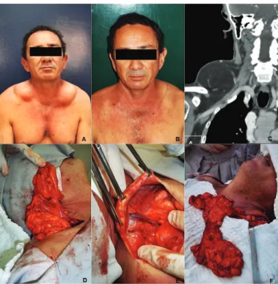
At physical examination, he showed good overall status, with bilateral supraclavicular mass of soft consistency, pain-less, measuring 10 × 6 cm to the left and 12 × 8 cm to the
right (Fig. 1A). The computed tomography (CT) conirmed
the presence of a symmetrical supraclavicular mass, whose density was compatible with fat (Fig. 1C). The mass was sur-gically removed, and the anatomopathological analysis of the specimen evidenced the presence of lipomatosis (Figs. 1D, 1E, 1F). To date, the patient has progressed well with-out recurrence, and with-outpatient follow-up has been main-tained (Fig. 1B).
Discussion
In spite of the uncertain etiology of MSL, its pathogenesis is believed to be related to the dysfunction of cyclic AMP in adipocytes and levels of catecholamines responsible for lip-olysis.3 Alcohol also appears to play a role in the adipocyte
hyperplasia process2 in genetically susceptible individuals
Multiple symmetrical lipomatosis (Madelung’s disease) 91
lipid oxidation effects. Other studies have also suggested the presence of mitochondrial inheritance through mutation of the maternal gene.3,5
The diagnosis is attained by patient history, physical examination, biopsy, and laboratory and imaging tests. Complementary tests are important to discard differential diagnoses such as multiple familial lipomatosis, sarcomas,
angiolipomas, lipoblastomas, neuroibromatosis, Dercum’s
syndrome, Hanhart syndrome, polydysplasia syndrome, lymphoproliferative disorders, and muscular dystrophies.2,3
CT is considered the imaging examination of choice for diagnosis, preoperative staging, and postoperative
fol-low-up, due to the overlap of indings with magnetic reso -nance imaging and its lower cost.3
The palliative treatment is surgical through lipectomy or liposuction, with good results, although recurrences are
common due to the dificulty of complete tumor excision, as
they are not encapsulated.2,3 Some studies have described a
clinical treatment with β2-agonist, in order to increase the adrenergic lipolysis and reduce the accumulation of fat, but
with questionable eficacy.2 Although weight loss and
alco-hol withdrawal appear to have no effect on disease progres-sion, these measures associated with surgery can decrease the recurrence rate,6 as a longitudinal study showed that the
amount of alcohol consumed is directly associated with the onset of the disease.3
The prognosis can be good if treated early; morbidity and mortality are more often associated with complications of alcoholism than with the fat deposits directly.3 The
le-sions hardly ever become malignant; however, they may evolve with compression of the larynx and trachea and de-velopment of neuropathies.2
Final comments
The identiication of this rare disease, the appropriate in -vestigation for the exclusion of differential diagnosis, and early surgical approach as observed in the present case, are important to prevent future complications.
Conlicts of interest
The authors declare no conlicts of interest.
References
1. Meyer TN, Meyer GPN. Caso atípico de lipomatose simétrica múltipla. Rev Soc Bras Cir Plast. 2007;22:64-6.
2. Vieira MV, Grazziotin RU, Abreu M, Furtado CD, Silveira MF, Furtado APA, et al. Lipomatose simétrica múltipla (doença de Madelung) – Relato de um caso. Radiol Bras. 2001;34:119-21. 3. Barbosa CC, Pires MTF, Guimarães MBS, Figueira RC, Nacif MS,
Lupi O. Lipomatose simétrica benigna: doença de Madelung. Relato de caso. Rev Bras Clin Med. 2010;8:165-9.
4. Busetto L, Sträter D, Enzi G, Coin A, Sergi G, Inelmen EM, et al. Differential clinical expression of multiple symmetric lipo-matosis in men and women. Int J Obes Relat Metab Disord. 2003;27:1419-22.
5. Lin F, Yang T. Madelung disease. CMAJ [serial online]. 2012, Jun 25. Available from: http://www.cmaj.ca/.
6. Ramos GHA, Trevizan GL, Pepe ES. Doença de Madelung. Rev Bras Otorrinolaringol. 2002;68:587-90.