Correspondence: Prof. Dr. Osvaldo Luiz Bezzon, Departamento de Materiais Dentários e Prótese, Faculdade de Odontologia de Ribeirão Preto, Universidade de São Paulo, Avenida do Café, S/N, 14040-904 Ribeirão Preto, SP, Brasil. Tel: 3602-4334. Fax: +55-16-3630-0999. e-mail: [email protected]
Effect of the Use of Die Spacer on the Marginal
Fit of Copings Cast in NiCr, NiCrBe and
Commercially Pure Titanium
Natércia Carreira SORIANI1
Mônica Barbosa LEAL1
Silvana Maria PAULINO2
Valéria Oliveira PAGNANO1
Osvaldo Luiz BEZZON1
1Department of Dental Materials and Prosthodontics, School of Dentistry of Ribeirão Preto, University of São Paulo, Ribeirão Preto, SP, Brazil
2School of Dentistry, University of Ribeirão Preto, Ribeirão Preto, SP, Brazil
The goal of this study was to evaluate the effect of using die spacers on the marginal fit of NiCr (M1) and NiCrBe (M2) alloys and commercially pure titanium (cpTi) (M3) copings cast by the lost wax technique. Using a metal matrix, 45 resin added extra hard type IV stone models were obtained for the fabrication of wax patterns under the following conditions: no die spacer (A), with one die spacer layer (B) and with two die spacer layers (C), with five repetitions for each condition (alloy x die). Each die was waxed and the wax patterns were invested as per manufacturer’s instructions. Three wax patterns were embedded in each casting ring, each corresponding to one of the conditions. Each coping, seated to the metal matrix by a seating pressure standardizing device (SPSD), was taken to an optical microscope for measurement of marginal discrepancy. The obtained data (μm) were analyzed statistically by ANOVA and Tukey’ test (α=5%). There was statistically significant difference (p<0.05) among the materials (M1=110.67; M2=130.33 and
M3=148.33). Regarding the use of the die spacer, there was a statistically significant difference (p<0.05) among the three conditions (A=162.00; B=131.06 and C=96.67). It was concluded that there is less marginal discrepancy with two die spacer layers.
Key Words: die spacer, marginal adaptation, cast crown, titanium, NiCr alloys.
INTRODUCTION
Over the past decades, the search for dental materials of good quality and safe to patient’s health has increased significantly. Biosafety has become a key factor for the choice of gold alloys. Alloys referred to as alternative, on the other hand, still face some resistance from prosthodontists. In spite of their lower cost, these alloys do not offer the proper biosafety for clinical use, as their formulation contains metals such as nickel and beryllium (1). The introduction of titanium cast by the lost wax technique has given the perspective of using a material with high biocompatibility and a cost not as high
as that of traditional gold alloys (2), despite the needed sophisticated technology for accurate casting (3).
satis-factory resistance to corrosion at room temperature (5). One of the critical factors associated to crown fabrication is fidelity to the cast copy, which must correctly reproduce tooth preparation (6-8). According to some authors (3,9), copings cast in commercially pure titanium (cpTi) have shown appropriate marginal fit. Other studies, however, have stated that, though considered satisfactory, titanium’s marginal fit is inferior to that of gold alloys (9).
Alternative alloys for crown fabrication, are, mostly, NiCr-based formulations that emerged from compositions initially used in removable prostheses. Their physical and mechanical properties are superior to those of gold alloys in prostheses with a large prosthetic space or in adhesive prostheses, and they have hardness, flexure strength, and modulus of elasticity comparable or slightly superior to those of gold alloys (4).
Nevertheless, the nickel in these alloys may trigger allergic reactions, in addition to having a certain carcinogenic potential, which implies on controlling its handling. Other base metals, such as beryllium, for instance, present in some formulations, also have a carcinogenic potential (1). It has been claimed that these base metal alloys, when used on implants, may trigger corrosion mechanisms by galvanic current (10).
In order to achieve success in the fabrication of fixed prostheses, an appropriate marginal fit is mandatory. Despite the technological advancements in terms of the improvement of casting techniques, die fabrication (11), waxing pattern (5) and coping fabrication (3,8,9,12), a discrepancy remains between the restoration’s margin and the cervical edge of the pre-pared tooth. This discrepancy will be filled with ce-ments that have different degrees of degradation in the oral environment. Clinical longitudinal studies, as well as the laboratorial evaluations judiciously carried out with base metal alloys, have shown that a discrepancy limit of up to 120.00 µm would assure clinical success when using these materials (13,14).
With the aim of reaching better results, some authors have recommended using crown seating techniques that include perforations, internal relief and die spacer (15). The acid or electrolytic attack not only harms the metal surface but also may result in an irregular relief. Therefore, it is safer to use die spacers because they promote internal relief close to the ideal and its application technique does not harm the metal surface, as it is applied before casting. In addition, it
promotes an internal relief for the luting agent, thus preventing the cement layer from interfering on the crown’s complete seating and helps compensating for the alloy’s casting shrinkage by adding to the thermal expansion of the refractory material (13,16). The internal relief promoted by die spacers has been shown to reduce the intrapulpar tensions and the force required at the moment of crown cementation (17,18), in addition to ensuring a better marginal fit and increasing the restoration’s retention in 25% (19,20).
The purpose of this study was to evaluate the effect of using die spacers on the marginal fit of metal copings cast in cpTi by the lost wax technique, in comparison to two alloy formulations commonly used in dentistry: NiCr and NiCrBe.
MATERIAL AND METHODS
For specimen preparation, a stainless steel matrix was fabricated in the Precision Workshop of the De-partment of Dental Materials and Prosthodontics of the School of Dentistry of Ribeirão Preto (USP, Brazil), with 7.0 mm in height, 7.0 mm in diameter, and a cervical finishing line with a 30° bevel (Fig. 1).
The matrix was molded with medium-viscosity polyether (Impregum soft; 3M ESPE, Seefeld, Ger-many) with the aid of a brass ring that acted as an individual impression tray, specially fabricated to stan-dardize the impression material’s thickness. The adhe-sive was applied to the ring and dried for 1 min, following the manufacturer’s instructions. The base and accelerator pastes were mixed together until a
homogeneous mass was obtained, according to the manufacturer’s recommendations. This material was applied with the aid of a syringe specific for impression material in order to prevent bubble formation. The brass ring was filled with polyether, placed on the matrix and the exceeding material removed.
After setting was completed, the mold was removed with the aid of an impression removal device (specifically designed for this purpose) that ensured mold removal parallel to the matrix’s long axis, avoiding discrepancies to any of its sides. A split device was then attached to the mold together with a Teflon ring that allowed for the standardization of the die’s base, where the plaster was poured. The models (dies) were constructed using resin added extra hard type IV stone (Polidental, São Paulo, SP, Brazil), mixed mechanically in vacuum and in the ratio recommended by the manu-facturer. After 30 min, the dies were manually removed. A total of 45 dies were obtained. Each die was used to make one single wax pattern. The wax patterns were assigned to 3 groups of 15 elements and cast with Verabond (NiCrBe) (Aalba Dent. Inc., Cordelia, CA, USA) and Verabond II (NiCr) (Aalba Dent. Inc.) alloys and cpTi (Dentaurum Inc. Pforzheim, Germany). Each 15-element group was then subdivided into 3 groups, to which the dies were randomly assigned: one group without die spacer application, another with a one die spacer layer application, and final one with a two die spacer layer application.
In the group without die spacer, dies received a thin sealer layer (Kota Ind. e Com., São Paulo, SP, Brazil). Pico-Fit (Renfert, Hilzingen, Germany) die spacer system was used. In the group with one die spacer layer, only a thin coat of golden varnish was applied covering all sides, except for the bevel, and then the sealer was applied. In the group with two layers, first a thin coat of the golden varnish was applied, then a coat of silver layer and, after drying, the sealer was applied.
The wax patterns were obtained by using inlay wax Schuler Dental (Ulm, Germany), liquefied in a plastificator and placed in droplets between the previ-ously sealed die and a brass ring previprevi-ously sealed with liquid vaseline and heated. The brass ring permitted the standardization of the copings’ thickness.
After wax cooling, the brass ring was removed and the wax margin was finished using a hollenback instrument by a previously calibrated operator.
The coping form wax patterns were invested in
Termocast investment (Polidental) for Verabond and Verabond II, and Rematitan Plus (Dentaurum) for cpTi, and vacuum mixed as per manufacturer’s instructions. In the same silicone ring, 3 wax patterns were invested, having either no, 1 or 2 die spacer layers, respectively. Next, the investment rings were taken to the Edgcon 5P oven (EDG, São Carlos, SP, Brazil) and submitted to thermal cycles for investment expansion. The heating cycle of the rings was specific for each investment, following manufacturer’s recommendations. After ring casting and cooling, the metal copings were divested, sandblasted with 100 μm aluminum oxide, under a pressure of 80 lib/pol2 in an airborne-particle machine
(Microjet III; EDG), machined and taken to the corre-sponding dies to check seating. Next, the discrepancy was determined with the copings seated on the metal matrix.
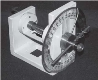
During measurement, it was noticed that the coping could move on the matrix, which would pro-duce unreliable measurements. Thus, a seating pres-sure standardizing device (SPSD) was created to allow the matrix-coping set to be taken to the microscope always with a standardized coping seating load (a patent application for this device has been submitted to the Industrial Property National Institute of Brazil- PI 0.505.036-7) (Fig. 2).
To determine the seating load, a matrix-coping set was first taken to a universal testing machine (Model DL 2000; Emic, São José dos Pinhais, PR, Brazil) and it was applied the compressive load. It was obtained a
load x displacement graphic. The displacement was interrupted at 3 kgf-load, therefore the 3 kgf-load was standardized for all specimens. After adjusting the SPSD, the matrix-coping set was taken to the Optical Microscope with linear measurements (Nikon Measurescope, Tokyo, Japan) to measure the marginal discrepancy.
Four fixed points were established to perform the measurements. Point 1 referred to the matrix stopping point, observing it by the base and rotating it clockwise. The other points were orthogonal among each other. Three readings were made of each point and the respective means were obtained.
RESULTS
The original data of this study resulted from the factorial product of 3 treatments (without die spacer, with one layer of die spacer, and with two layers of die spacer), 3 metal alloys (Verabond, Verabond 2, and cpTi), 4 reference points (buccal, lingual, mesial, and
distal surfaces), 3 readings of each point to obtain the respective means, and 5 repetitions for each condition (treatment X metal bond), adding to a total of 540 marginal discrepancy measurements. The mean dis-crepancy of the 4 points evaluated for each experimental condition represented each coping’s discrepancy, re-sulting in 9 original values, listed in Table 1.
Having verified the normal distribution of the sample data, analysis of variance - square root of data, showed showed statistically significant difference be-tween absence and presence of the die spacer layer (p<0.001) as well as among the materials (p=0.045), and no statistically significant difference for the interac-tion between die spacer and material (p=0.471).
Table 1. Cervical discrepancies measurements (μm).
Materials Conditions
No die 1 die spacer 2 die spacer
spacer layer layers
Verabond 172.0 136.0 84.2
(NiCrBe)
Verabond II 144.4 117.8 69.8
(NiCr)
cpTi 169.9 139.4 136.0
Table 2. Tukey's test.
Source of variation: die spacer
No die spacer 162.00000a
One layer 131.06667ab
Two layers 96.66667b
Different letters indicate statistically significant difference at 1%. p critical value: 45.3816.
Table 4. Calculated sample means (original values in μm).
Interaction alloys x die spacer Means
NiCrBe x no die spacer 172.00000
cpTi x no die spacer 169.60000
NiCr x no die spacer 144.40000
cpTi x 1 layer 139.40000
cpTi x 2 layers 136.00000
NiCrBe x 1 layer 136.00000
NiCr x 1 layer 117.80000
NiCrBe x 2 layers 84.20000
NiCr x 2 layers 69.80000
Table 3. Tukey's test.
Source of variation: materials
NiCrBe 130.73333ab
NiCr 110.66667 b
cpTi 148.33333a
Tukey’s test was performed and revealed a statistical significance (p<0.05) for the variation factors (die spacers and materials) (Tables 2 and 3), and no statistical significance for the interaction, of which values are shown in Table 4, from the highest to the lowest discrepancy values.
DISCUSSION
In this study, it could be observed that there was no significant difference for the interactions among the analyzed variation factors, which means that the use of die spacers did not interfere with the result of a specific material. On the other hand, the statistical analysis clearly showed that the die spacer was a key variation factor for reduction of marginal discrepancy of the 3 evaluated materials, given that the use two layers of die spacer provided a significantly different value (96.67 µm) compared to no use of a die spacer (162.00 µm). The use of only one layer of die spacer produced an intermediate value (131.07 µm) with no significant difference in relation to the higher and lower values. These results are consistent with those of previous studies, and highlight the importance of using this technical resource to ensure a lower discrepancy of prosthetic castings fabricated with alternative alloys (3,17,19).
When the material variation factor was evalu-ated, it was observed that the value obtained with cpTi (148.33 µm) showed a statistically significant difference in relation to the value obtained with the NiCr alloy (110.67 µm). The discrepancy obtained with the NiCrBe alloy (130.73 µm) did not differ significantly from the higher and lower values.
The presence of Be in NiCr alloys creates a Ni-Be eutectic phase, with a lower melting point than the other phases that compose the material. This makes the metal alloy more fluid, with a lower melting point, reducing the casting shrinkage. Therefore, it could be assumed that, with this material, a lower cervical discrepancy would be obtained, although it is necessary to consider that there was no significant difference in terms of the value obtained with the alloy without Be. On the other hand, this study did not intend to control the casting temperature of the evaluated materials, which is possible with the induction machine. Casting of NiCr and NiCrBe alloys was determined by visual inspection of the material inside the crucible when the centrifuge
was emptied. It is possible that controlling casting temperature may be advantageous because of the lower casting temperature of the Be-containing alloy.
Data on Table 4 show that, with the use of 2 die spacer layers, the lowest discrepancy values were obtained with the NiCr and NiCrBe alloys (69.8 µm and 84.2 µm). With the NiCr alloy, the use of a single layer of die spacer produced a lower discrepancy value (117.80 µm) than the one considered as the clinical discrepancy limit (120.00 µm). On the other hand, with cpTi, even with the association of 2 die spacer layers, the value of 120.00 µm was not achieved, indicating the need for further improvement of the technique for obtaining prosthetic castings with this material.
It is important to comment that the proposal of evaluating the use of die spacer determined that stone dies should be obtained, as done in clinical conditions. Some castings that had excellent seating on the die showed an unsatisfactory seating on the metal matrix, repeating a condition that sometimes occurs in daily practice and that certainly causes failure of impression. No fitting by internal relief was done to improve coping seating to the metal matrix. Casting seating on the die was performed only by removing eventual casting nodes, which means that no additional relief was done to that provided by the spacer. As the stone dies were randomly assigned to the experimental groups, it can be affirmed that such circumstances did not interfere with the results. Nevertheless, some specimens were discarded due to an eviden lack of fitting to the matrix. Regarding the superiority of NiCr and NiCrBe alloys in relation to cpTi, lower discrepancy levels for these alternative materials should undergo further studies that might broaden the knowledge of the details involved in the casting process by the lost wax technique.
Based on the obtained results, the lowest cervical discrepancy was obtained using 2 die spacer layers (96.67 µm) with a significant difference from the discrepancy obtained without die spacer (162.00 µm). The value obtained using a single die spacer layer (131.07 µm) was intermediary and did not differ significantly from the higher and lower values.
the other tested materials.
It is important that other aspects of the study, such as the variation of the material refractory, casting temperature and cemented copping removal strenght, are further addressed.
ACKNOWLEDGEMENTS
The authors would like to thank FAPESP for financial support (grant # 05/53469-0).
RESUMO
O objetivo deste trabalho foi avaliar o efeito do uso de espaçadores na adaptação marginal de “copings” de ligas de NiCr (M1) e NiCrBe (M2) e titânio (M3) fundidos pela técnica de cera perdida. A partir de uma matriz metálica, foram obtidos 45 troquéis de gesso resinado tipo IV para confecção dos padrões de cera nas seguintes condições: sem a presença de espaçador (A), com uma camada de espaçador (B) e com duas camadas de espaçador (C), com cinco repetições para cada condição (liga x espaçador). Foi realizado o enceramento de cada troquel e os padrões de cera foram incluídos no revestimento indicado pelo fabricante. Em cada anel de fundição foram incluídos 3 padrões de cera, sendo cada um correspondente a cada condição. Cada “coping”, adaptado à matriz metálica por meio de um dispositivo padronizador da pressão de assentamento, foi levado ao microscópio óptico para aferição das medidas da desadaptação marginal. Os dados obtidos (μm) foram submetidos à análise estatística por ANOVA e teste de Tukey (α=5%). Entre os materiais houve diferença estatisticamente significante (M1=110,67, M2=130,33 e M3=148,33). Em relação ao fator espaçador, houve diferença estatisticamente significante entre as três condições (A=162,00; B=131,06 e C=96,67). Conclui-se que a presença de duas camadas de espaçador propicia menor desadaptação marginal.
REFERENCES
1 . Bezzon OL. Allergic sensivity to several base metals: a clini-cal report. J Prosthet Dent 1993;69:243-244.
2 . Berg E. Dentists opinion on aspects of cast titanium restora-tions. J Dent 1997;25:113-117.
3 . Fragoso WS, Henriques GE, Contreras EF, Mesquita MF. The influence of mold temperature on the fit of cast crowns with commercially pure titanium. Braz Oral Res 2005;19:139-143.
4 . Anusavice KJ. Phillips’ science of dental materials. 11th ed. Philadelphia: WB Saunders Co.; 2003.
5 . Limkangwalmongkol P, Chiche GJ, Blatz MB. Precision of fit
of two margin designs for metal-ceramic crowns. J Prosthodont 2007;16:233-237.
6 . Witkowski S, Komine F, Gerds T. Marginal accuracy of titanium copings fabricated by casting and CAD/CAM tech-niques. J Prosthet Dent 2006;96:47-52.
7 . Stoll R, Fischer C, Springer M, Stachniss V. Marginal adapta-tion of partial crowns cast in pure titanium and in a gold alloy – an in vivo study. J Oral Rehabil 2002;29:1-6.
8 . Wolf BH, Walter MH, Boening KW, Schmidt AE. Margin quality of titanium and high-gold inlays and onlays – a clinical study. Dent Mater 1998;14:370-374.
9 . Al Wazzan KA, Al Nazzawi AA. Marginal and internal adap-tation of commercially pure titanium and titanium-alumi-num-vanadium alloy cast restorations. J Contemp Dent Pract 2007;8:19-26.
10. Watanabe I, Watanabe E, Nakajima H, Atsuta M, Okabe T. Marginal accuracy in casting titanium fixed partial dentures. J Dent Rees 1997;76:773-779.
11. Lombard’s P, Carbineer A, Malarkey ME, Toothier RW. Dimensional accuracy of castings with ringlets and metal ring investment systems. J Prosthet Dent 2000;84:27-31. 12. Milan FM, Consani C, Sobrinho LC, Sinhoreti MAC,
Souza-Neto MD, Knowles JC. Influence of casting methods on marginal and internal discrepancies of complete cast crowns. Braz Dent J 2004;15:127-132.
13. McLean JW, Fraunhofer JA von. The estimation of cement film thickness by an in vivo technique. Br Dent J 1971;131:107-111.
14. Leong D, Chai J, Lautenschlager E, Gilbert J. Marginal fit of machine-milled titanium and cast titanium single crowns. Int J Prosthodont 1994;7:440-447.
15. Emtiaz S, Goldstein G. Effect of die spacers on precementation of complete-coverage restorations. Int J Prosthodont 1997;10:131-135.
16. Psillakis JJ, McAlarney ME, Wright RF, Urquiola J. Effect of evaporation and mixing technique on die spacer thickness: a preliminary study. J Prosthet Dent 2001;85:82-87. 17. Cherkasski B, Wilson PR. The effect of oscilation, low
seat-ing force and dentine treatment on pulpward pressure trans-mission during cementation: a laboratory study. J Oral Rehabil 2003;30:957-963.
18. Carter SM, Wilson PR. The effect of die-spacing on crown retention. Int J Prosthodont 1996;9:21-29.
19. Passon C, Lambert RH, Lambert RL, Newman S. The effect of multiple layers of die spacer on crown retention. Oper Dent 1992;17:42-49.
20. Oliveira AB, Saito T. The effect of die spacer on retention and fitting of complete cast crowns. J Prosthodont 2006;15:243-249.