• Nenhum resultado encontrado

Braz. Dent. J. vol.14 número2

N/A
N/A
Protected

Academic year: 2018

Share "Braz. Dent. J. vol.14 número2"

Copied!
4
0
0

Texto

(1)

Braz Dent J 14(2) 2003

132 C.M. Kussano et al.

Evaluation of Shear Bond Strength of Composite to

Porcelain According to Surface Treatment

Claudia Minami KUSSANO Gerson BONFANTE José Gilmar BATISTA João Henrique Nogueira PINTO

Prosthodontics Department, Hospital for Rehabilitation of Craniofacial Anomalies, University of São Paulo, Bauru, SP, Brazil

This study evaluated the shear bond strength of porcelain/composite using 40 metal + porcelain + composite cylindrical specimens divided into 4 groups, according to porcelain surface treatment: 1) no treatment, 2) mechanical retentions performed with diamond burs, 3) etching with phosphoric acid+silane, and 4) etching with hydrofluoric acid+silane. After being stored in distilled water at room temperature for one week, the specimens were submitted to a shear force (load) and the data were analyzed statistically (ANOVA). The means (in Mpa) of the groups were: 4.71 (group 1); 4.81 (group 2); 11.76 (group 3); 11.07 (group 4). There were no statistically significant differences between groups 1 and 2 and between groups 3 and 4.

Key Words: porcelain/composite bond strength, repair, porcelain surface etching.

Correspondence: Dra. Claudia Minami Kussano, Av. Orlando Ranieri 7-108, bloco 45, apto 32, Jardim Marambá, 17047-001 Bauru, SP, Brasil. Tel: +55-14-230-2438. e-mail: claudiamk@terra.com.br

ISSN 0103-6440

INTRODUCTION

Metal-ceramic crowns are a restorative treat-ment alternative which combines metal resistance and porcelain esthetics, tending to last for many years if made according to specific technical rules. Factors such as impact and fatigue, occlusal forces, and incompat-ibility between metal and porcelain physical properties may result in porcelain fracture, frequently of a cohe-sive nature (1-3). Due to the inherently brittle nature of ceramic restorative materials, failure under intraoral conditions of metal-ceramic restorations is not uncom-mon (4). The majority (65%) of the failures have been observed in the anterior region, whereas 35% were in the posterior region. Sixty percent of the failures oc-curred at the labial, 27% at the buccal, 5% at the incisal, and 8% at the occlusal regions. These fractures were mainly in the maxilla (75%), predominantly at the labial surface (5). Thus, it is necessary to assess the possible causes of fracture, so that the most suitable treatment can be recommended. Depending on the extension of the area to be restored, cost and time available may range from the making of a new

prosthe-sis, facet (6), overcasting (7)to a composite repair (8-10).

According to Kato et al. (11), the bond strength to porcelain was predominantly influenced by the type of conditioning agent rather than the type of luting agent.

Usually, when performing repair with composite on porcelain, the surface must be prepared with burs, sandblasted with aluminum oxide, etched and silaned. The increase in porcelain surface roughness by means of diamond burs and discs contributes to the repair’s adhesive resistance, and the use of acids, by creating microretentions, improves adhesion, producing cohe-sive failures if used with silane agents (12). Research shows that sandblasting with 50-µm aluminum oxide is a better method for preparing the surface than bur performed retentions(13). Nevertheless, sole sandblast-ing is not sufficient to improve the bond of composite-porcelain interface (4,14).

In one of the first studies on porcelain repair with composite, utilizing a silane agent, it was verified that this technique might be a clinical solution to fractured

(2)

Braz Dent J 14(2) 2003

Shear bond strength and surface treatment 133

porcelain repairs (15). Silanization improved repair resistance when hydrofluoric acid was previously uti-lized (16). However, there is no consensus on the best porcelain treatment method to be applied in different clinical situations.

Thus, aiming at further clarifying this issue, the present study evaluated the porcelain-composite bond strength, employing several methods for surface treat-ment associated with an adhesive system prior to resin application.

MATERIAL AND METHODS

Forty metal + porcelain + composite specimens were made using a stainless steel matrix, comprised of

a metallic base over which a 22.0-mm high X 30-mm diameter cylinder was adapted, presenting a central perforation of 6.5-mm deep by 6.0-mm diameter sur-face, determining specimen dimensions. The opposite surface had a 5.0-mm diameter screw, utilized for specimen removal following condensation and stabili-zation of the set during shear tests. The matrix had spacing discs of 6.0 mm diameter by 1.5 mm height, which adapted to the central perforation, allowing thick-ness standardization of the metal (2 discs; 3.0 mm height X 6.0 mm diameter), porcelain (1 disc; 1.5 mm height X 6.0 mm diameter) and composite (no disc; 1.5 mm height X 6.0 mm diameter). The matrix and spac-ing disc are shown in Figure 1.

To obtain the wax pattern, 2 discs were posi-tioned in the interior of the matrix, corresponding to porcelain and composite portions. Fused wax was poured and, after cooling, removed with the aid of the screw. The patterns were cast in Ni-Cr alloy (Durabond Uni-versal, São Paulo, SP, Brazil) and polished with aluminum oxide abrasive burs at low rotation until their complete fitting in the interior of the matrix. One of the surfaces was sandblasted with 50-µm aluminum oxide particles (Trijato Odonto Larcon, Maringá, PR, Brazil), after which the metallic discs were cleaned and degreased in pressurized water steam (Vaporeto, Polti, Italy), for 1 min. After drying, these surfaces were kept free from manual contact. Conventional application and opaque firing in paste, in two layers (Opaquer, IPS Classic, Ivoclar), and specimen porcelain was done according to manufacturer instructions. Figure 2 shows the manufacturing phase.

The ceramic surface was made uniform by

abra-Figure 1. Stainless steel matrix used for making the specimens. A) Base. B) Cylinder. C) Screw. D) Frontal perforation. E) Spacing disc.

Figure 2. Manufacturing phase. F) Wax pattern. G) Cast pattern.

(3)

Braz Dent J 14(2) 2003

134 C.M. Kussano et al.

sion with a 2214 KG Sorensen diamond bur prior to surface treatment. The specimens were randomly di-vided into 4 groups of 10 specimens each. Group 1: Control. Application of one layer of adhesive (Scotchbond Mult Purpose Plus, 3M, Sumaré, SP, Bra-zil), light-cured for 20 s, and insertion of composite in small portions (Z100, 3M), light-cured for 40 s, accord-ing to manufacturer instructions. Group 2: Preparation of mechanical retentions with a wheel-shaped diamond bur (KG Sorensen 1052, Barueri, SP, Brazil), adhesive application, light curing for 20 s, insertion of composite and light curing for 40 s. Group 3: Application of 35% phosphoric acid gel (Scotchbond Mult Purpose Plus, 3M) over porcelain surface for 1 min, washing and drying, application of silane agent in 2 layers (Ceramic Primer, Scotchbond Mult Purpose Plus, 3M), allowing drying. The procedure to apply the bond and resin agent was similar to that in the previous group. Group 4: The only difference between this group and group 3 was the application of 10% hydrofluoric acid gel (Dentsply, Rio de Janeiro, Brazil) for 4 min, in lieu of phosphoric acid. The specimens were subsequently stored in dis-tilled water, at room temperature, for 7 days, and then submitted to shear tests in a universal assay machine (Kratos, Dinamômetros Kratos Ltda, São Paulo, SP, Brazil) with a 0.5-mm/min speed. The active tip was applied on the composite-porcelain interface. For the tests, the specimens were placed in the matrix, expos-ing only the composite portion (Figure 3).

The data obtained were recorded and submitted to analysis of variance (ANOVA) (p<0.05). Individual comparisons were made with the Tukey test (p<0.05).

RESULTS

The means and standard deviations for the 4 groups are reported in Table 1. By comparing the results of groups 1 and 2, one observes that the perfor-mance of mechanical retentions on the porcelain surface added little to increase the bond strength of composite with porcelain. Individual comparisons performed by the Tukey test showed statistically significant differ-ences when using conditioning with either phosphoric (group 3) or hydrofluoric acid (group 4), associated to silane agent and adhesive in the comparison to the control group (use of adhesive only) and to group 2 (mechanical retention). However, the differences

be-tween the types of acids were not significant, nor were the differences between groups 1 and 2 statistically significant.

DISCUSSION

The clinical success of porcelain repair systems is almost entirely dependent on the integrity of the bond between porcelain and composite resin. The bond is achieved either by chemical or mechanical methods. Etching procedures have been used to facilitate bond-ing between porcelain and resin, creatbond-ing a porous surface on the porcelain that leads to a retentive bond (4).

The combination of sandblasting with acids on porcelain silanization has provided the best results. When failure involves only the porcelain, the acid-conditioning is effective in increasing micromechanical retention (9); but, it was observed that silane priming considerably improved bond strength to the porcelain of the composite material (17).

Hydrofluoric acid is not necessary to obtain resistant adhesion between composite and porcelain if the silane agent, which reacts with OH groups on the porcelain surface, is used. Silane agents appear to be the essential components for a porcelain repair proce-dure, by modifying its surface structure, rendering it more reactive with the composite, enabling chemical adhesion in both inorganic/porcelain and organic/com-posite surfaces. Theoretically, the silane bond should provide a stable repair of the fractured porcelain (18). Lacy et al. (12) observed that when silane was not applied the composite bond strength to porcelain was relatively weak, regardless of the type of surface treat-ment, with the failure occurring at the interface. The

(4)

Braz Dent J 14(2) 2003

Shear bond strength and surface treatment 135

values were higher with silane application after etch-ing. Thus, the findings suggests that bonding to porcelain is mostly chemical and not mechanical (19). The results of the present study cooborate this. Leibrock et al. (20) did not observe statistically significant differences in bond strength following thermocycling and mechanical overloading among the systems which used phosphoric or hydrofluoric acid. Owing to its chemically aggres-sive nature, hydrofluoric acid should be avoided and substituted by phosphoric acid, which, although ag-gressive, is not as noxious to tissues but produces the same effects if associated with the silane agent.

It is not possible to state whether the lower values found in this study are compatible with the clinical success of a composite repair, but quite prob-ably, the application of silane is capable of contributing to the longevity of this sort of procedure.

RESUMO

Este estudo avaliou a resistência ao cisalhamento da união porcelana/resina, usando 40 corpos-de-prova cilíndricos de metal + porcelana + resina composta quer foram divididos em 4 grupos, de acordo com o tratamento superficial da porcelana: 1) sem tratamento, 2) retenções mecânicas feitas com ponta diamantada, 3) ataque ácido com ácido fosfórico + silano, 4) ataque ácido com ácido fluorídrico + silano. Após uma semana de armazenamento em água destilada à temperatura ambiente, os espécimes foram submetidos à carga de cisalhamento e os dados analisados estatisticamente (ANOVA). As médias dos grupos foram em MPa: 4.71 (grupo 1); 4.81 (grupo 2); 11.76 (grupo 3) e 11.07 (grupo 4). Não houve diferenças estatisticamente significantes entre os grupos 1 e 2 e entre os grupos 3 e 4.

REFERENCES

1. Zhukovsky L, Godder B, Settembrini L, Scherer W. Repairing porcelain restorations intraorally: techniques and materials. Comp Cont Educ Dent 1996;17:18-28.

2. Pameijer CH, Louw NP, Fischer D. Repairing fractured porce-lain: how surface preparation affects shear force resistance. J Am Dent Assoc 1996;127:203-209.

3. Chung KH, Hwang YC. Bonding strengths of porcelain repair systems with various surface treatments. J Prosthet Dent 1997;78:267-274.

4. Kelsey WP, Latta MA, Stanislav CM, Shaddy RS. Comparison of composite resin-to-porcelain bond strength with three adhesives. Gen Dent 2000;48:418-421.

5. Özcan M, Niedermeier W. Clinical study on the reasons for and location of failures of metal-ceramic restorations and survival of repairs. Int J Prosth 2002;15:299-302.

6. Noel LG, Michell WC. Porcelain veneer repair of prostheses. Gen Dent 1997;45:182-185.

7. Matsumura H, Atsuta M. Repair of an eight-unit fixed partial denture with a resin-bonded overcasting: a clinical report. J Prosthet Dent 1996;75:594-596.

8. Denehy G, Bouschlicher M, Vargas M. Intraoral repair of cos-metic restorations. Dent Clin N Am 1998;42:719-937.

9. Latta MA, Barkmeier WW. Approaches for intraoral repair of ceramic restorations. Comp Cont Educ Dent 2000;21:635-644. 10. Margeas RC. Salvaging a porcelain-fused-to-metal bridge with

intraoral ceramic repair. Comp Cont Educ Dent 2002;23:952-956.

11. Kato H, Matsumura H, Ide T, Atsuta M. Improved bonding of adhesive resin to sintered porcelain with the combination of acid etching and a two liquid silane conditioner. J Oral Rehabil 2001;28:102-108.

12. Lacy AM, La Luz J, Watanabe LG, Dellinges M. Effect of porce-lain surface treatment on the bond to composite. J Prosthet Dent 1988;60:288-291.

13. Wolf DM, Powers JM, O’Keefe KL. Bond strength of composite to etched and sandblasted porcelain. Am J Dent 1993;6:155-158. 14. Kupiec KA, Wuertz KM, Barkmeier WW, Wilweding TM. Evalu-ation of porcelain surface treatment and agents for composite porcelain repair. J Prosthet Dent 1996;76:119-124.

15. Newburg N, Pameijer CH. Composite resin bonded to porcelain with silane solution. J Am Dent Assoc 1978;96:288-291. 16. Swift EJ, Cloe BC, Boyer DB. Effect of a silane coupling agent

on composite repair strengths. Am J Dent 1994;7:200-202. 17. Chen JH, Matsumura H, Atsuta M. Effect of etchant, etching

period, and silane priming on bond strength to porcelain of composite resin. Oper Dent 1998;23:250-257.

18. Shaverdi S, Canay S, Sahin E, Bilge A. Effects of different surface treatment methods on the bond strength of composite resin to porcelain. J Oral Rehabil 1998;25:699-705.

19. Berry T, Barghi N, Chung K. Effect of water storage on the silanization in porcelain repair strength. J Oral Rehabil 1999;26:459-463.

20. Leibrock A, Degenhart M, Behr M, Rosentritt M, Handel G. In vitro study of the effect of thermo- and load-cycling on the bond strength of porcelain repair systems. J Oral Rehabil 1999;26:130-137.

Imagem

Figure 2. Manufacturing phase. F) Wax pattern. G) Cast pattern.
Table 1. Shear resistance (in MPa) of the 4 analyzed groups.

Referências

Documentos relacionados

This study evaluated the effect of chemical and mechanical surface treatments for cast metal alloys on the bond strength of an indirect composite resin (Artglass) to commercially

The aim of this study was to evaluate the shear bond strength (SBS) of metallic orthodontic brackets bonded to bovine teeth using light-activated or chemically activated composite

This study evaluated the masking ability of different porcelain thicknesses and combination of enamel and/or dentin porcelain layers over simulated background dental substrates

materials were based on literature data (Table 1); and GIE - mechanical properties of resin cement, composite resin core, metal coping and porcelain were determined by the

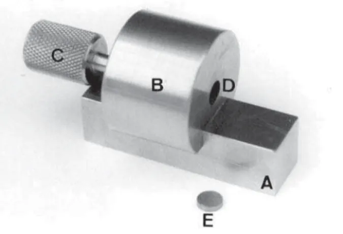
This study evaluated the effects of three metal primers and one multi-mode adhesive system on the shear bond strength (SBS) of a flowable composite resin to nickel-chrome metal

 Normalização contabilística - neste último grupo, estendido por sete questões, o nosso objetivo é saber se o respondente conhecia o SNC-ESNL; em que medida concorda com a

Foram incluídos todos os estudos encontrados na estratégia de busca que preencheram os critérios de inclusão, listados a seguir: a) estudos primários que realizaram ensaios clínicos

Conforme as respostas de grande parte das participantes, a vida sexual também não está satisfatória e isso mostra que a qualidade de vida após uma gravidez precoce e