Plasma rich in growth factors
and bone formation: a radiological
and histomorphometric study in
New Zealand rabbits
Abstract: A radiographic and histomorphometric study was conducted on the inluence of autologous plasma rich in growth factors (PRGF) upon bone healing in surgically created defects in rabbits. Radiographically, bone regeneration was signiicantly greater with the use of PRGF after one month (p = 0.005), though no differences were recorded after the second month. In the histomorphometric analysis one month after sur-gery, the defects illed with autologous bone plus PRGF showed a greater percentage of neoformed bone (35.01 ± 5.31) than the control defects (22.90 ± 12.23), though the differences were not signiicant. Two months after surgery, the defects illed with autologous bone showed greater re-generation (46.04 ± 10.36%) than the control defects (30.59 ± 5.69%), though the differences were not signiicant. The application of PRGF in the bone defects produced in New Zealand rabbits exerted a limited ef-fect on local bone formation.
Descriptors: Bone and bones; Intercellular signaling peptides and proteins; Rabbits; Radiography; Histology.
Francisco Molina-Miñano(a) Pía López-Jornet(b)
Fabio Camacho-Alonso(c) Vicente Vicente-Ortega(d)
(a) DDS; (b)PhD, MD, DDS; (c)PhD, DDS
– Department of Oral Medicine, School of Medicine and Dentistry, University of Murcia, Spain.
(d) PhD, MD, Service of Pathology, School
of Medicine and Dentistry, University of Murcia, Spain.
Corresponding author:
Pía López-Jornet
Clínica Odontológica Universitaria Hospital Morales Meseguer Adv. Marques de los velez, s/n Murcia 30008 - Spain E-mail: [email protected]
Introduction
Platelet-rich preparations constitute a relatively new biotechnology for the stimulation and accel-eration of tissue healing and bone regenaccel-eration.1-3
Platelet-rich plasma (PRP) is deined as a concen-tration of autologous platelets in a small volume of plasma, and is considered to be a rich source of au-tologous growth factors. It was irst introduced by Marx et al.3 (1998) in combination with autologous
bone grafts for the reconstruction of mandibular defects. The contribution of PRP formulations to the bone healing process is thought to be based on the growth factors (GFs) contained in such formula-tions. In 1999, Anitua4 proposed the use of plasma
rich in growth factors (PRGF), where the platelets contain growth factors such as TGF-B1, VEGF, and IGF. These proteins intervene in functions such as directed cell migration (chemotaxis) and in cellular differentiation and proliferation, all of which are key events in repair and regeneration processes.
The biological effects of the different GFs cannot yet be deined in terms of a speciic concentration. As a result, it is not known how much of a given growth factor is needed to accelerate the bone heal-ing process.5,6
Marx et al.3 (1998) used PRP together with
au-tologous bone grafts in mandibular reconstructions, and showed the radiographic density to be greater in grafts to which PRP had been added than in grafts without added PRP, after 6 months. The same study found the radiographic maturation rate to be 1.62-2.16 times greater in the PRP-added grafts than in the grafts without added PRP. Afterwards, a num-ber of studies7-9 have been published on the use of
PRP to enhance many different combinations of au-tologous and/or exogenous graft materials.
Aghaloo et al.10 (2002) performed bicortical
de-fects in rabbit skulls, and evaluated the efde-fects of PRP upon regeneration of the lesions. No signiicant ra-diographic or morphometric differences were found in relation to bone formation. In another experimen-tal model in dogs, Gerard et al.11 (2006) reported
that PRP appeared to enhance early autologous graft healing. However, after two months this effect was no longer signiicant, and PRP did not change the rate at which new bone was formed. Furthermore,
no increase in trabecular density was observed in the PRP-added grafts. The controversy found in the liter-ature regarding the use of this technique is probably related to a lack of standardization in the different PRP formulations, and in the protocols, experimen-tal models and surgical techniques employed.11-13
The present study comprises a radiographic and histomorphometric analysis of the inluence of PRGF and autologous bone grafting upon bone healing in surgically created defects in New Zealand rabbits, after one and two months.
Material and Methods
The study involved 20 adult male albino New Zealand rabbits, with a mean weight of 3,662 g, and was carried out in the laboratory of experimen-tal surgery of the University of Murcia (Spain), fol-lowing approval from the local ethics board. The experiments were conducted in accordance with the guidelines laid down by the European Com-munities Council Directive of November, 24 1986 (86/609/EEC). The animals were anesthetized with a mixture of 60% ketamine and 40% xylazine. Fol-lowing disinfection of the zone and under aseptic conditions, we performed an incision in the antero-internal proximal zone of the tibia, since this access is scantly traumatic and causes limited bleeding. The muscle layers were dissected until the perioste-um was reached. Four 4-mm diameter defects were created with a trephine bur under copious irrigation. Two bone defects were made in each hind tibia: in the right tibia, one defect was used as control, while PRGF was applied to the other. In the left tibia, the previously crushed autologous bone obtained from the right tibia was used to ill both osteotomy defects, with the addition of PRGF in one of the defects. The wounds were closed with resorbable sutures (Labo-ratorios Lorca Marín, S.A. Murcia, Spain).
for 8 minutes, thereby separating the different blood phases. The plasma poor in growth factors (PPGF) located in the upper part of the plasma in the tube was eliminated with 500 µl pipettes. PPGF has the least amount of platelets in the plasma. The PRGF (0.5 ml) was located on top of the white cells series. The PRGF was separated with 500 µl pipettes and transported to an independent tube. Next, the PRGF was activated using 50 µl of 10% calcium chloride.
The mixture of PRGF and calcium chloride was left at room temperature for 10 minutes, until a consistent and easy to handle gelatinous layer was formed. This layer was close to the white cells.
The gelatinous layer was then mixed with autol-ogous bone and placed in the bone defects.
The 20 animals were randomized to two groups: Group 1 (n = 10) (sacriiced by CO2 inhalation after one month) and Group 2 (n = 10) (sacriiced after two months). All animals were subjected to the pro-cedure described above, which was followed by ra-diographic, histological and morphometric analyses. The radiographic evaluation was carried out with an intraoral X-ray apparatus (60 kVp, 7 mA, 0.05 s), generating and processing the images with a radiovisiographic system (Trophy, Eastman Kodak Co., Rochester, NY) and personal computer using Trophy Windows v.5.06 software. The osteotomy defects were classiied and scored according to Pry-or14 as follows:
Grade 1 - No/limited bone illing: < 25% of the •
defect width was illed with bone;
grade 2 - Partial bone illing: > 25% and
• ≤ 90%
of the defect width was illed with bone;
grade 3 - Complete bone illing: > 90% of the de-•
fect width was illed with bone.
For the light microscopy analysis, the tissues were decalciied in 4.13% EDTA for 30 days, cut in half and processed for embedding in parafin. The 5 µm–thick sections were then stained with hema-toxylin-eosin and the Masson trichromic stain.
To perform the histomorphometric analysis, light micrographs (x6 magniication) of the biopsy sections were obtained with a digital camera and analyzed using MIP-4 histomorphometry software (Digital Image, Barcelona, Spain). The amount of new bone formed was calculated as a percentage of
the total area of the original defect.
Evaluation of the radiographic images and histo-morphometric results was carried out by an investi-gator blinded to the treatment involved in each case. The data were analyzed using the SPSS v.12.0 statistical package (SPSS Inc., Chicago, IL, USA). A descriptive study was made of each variable. The Kolmogorov-Smirnov normality test and Levene variance homogeneity test were applied, and if the data showed a skewed distribution, analysis was carried out using a nonparametric ranking test. Sta-tistical signiicance was accepted for p ≤ 0.05.
Results
Of the 20 rabbits included in the study, 2 were lost to the effects of analysis because of fracture of the operated limb (due to unknown causes). In ad-dition, 6 specimens were damaged in the course of sample manipulation and sectioning, and 6 were damaged during histological processing. A inal to-tal of 50 samples could be evaluated.
Evaluation of the standardized digital radio-graphic images showed that one month after surgery, most of the defects in which PRGF was applied were either partially (50%) or completely illed (37.5%), while in the control group most of the defects re-mained partially vacant (62.5%). The differences were statistically signiicant. After two months, the PRGF treated defects showed a greater percent-age of radiographically manifest bone regeneration (66.67%), though without statistically signiicant differences (Table 1).
In the histomorphometric analysis one month af-ter surgery, the defects illed with autologous bone plus PRGF showed a greater percentage of neo-formed bone (35.01 ± 5.31) than the control defects (22.90 ± 12.23), though the differences were not signiicant. Two months after surgery, the defects illed with autologous bone showed greater regen-eration (46.04 ± 10.36%) than the control defects (30.59 ± 5.69%), though once again the differences were not signiicant (Table 2, Figures 1 and 2).
Discussion
study, autologous grafting was associated to PRGF in the New Zealand rabbit.
The PRGF used in this study was obtained fol-lowing the protocol described by Anitua4 (1999).
PRGF was chosen because the activator is calcium chloride, which eliminates the risk of immune re-actions and the transmission of diseases associated with the use of exogenous bovine thrombin. Fur-thermore, PRGF can be obtained in a single centrif-ugation step at 460 g for 8 minutes. In contrast, the double centrifugation technique used to obtain PRP requires a greater blood volume (minimum 50 ml), which is unfeasible in rabbits.
PRGF offers a number of advantages: it allows the simultaneous action of multiple growth factors,
and is an autologous product. PRGF also increases tissue vascularization. The product is biocompatible, effective, and safe, and is reabsorbed by the body within days after initiating local regeneration.1-4 de
Obarrio et al.15 (2000) considered not only the
posi-tive effects of PRP resulting from the release of GFs but also its physical and chemical properties.
Gerard et al.11 (2006), in an experimental model,
found PRP to increase bone formation and remodel-ing in the irst and second months, though these ben-eicial effects decreased after three and 6 months.
Our results coincide with those reported by Agh-aloo et al.10 (2002), who found no statistically
signif-icant radiographic or histomorphometric differences in bone formation as a result of the addition of PRP. Table 1 - Radiographic analysis of bone regeneration. Pearson’s chi-squared test.
Observation
period month Group Total
Radiographic bone regeneration
n (%) p-value
Grade 1 Grade 2 Grade 3
1
PRGF 8 1 (12.50) 4 (50) 3 (37.50)
0.005
Control 8 5 (62.50) 3 (37.50) 0 (0)
Autologous bone + PRGF 5 0 (0) 3 (60) 2 (40)
Autologous bone 5 3 (60) 2 (40) 0 (0)
2
PRGF 6 0 (0) 2 (33.33) 4 (66.67)
0.006
Control 6 2 (33.33) 4 (66.67) 0 (0)
Autologous bone + PRGF 6 0 (0) 3 (50) 3 (50)
Autologous bone 6 2 (33.33) 4 (66.67) 0 (0)
Radiographic evaluation scored14: Grade 1 - No/limited bone filling: < 25% of the defect width was filled with bone; Grade 2 - Partial bone filling: > 25%
and ≤ 90% of the defect width was filled with bone; Grade 3 - Complete bone filling: > 90% of the defect width was filled with bone.
Note: 14 samples from the one month observation period, 2 from the PRGF group, 2 from the control group, 5 from the Autologous bone + PRGF group, and 5 from the autologous bone group were lost during sample preparation. 16 samples from the two month observation period, 4 from the PRGF group, 4 from the control group, 4 from the Autologous bone + PRGF group, and 4 from the autologous bone group were lost during sample preparation.
Observation
period month Group
Histomorphometry
mean ± sd p-value
1
PRGF 28.58 ± 11.22
0.658
Control 22.90 ± 12.23
Autologous bone + PRGF 35.01 ± 5.31
Autologous bone 34.61 ± 16.93
2
PRGF 41.02 ± 9.15
0.648
Control 30.59 ± 5.69
Autologous bone + PRGF 40.63 ± 14.74
Autologous bone 46.04 ± 10.36
Our study intervals of one and two months are simi-lar to those published by other authors.14,16
Investi-gators such as Zechner et al.17 (2003) suggest a
time-dependent action on the part of PRP, with a favoring of bone regeneration in the early stages of healing. In our study, certain limitations were imposed by the use of the tibial bone in rabbits instead of the skull. In effect, the defects were limited to 4 mm in diameter, in order to avoid the risk of fracture.
The data found in the literature regarding bone formation and maturation under the inluence of PRP are contradictory. PRP has been combined with autologous bone or bone substitutes such as in-organic bone mineral and in-organic bone matrix. It remains questionable whether or not these combina-tions enhance the biological activity of PRP.12
According to Pryor et al.14 (2006), few studies
have used radiographic evaluations in experimental animal models based on triple-point scoring systems such as that used in our study. In effect, most stud-ies make use of dichotomic scores or visual measures based on different tones of gray.18,19 In our study, the
radiographic analysis showed important variability in the results obtained. In effect, with the scoring system used, signiicant differences were recorded in radiographic bone density when applying PRGF to
the surgical defects, one month after surgery. How-ever, the differences after two months were no lon-ger signiicant. Pryor et al.14 (2006) indicated that
while radiography is able to differentiate mineral-ization patterns, the results should not be overesti-mated, and histological conirmation is required.
However, in the histomorphometric study, the application of autologous bone either alone or in combination with PRGF did not yield differences in terms of osteogenesis at either of the two time points considered despite a tendency towards increased bone formation compared with the controls.
On the other hand, the group led by Choi et al.20
(2004) has suggested that the addition of PRP does not enhance bone formation in autologous bone grafts.
Consensus is lacking as regards the “optimum concentration of the platelet concentrate”, despite the existence of a number of studies comparing dif-ferent PRP production systems.12,13 A number of
as-pects must be taken into account in relation to the preparation of PRP. Although the optimum plate-let concentration in PRP has not been established, Weibrich et al.13 (2004) suggests that while
con-centrations below 1,000,000/µl exert suboptimum effects, higher concentrations paradoxically could
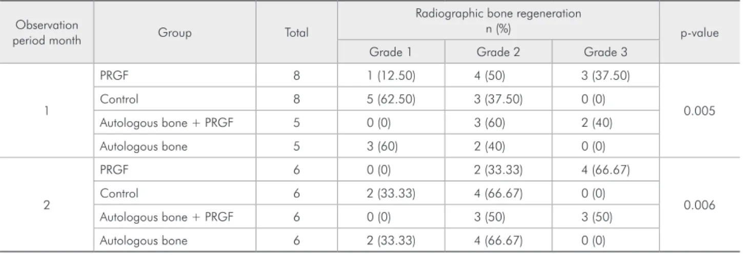
Figure 2 - PRGF treated defect, two months after surgery. (A) Larger-size bone trabeculae (x200, hematoxylin-eosin). (B) Detail of ossification (x220, Masson trichromic stain).
B A
B
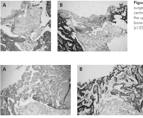
A Figure 1 -surgery. (A) PRGF treated defect, one month after Filled defect showing bone marrow at
exert inhibitory effects. Other authors consider the concentration of receptors in the target tissue to be more important than the concentration of growth factors in PRP.12
Conclusions
In conclusion, while PRGF either alone or in
combination with autologous bone appears to have increased new bone formation in our experimental model, statistical signiicance was not observed. Thus, the application of PRGF in bone defects pro-duced in New Zealand rabbits presented only a lim-ited potential in terms of local bone formation.
References
1. Anitua E, Sánchez M, Orive G, Andía I. The potential impact of the preparation of plasma rich in growth factors (PRGF) in different medical fields. Biomaterials. 2007;28(31):4551-60. 2. Anitua E, Sánchez M, Nurden A, Nurden P, Orive G, Andía
I. New insights into and novel applications for platelet-rich fibrin therapies. Trends in Biotechnol. 2006;24(5):227-34. 3. Marx RE, Carlson ER, Eichstaedt RM, Schimmele SR, Strauss
JE, Georgeff KR. Platelet-rich plasma: growth factor enhance-ment for bone grafts. Oral Surg Oral Med Oral Pathol Oral Radiol Endod. 1998;85(6):638-46.
4. Anitua E. Plasma rich in growth factors: preliminary results of use in the preparation of future sites for implants. Int J Oral Maxillofac Implants. 1999;14(4):529-35.
5. Steed DL. The role of growth factors in wound healing. Surg Clin North Am. 1997;77(3):575-86.
6. Marx RE. Platelet-rich plasma (PRP): what is PRP and what is not PRP? Implant Dent. 2001;10(4):225-8.
7. Zhu SJ, Choi BH, Jung JH, Lee SH, Huh JY, You TM et al. A comparative histologic analysis of tissue-engineered bone using platelet-rich plasma and platelet-enriched fibrin glue. Oral Surg Oral Med Oral Pathol Oral Radiol Endod. 2006;102(2):175-9.
8. Lindeboom JA, Mathura KR, Aartman IHA, Kroon FHM, Milstein DMJ, Ince C. Influence of the application of platelet enriched plasma in oral mucosal wound healing. Clin Oral Impl Res. 2007;18(1):133-9.
9. Wiltfang J, Kloss FR, Kessler P, Nkenke E, Schultre-Mosgau S, Zimmermann R et al. Effects of platelet-rich plasma on bone healing in combination with autogenous bone and bone substitutes in critical-size defects. An animal experiment. Clin Oral Impl Res. 2004;15(2):187-93.
10. Aghaloo TL, Moy PK, Freymiller EG. Investigation of plate-let-rich plasma in rabbit cranial defects: A pilot study. J Oral Maxillofac Surg. 2002;60(10):1176-81.
11. Gerard D, Carlson ER, Gotcher JE, Jacobs M. Effects of platelet-rich plasma on the healing of autologous bone
grafted mandibular defects in dogs. J Oral Maxillofac Surg. 2006;64(3):443-51.
12. Messora MR, Nagata MJH, Dornelles RC, Bomfim SR, Furlaneto FA, de Melo LG et al. Bone healing in critical-size defects treated with platelet-rich plasma: a histologic and histometric study in rat calvaria. J Periodontal Res. 2008;43(6):723-9.
13. Weibrich G, Hansen T, Kleis W, Buch R, Hitzler WE. Effect of
platelet concentration in platelet-rich plasma on peri-implant bone regeneration. Bone. 2004;34(4):665-71.
14. Pryor ME, Susin C, Wikesjö UME. Validity of radiographic evaluations of bone formation in a rat calvaria osteotomy defect model. J Clin Periodontol. 2006;33(6):455-60. 15. de Obarrio JJ, Araúz-Dutari JI, Chamberlain JM, Croston A.
The use of autologous growth factors in periodontal surgical therapy: platelet gel biotechnology - Case reports. Int J Peri-odontics Restorative Dent. 2000;20(5):486-97.
16. Fuerst G, Gruber R, Tangl S, Sanroman F, Watzek G. Ef-fects of fibrin sealant protein concentrate with and without platelet-released growth factors on bony healing of cortical mandibular defects. An experimental study in minipigs. Clin Oral Implants Res. 2004;15(3):301-7.
17. Zechner W, Tangl S, Tepper G, Fürst G, Bernhart T, Haas R
et al. Influence of platelet-rich plasma on osseous healing of dental implants: A histologic and histomorphometric study in minipigs. Int J Oral Maxillofac Implants. 2003;18(1):15-22. 18. Cacciafesta V, Dalstra M, Bosch C, Melsen B, Andreassen TT.
Growth hormone treatment promotes guided bone regenera-tion in rat calvarial defects. Eur J Orthod. 2001;23(6):733-40.
19. Hollinger JO, Schmitz JP, Mizgala, JW, Hassler C. An evalua-tion of two configuraevalua-tions of tricalcium phosphate for treating craniotomies. J Biomed Mater Res. 1989;23(1):17-29. 20. Choi BH, Im CJ, Huh JY, Suh JJ, Lee SH. Effect of