Influence of anatomic reference on the
buccal contour of prosthetic crowns
Abstract: During clinical practice, when performing prosthetic rehabili-tation with single crowns, improper reproduction of the dental contour by the dental laboratory is a common occurrence. Therefore, the present study evaluated the idelity of the reproduction of the buccal contour in an upper left canine performed by three Dental Prosthesis Technicians (DPT) using the indirect laminate veneer technique. First, the DPTs con-fected the veneers based on a model obtained from the upper arch of a dental dummy, containing a replica of an upper left canine with a pros-thetic preparation for a laminate veneer. Then, the same DPTs received other identical models, now with the replica of the upper left canine with no preparation, to be used as an anatomical reference for confecting the laminate veneers. The laminate veneers were then bonded to the plaster models and had their buccal contour individually measured. Measure-ments were also made of the buccal contour of the reference canine. The data were analyzed by ANOVA and the t-test (p = 0.05). Results showed 100% of buccal overcontour when the laminate veneers were compared to the reference canine, regardless of which DPT confected the veneer and regardless of using or not the anatomical reference. The DPTs who participated in the present study were unable to acomplish a faithful ana-tomical reproduction of the buccal contour, creating an overcontour in all samples. This situation may be responsible for increasing the prob-ability of periodontal and esthetic harm in clinical practice.
Descriptors: Dental restoration failure; Dental crowns; Dental prosthesis.
Flávia Sabrina Queirós Vasconcelos(a)
Ana Christina Claro Neves(b) Laís Regiane da Silva-Concílio(b) Leonardo Gonçalves Cunha(b) Sigmar de Mello Rode(c)
(a) MSc, Graduate Student; (b)PhD, Professor; (c)PhD, Full Professor – Graduate Program in Prosthodontics, Dentistry Course, University of Taubaté, Taubaté, SP, Brazil.
Corresponding author:
Laís Regiane da Silva-Concílio Rua Expedicionário Ernesto Pereira, 110 Taubaté - SP - Brazil
CEP: 12020-330
E-mail: [email protected]
Introduction
Correct reconstruction of the dental anatomy is one of the main objectives of dental restorative treat-ment. Restoring the form and function of the tooth allows proper functioning of the temporomandibu-lar joint structures, resulting in health and improv-ing the quality of life of the patient. It additionally facilitates mastication and oral hygiene, preserving periodontal physiology and propitiating clinical lon-gevity of the restoration.1-5
Inadequate reproduction of the dental contour is frequently associated with esthetic drawbacks, in addition to periodontal damage.3,4 The periodontal
tissue is frequently colonized by agents associated with periodontal pathologies. Therefore, a correct contour of the restoration is essential for periodon-tal health.6,7
Preservation of periodontal health depends, among other factors, on the dental preparation, which must create enough space for the restorative material,4,8 thus preventing overcontour of the
res-toration. A prosthetic element with overcontour can cause gingival inlammation, increasing the degree of severity of periodontal illness and promoting loss of supporting bone in the adjacent regions of the surfaces with excess material.8
Natural teeth, with rare exceptions caused by individual variation,9 present rectilinear contour in
the gingival margin and an arched coronary contour from the cementoenamel line towards the occlusal aspect.9 Aiming at preserving the natural contour of
the tooth during reproduction, there are some fac-tors that cannot be neglected by the dentist or by the dental prosthetic technician (DPT), such as a standard confection of the prosthetic crown.10-14 A
previous study observed the importance of using the homologous natural tooth as a guide for the con-fection of the contour of crowns, thus producing a restoration with a morphology similar to that of the natural tooth being reproduced.12,15-18
Therefore, the aim of the present study was to compare the buccal contour of laminate veneers confected by DPTs during the laboratorial step of prosthodontic rehabilitation, using or not a refer-ence tooth for guidance. The tested hypothesis was that use of the reference tooth provides a signiicant
improvement of the buccal contour of the laminate veneers confected by the DPTs.
Material and Methods
Obtaining the replica and performing the preparation
For the present study, an artiicial tooth corre-sponding to the upper left canine was selected from a dental dummy (Dental Sem Limites, São Paulo, SP, Brazil). The tooth was removed from the dum-my and a irst impression was taken using a poly-vinyl siloxane impression material (Adsil, Vigodent, Petrópolis, RJ, Brazil). This preliminary impression was illed using dental stone type IV (Herostone, Vi-godent, Petrópolis, RJ, Brazil) to obtain a replica of the artiicial canine (control).
After that, a laminate veneer preparation was performed on the replica9 with a depth of 1.0 mm
using a bur especially designed for laminate prepa-rations (FG2309, Swiss Dental Products, Grancia, Switzerland). A chamfer was created on the cervical portion of the tooth using a diamond bur (FG2135, Swiss Dental Products, Grancia, Switzerland) and its limit was standardized at the gingival margin, simulating a clinical condition of no invasion of the biological space. The prepared replica was posi-tioned on the dental dummy in the upper left canine position. After that, impressions using a stock tray (Vernes S3, Tecnodent Ind. Com. Ltda., São Paulo, Brazil) and polyvinyl siloxane (Adsil, Vigodent, Petrópolis, RJ, Brazil) of the total upper dental arch were taken. From these impressions, 24 total plas-ter models with the prepared replica were obtained using dental stone type IV (Herostone, Vigodent, Petrópolis, Brazil).
Three dental prosthesis technicians (DPT) were selected to confect the laminate veneers. All of them had the same educational degrees and time of pro-fessional experience. The veneers were performed in two different conditions, as follows:
Using no reference:
• In the no reference
con-dition, each DPT received just the four models with the prepared replica, with no guidance as to how confect the laminate veneer.
Using a reference tooth:
• In the reference
other four models, which were identical to those used in the initial condition, and an artiicial up-per left canine with no preparation (control), to be used as an anatomical reference for the con-fection of the laminate veneer.
All laminate veneers were confected with an in-direct restorative composite (Artglass, Heraeus Kul-zer, Hanau, Germany) and bonded to the prepared teeth using a universal cyanoacrylate base adhesive, (Loctite Henkel, Itapevi, SP, Brazil) by a single den-tist. One layer of the adhesive was applied using a microbrush (Microbrush International, Grafton, WI, United States) on the inner surface of the lami-nate veneers and held in place with inger pressure for 30 seconds.
Measurement of the buccal contour of all sam-ples was performed using a contact proilometer (Hyper KN810, Mitutoyo, Kawasaki, Japan). The readings were made on the laminate veneers bonded to the prepared replica and on the artiicial upper left canine with no preparation (control). The sam-ples were individually positioned in an acrylic resin (Classico, São Paulo, SP, Brazil) device, confected for the present study, consisting of a negative impression mold of the lingual aspect of the canine in order to
standardize sample position while performing read-ings with the proilometer. The measurements were performed vertically, using the gingival margin as a reference for the zero point. The values measured by the contact proilometer were recorded at zero, 0.5, 1.0, 1.5, and 2.0 mm from the gingival margin.
The inter- and intra-group results were statisti-cally analyzed, irst observing a normal distribution by the Kolmogorov-Smirnorv test (adherence Lil-liefors). After that, one-way ANOVA and the post hoc Student t-test (α = 0.05) were used.
Results
The mean values (µm) of overcontour at the positions zero, 0.5, 1.0, 1.5, and 2.0 mm from the gingival margin for the unprepared artiicial teeth (control) and for the laminate veneers (regardless of using or not an anatomical reference) are shown in Table 1. All laminate veneers presented statistically higher mean values of overcontour when compared to the unprepared artiicial teeth, regardless of the position measured and of the DPT.
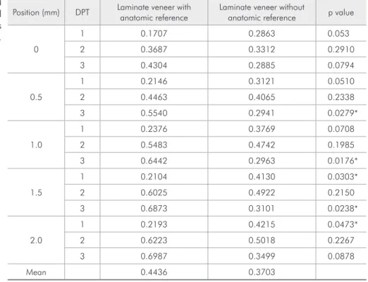
Table 2 shows the mean values of buccal contour when using or not using the reference tooth during the confection of the laminate veneers. For DPT 1,
Position (mm) DPT Control Laminate veneer p value
Mean SD
0
1
0.00
0.2285 0.1092 < 0.001*
2 0.3499 0.1307 < 0.001*
3 0.3594 0.1661 < 0.001*
0.5
1
0.05
0.2633 0.0813 < 0.001*
2 0.4264 0.1040 < 0.001*
3 0.4240 0.1965 < 0.001*
1.0
1
0.09
0.3072 0.1037 < 0.001*
2 0.5112 0.1092 < 0.001*
3 0.4702 0.2320 < 0.001*
1.5
1
0.14
0.3117 0.1138 < 0.001*
2 0.5473 0.1297 < 0.001*
3 0.4987 0.2482 < 0.001*
2.0
1
0.18
0.3204 0.1368 < 0.001*
2 0.5620 0.1370 < 0.001*
3 0.5243 0.2554 < 0.001*
*statistically significant at the level of 5% (Student’s t-test). DPT: Dental prosthetic technician.
when the veneers were confected without the ana-tomical reference tooth, statistically higher mean values of overcontour were observed at positions 1.5 and 2.0 mm. For DPT 2, no difference was observed when using or not using the reference tooth, at all positions. However, for DPT 3, when the reference tooth was used, statistically higher mean values of overcontour were observed at positions 0.5, 1.0 and 1.5 mm.
Discussion
Evaluating variations in dental contour is an important aspect of dental research4,12 because the
presence of overcontour in restorations is an iat-rogenic factor that propitiates gingival
inlamma-tion3,4,10 and dental breaking, compromising
estheti-cally the restored element.1,4
Aiming at eliminating the possibility of over-contour by the DPT related to lack of space for the restorative material, and also at reducing the prob-ability of fracture of the restoration, a wear to the depth of 1 mm during tooth preparation was ac-curately performed to create enough space for the veneer.12,17,19 However, overcontour occurred in all
laminate veneers confected in the present study, re-gardless of which DPT performed the procedure and of the use or not of an anatomical reference tooth (Table 1). Similar results were observed in previous studies, in which most of the restorations presented an increased volume when compared to the original tooth.10,17
An interesting inding of the present study was that when the values of buccal contour using or not an anatomic reference were compared (Table 2), it was observed that the veneers using the reference presented higher means of overcontour. Thus, the ideal anatomical outline that was supplied for con-fecting the veneers seems to have been neglected. Signiicant differences were observed at positions 0.5, 1.0 and 1.5 mm for DPT 3, and at positions 1.5 and 2.0 mm for DPT 1. Interestingly, the values of overcontour observed for DPT 3 were higher when the anatomical reference was used (Table 2). There-fore, these results suggest that DPTs are not usually able to adequately reproduce the dental contour dur-ing a prosthetic rehabilitation. This situation may be related to a lack of knowledge about the effects of over- or undercontour on the periodontal tissue.3,5,10
Position (mm) DPT Laminate veneer with anatomic reference
Laminate veneer without
anatomic reference p value
0
1 0.1707 0.2863 0.053
2 0.3687 0.3312 0.2910
3 0.4304 0.2885 0.0794
0.5
1 0.2146 0.3121 0.0510
2 0.4463 0.4065 0.2338
3 0.5540 0.2941 0.0279*
1.0
1 0.2376 0.3769 0.0708
2 0.5483 0.4742 0.1985
3 0.6442 0.2963 0.0176*
1.5
1 0.2104 0.4130 0.0303*
2 0.6025 0.4922 0.2150
3 0.6873 0.3101 0.0238*
2.0
1 0.2193 0.4215 0.0473*
2 0.6223 0.5018 0.2267
3 0.6987 0.3499 0.0878
Mean 0.4436 0.3703
*statistically significant at the level of 5% (Student’s t-test). DPT: Dental prosthetic technician.
It may also be explained by an attempt at increasing the thickness of the veneer to improve the strength of the restoration, thus reducing the possibility of fracture of the prosthetic component.4
It must be pointed out that coronary contour may vary in different regions of the mouth, and also in a same tooth. Therefore, it is necessary to use the natural outline as a model during the reconstruction of the tooth contour,5 aiming at obtaining a
satis-factory rehabilitation. Nevertheless, this situation was not observed in the present study since 100% of the veneers presented overcontour, even when a morphological reference was used during the sculp-ture of the laminate veneers.
Access to hygienic cleaning is a basic factor for the maintenance of periodontal health. In some cas-es, this is even more relevant than the restorative con-tour.20,21 A previous study concluded that a healthy
periodontium supports overcontour or undercontour of up to 1.0 mm during four months.22 All of the
ve-neers in the present study presented overcontour val-ues below 1.00 mm. However, in order to evaluate the consequences of this level of overcontour, longi-tudinal clinical studies would be necessary.
Also, the method used to measure overcontour is of signiicant importance,4,12 because it has a
signii-cant inluence on the compliance of the results. In
the present study, a contact proilometer was used because this device allows the standardization of the sample position at the moment of the measurement of each sample, thus increasing the conidence of the results obtained.
The hypothesis tested in the present study was not validated. Even when using a reference tooth as guidance during prosthetic confection, all the ve-neers presented overcontour. Thus, dentists should always supply the DPT with as much information as possible regarding the form and position of the natural tooth in the arch, so that this information can be used as guidance during the sculpture of the prosthetic restoration. The only way to modify the results observed in the present study is to establish a closer communication between dentist and DPT. Furthermore, the dentist should bear in mind that he is responsible for making a correct prosthetic prepa-ration, thus allowing a correct indirect restoration to be made by the DPT, and also for not accepting res-torations made with inadequate coronary contour.
Conclusion
Within of the limits of the present study, it could be concluded that 100% of the laminate veneers pre-sented overcontour, regardless of the use of an ana-tomic reference during the sculpture of the veneers.
References
1. Kissov HK, Popova EV, Katsarov SG. Position of crown margin in relation to the tooth preparation line. Folia Med. 2008;50(2):57-62.
2. Davis MV. The importance of contour on full coverage res-torations. Pract Periodontics Aesthet Dent. 1992;4(1):17-23. 3. Kohal RJ, Gerds T, Strub JR. Effect of different crown con-tours on periodontal health in dogs: Clinical results. J Dent. 2003;31(6):407-13.
4. Becker CM, Kaldahl WB. Current theories of crown contour, margin placement, and pontic design. 1981. J Prosthet Dent. 2005;93(2):107-15.
5. Yu H, Li Q, Hu J, Wang Y. An improved method to analyse tooth and restoration contour using image analysis: applica-tion in the maxillary anterior teeth in Chinese populaapplica-tion. Arch Oral Biol. 2008;53(6):503-8.
6. Broadbent JM, Williams KB, Thomson WM, Williams SM. Dental restorations: a risk factor for periodontal attachment loss? J Clin Periodontol. 2006;33(11):803-10.
7. Matthews DC, Tabesh M. Detection of localized tooth-related factors that predispose to periodontal infections. Periodontol 2000. 2004;34(1):136-50.
8. Gilmore N, Sheiham A. Overhanging dental restorations and periodontal disease. J Periodontol. 1971;42(1):8-12. 9. Schillingburg HT, Hobo S, Whitsett LD, Jocobi R, Brackett
SE. Fundamentals of fixed prosthodontics. 3rd ed. Chicago
(IL): Quintessence Publishing Co.; 1997.
10. Kissov HK, Todorova BP, Popova EV. Correlation between overcontouring of fixed prosthetic constructions and accu-mulation of dental plaque. Folia Med. 2001;43(1-2):80-3. 11. Khalaf K, Elcock C, Smith RN, Brook AH. Fluctuating dental
asymmetry of multiple crown variables measured by an image analysis system. Arch Oral Biol. 2005;50(2):249-53. 12. Alhouri N, Watts DC, McCord JF, Smith PW. Mathematical
13. Hahn P, Gustrav M, Hellwig E. An in vitro assessment of the strength of porcelain veneers dependent on tooth preparation. J Oral Rehabil. 2000;27(12):1024-9.
14. Burch JG. Miller JB. Evaluating crown contours of a wax pattern. J Prosthet Dent. 1973;30(4):454-8.
15. Koidis PT, Burch JG, Melfi RC. Clinical crown contours: contemporary view. J Am Dent Assoc. 1987;114(6):792-5. 16. Croll BM. Emergence profile in natural tooth contour. Part
I: photographic observations. J Prosthet Dent. 1989;62(1):4-10.
17. Meijering AC, Peters MC, DeLong R, Pintado MR, Creugers NH. Dimensional changes during veneering procedures on discoloured teeth. J Dent. 1998;26(7):569-76.
18. Padbury Jr A, Eber R, Wang HL. Interactions between the gingiva and the margin of restorations. J Clin Periodontol. 2003;30(5):379-85.
19. Croll BM. Emergence profile in natural tooth contour. Part II: clinical considerations. J Prosthet Dent. 1990;63(4):374-9. 20. Tjan AHL, Freed H, Miller GD. Current controversies in axial
contour design. J Prosthet Dent. 1980;44(5):536-9.
21. Kohal RJ, Pelz K, Strub JR. Effect of different crown contours on periodontal health in dogs. Microbiological results. J Dent. 2004;32(2):153-9.