Micro-morphological changes prior to
adhesive bonding: high-alumina and
glassy-matrix ceramics
Abstract: The aim of this study was to qualitatively demonstrate surface micro-morphological changes after the employment of different surface conditioning methods on high-alumina and glassy-matrix dental ceram-ics. Three disc-shaped high-alumina specimens (In-Ceram Alumina, INC) and 4 glassy-matrix ceramic specimens (Vitadur Alpha, V) (diameter: 5 mm and height: 5 mm) were manufactured. INC specimens were sub-mitted to 3 different surface conditioning methods: INC1 - Polishing with silicon carbide papers (SiC); INC2 - Chairside air-borne particle abrasion (50 µm Al2O3); INC3 - Chairside silica coating (CoJet; 30 µm SiOx). Vita-dur Alpha (V) specimens were subjected to 4 different surface condition-ing methods: V1 - Polishing with SiC papers; V2 - HF acid etching; V3 - Chairside air-borne particle abrasion (50 µm Al2O3); V4 - Chairside silica coating (30 µm SiOx). Following completion of the surface conditioning methods, the specimens were analyzed using SEM. After polishing with SiC, the surfaces of V specimens remained relatively smooth while those of INC exhibited topographic irregularities. Chairside air-abrasion with either aluminum oxide or silica particles produced retentive patterns on both INC and V specimens, with smoother patterns observed after silica coating. V specimens etched with HF presented a highly porous surface. Chairside tribochemical silica coating resulted in smoother surfaces with particles embedded on the surface even after air-blasting. Surface condi-tioning using air-borne particle abrasion with either 50 µm alumina or 30 µm silica particles exhibited qualitatively comparable rough surfaces for both INC and V. HF acid gel created the most micro-retentive surface for the glassy-matrix ceramic tested.
Descriptors: Air abrasion, dental; Hydroluoric acid; Acid etching, dental; Ceramics.
Marco Cícero Bottino(a)
Mutlu Özcan(b)
Paulo Guilherme Coelho(c)
Luiz Felipe Valandro(d)
José Carlos Bressiani(e)
Ana Helena Almeida Bressiani(e)
(a)Department of Materials Science and
Engineering, University of Alabama at Birmingham, Birmingham, USA.
(b)Department of Dentistry and Dental
Hygiene, University Medical Center Groningen, University of Groningen, Groningen, The Netherlands.
(c)Department of Biomaterials and
Biomimetics, New York University College of Dentistry, New York, USA.
(d)Department of Restorative and Prosthetic
Dentistry, School of Dentistry, Federal University of Santa Maria, Santa Maria, Brazil.
(e)Materials Science and Technology Center,
Institute for Energy and Nuclear Research (IPEN), São Paulo, Brazil.
Corresponding author:
Marco Cícero Bottino
Materials Science and Engineering University of Alabama at Birmingham BEC 254 - 1530 3rd Avenue South
Birmingham - AL - USA 35294-4461
E-mail: [email protected]
Introduction
With the addition of crystals such as quartz and aluminum oxide as reinforcing components, me-chanical properties of dental ceramic materials have signiicantly improved.1 New ceramic systems have been introduced and recommended as substitutes for metalloceramic restorations. One such ceramic system is In-Ceram (Vita-Zahnfabrik), an alumi-nous ceramic (~ 80 wt%) iniltrated with glass. This system has demonstrated lexural strength three to four times higher compared to other ceramics.2 A restoration made of In-Ceram has an aluminum-sin-tered core that is later veneered with a glass ceramic (Vitadur Alpha, Vita-Zahnfabrik). This veneering ceramic is composed of oxidized crystals of alumi-num, dispersed throughout its vitreous amorphous matrix.2
A key determinant for the clinical success of re-inforced ceramic restorations is the achievement of reliable bond strength between luting agent and in-ternal surfaces of the restoration. In case of chipping or fracture of ceramic restorations, a reliable repair strength with composite is desirable.3 However, in order to maximize the adhesion between new ce-ramic restorative systems and resin cements or re-pair composites, various procedures for the prepara-tion of the ceramic surface have been reported.3-26
The purpose of this study was to qualitatively demonstrate micro-morphological changes in two commonly used ceramic materials after various sur-face treatment methods by means of Scanning Elec-tron Microscopy (SEM) that would improve our un-derstanding of surface changes that would affect the bond strength results reported in dental literature.
Material and Methods
Specimen preparation
Disc-shaped specimens of high-alumina (INC) (In-Ceram Alumina, Vita-Zahnfabrik, Bad Säck-ingen, Germany) (n = 3) and glassy-matrix (V) (Vitadur Alpha, Vita-Zahnfabrik, Bad Säckingen, Germany) (n = 4) were obtained according to the manufacturer’s instructions. A custom designed separable stainless steel mold (height: 5 mm, diam-eter: 5 mm) was used for the standardization of the specimens.
Prior to surface treatment procedures, both sides of each specimen were wet ground inished in a polishing machine using a series of silicon carbide (SiC) abrasive papers in sequence (No. 240, 320, 400, 600 grit, Buehler, Lake Bluff, IL, USA) for 15 seconds under water irrigation at 300 rotations per minute (rpm) to obtain standardized lat and smooth surfaces. The specimens were then cleaned in an ultrasonic bath (Vitasonic) (Vita Zahnfabrik, Bad Säckingen, Germany) containing isopropanol for 3 minutes and were air-dried (Method 1 - con-trol). Subsequently, surface conditioning methods and their combinations were applied on the speci-men surfaces. The specispeci-mens in each ceramic group were randomly assigned to one of the 4 surface con-ditioning methods for V and 3 methods for INC:
Surface conditioning methods
Method 2: HF acid gel (9.5%) (Ultradent® Por-celain Etch, South Jordan, UT, USA) was applied for 90 s, rinsed with distilled water for 20 seconds, and air-dried for 10 seconds according to the manu-facturer’s recommendations. It should be noted that HF acid gel application was excluded for INC speci-mens since this surface treatment was found not to be effective on changing surface morphology in this ceramic due to its microstructure and composi-tion.13,18
Method 3: Chairside airborne particle abrasion with 50 µm Al2O3 particles (Korox®, Bego, Bremen, Germany) was applied using an intraoral air abra-sion device (Microetcher) (Danville Inc., Danville Inc, San Ramon, CA, USA) from a distance of ap-proximately 10 mm, in circular motions perpendic-ular to the disk surface at a pressure of 2.8 bars for 14 seconds and air dried.
Method 4: In this method, silica coating process was achieved using the same intraoral abrasion de-vice under the same conditions as in Method 3, but this time 30 µm SiOx (CoJet-Sand®) (3 M Dental Products, St. Paul, MN, USA) particles were used.
Surface morphology evaluation
Eind-hoven, The Netherlands) at 1,500 X magniication with an acceleration voltage of 15 kV and working distance of 8 mm, after sputtering the ceramic sur-faces with a gold palladium alloy to a layer of ap-proximately 30 nm in thickness.
Results
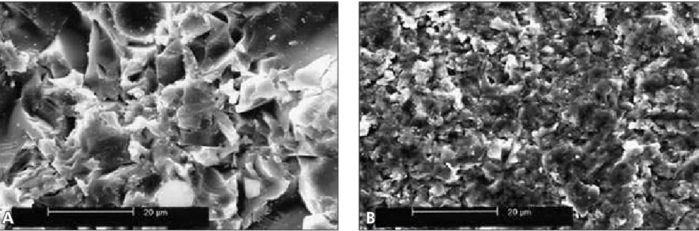
Wet ground inishing of the ceramic surfaces for 15 seconds using SiC abrasive papers resulted in smooth surfaces for V (Figure 1A). In contrast, INC surfaces presented more pronounced irregularities (Figure 1B).
V ceramic surfaces etched with HF acid gel pre-sented a porous surface pattern comparable to three-dimensional dendrites (Figure 2).
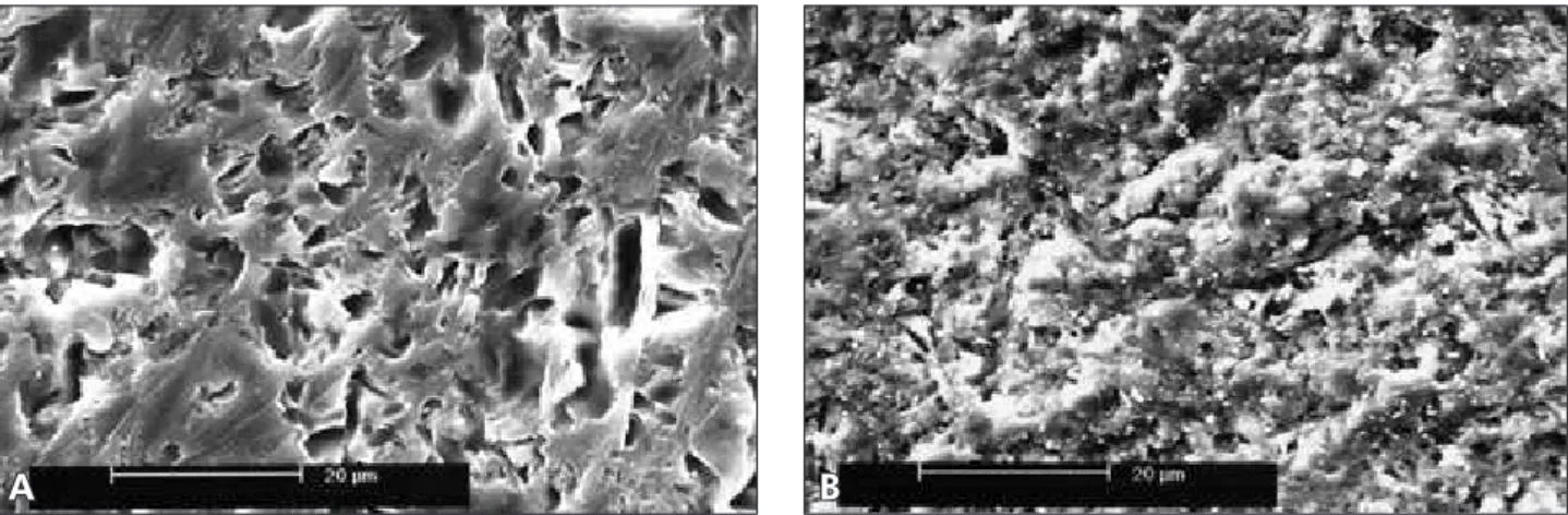
In the case of chairside air-abrasion procedure with 50 µm Al2O3, both V and INC ceramic sur-faces exhibited similar rough surface patterns that presented incorporation of sand particles on their surfaces (Figures 3A-B).
The use of chairside type of silica coating (Co-Jet System) with smaller silica particles resulted in smoother surfaces compared to surfaces obtained with 50 µm Al2O3. V and INC did not present re-markable differences in the qualitative surface to-pography after the use of both chairside abrasion methods (Figures 4A-B).
Discussion
In this study, an etchable and a non-etchable ce-ramic, namely V – a feldspathic aluminous cece-ramic,
and INC – high alumina, were selected for surface micro-morphology evaluation after various condi-tioning methods used prior to cementation or for repair purposes.
SEM evaluation after the SiC papers employment (Figures 1A-B) demonstrated different surface rough-ness patterns for the two ceramics tested. This dif-ference in surface roughness appearance may be at-tributed to differences between surface hardness and microstructure composition of the two ceramics.
Several investigations have considered HF acid etching followed by silane application a crucial and an effective method of surface conditioning for feldspathic ceramics, silica-based ceramics, and
Figure 1 - Surfaces of wet ground finished (A) V and (B) INC ceramics using SiC abrasive papers in sequence for 15 seconds under water irrigation. Note the rougher surface at the INC than at the V ceramic (original magnification 1,500 X).
A B
low-fusing ceramics, providing the rationale for this mode of treatment for the V ceramic evaluated in the present study.8,9,19,20 SEM micrographs of the V ceramic surface (Figure 2) treated with HF acid gel presented a porous surface pattern comparable to three-dimensional dendrites previously described by Phoenix, Shen20 (1995) or to a three-dimension-al lattice of voids and channels reported by Tylka, Stewart3 (1994). It is important to point out that HF acid etching was not used in the present study for In-Ceram ceramic, since it does not suficiently etch the surface, providing small amount of increase in the adhesion of resin cements to this ceramic.18,19 HF acid gels of approximately 10% have been con-sidered the most commonly used agent. However, this acid solution is known to be highly toxic,
caus-tic, and extremely deleterious when in contact with human tissues.5
Phoenix, Shen20 (1995) suggested that 9.5% HF acid etching resulted in the lowest contact angle val-ues when compared with other acid solutions and after aluminum oxide air-abrasion. The results of previous studiesdemonstrated that HF acid etching used as surface treatment yields the highest bond strength values, since surface topography changes provided by this surface treatment allowed better wettability of the silane due to the higher surface energy of the etched surface.3,7,11,18-21
Kern, Thompson13 (1995) have shown that the isolated use of air abrasion in In-Ceram ceramic surface does not produce satisfactory results when used in combination with dual-cured Bis-GMA
Figure 3 - Typical SEM views of (A) V and (B) INC ceramic surfaces with abundant sand particles (clearer areas) after 50 µm Al2O3 air-abrasion followed by air blasting (original magnification 1,500 X).
Figure 4 - Representative SEM micrographs of (A) V and (B) INC ceramic surfaces after chairside silica coating (30 µm SiOx, CoJet System). Note the similar rough pattern presented for both ceramic surfaces (original magnification 1,500 X).
A B
resin cement. As an alternative, Kern, Thompson13 (1995) attempted to add a silane agent to the treat-ment. Despite the silane addition, bond strength values luctuated due to the lack of silica on the In-Ceram surface.In the same study, aluminum oxide air-abrasion combined with a phosphate monomer (10-methacryloyloxydecyl-dihydrogen-phosphate, MDP) resin cement demonstrated signiicantly high-er bond strength compared to the use of conven-tional Bis-GMA. This phenomenon was attributed to the stable bond between phosphate ester groups of the monomer and metal oxides.
Based on the results obtained utilizing SEM (Figures 3A-B), it was possible to qualitatively show that the ceramic surfaces following air-abrasion with aluminum oxide presented sharp edges and fragments of abrasive agent after air blasting. These observations support the indings of Blixt et al.6 (2000), who observed the same phenomenon while utilizing a larger particle size as chairside air abra-sion media. According to Phoenix, Shen20 (1995), the sharp points observed were due to the microcracks produced by the impact of aluminum oxide parti-cles on the ceramic surface. Even though employing abrasive particles might produce intermediate wet-ting and contact angle values, according to Rou-let et al.21 (1995), the resulting surfaces were most likely not ideal for bonding since sharp irregulari-ties might serve as stress concentration points which could lead to fracture within the ceramic material. The advantage of the airborne particle abrasion pro-cedure over mechanical roughening with diamond burs has already been demonstrated.26 The abrasive process removes loose contaminated layers and the resulting roughened surface provides some degree of mechanical interlocking or “keying” with adhesive agents. It can be argued that the increased rough-ness also forms a larger surface area for the bond. While these mechanisms explain some of the gener-al characteristics of adhesion to roughened surfaces, it may also introduce physico-chemical changes that affect surface energy and wettability.
Silane coupling agents with the general chemical formula X-(CH2)3 Si-(OR)3 are capable of forming chemical bonds with both organic and inorganic surfaces.16,18,26 The chemical coupling is achieved by
the reaction of 10-methacryloxypropyltrimethoxysi-lane (MPS) to the silicon oxide phase in the ceram-ic. Particularly with feldspathic ceramics, HF acid etching combined with silane coupling agents yield-ed satisfactory results with respect to bond strength, and also revealed results superior to those of surface treatments with aluminum oxide air-abrasion.4,21 It is important to apply the silane coupling agent in a homogeneous monolayer since uncoated regions may eventually suffer from lack of resin adhesion.17 On the other hand, thick layers of silane will require longer hydrolysis and condensation time and there-fore act as a weak link between the ceramic and the resin cement.
In tribochemical silica-coating systems, mechan-ical energy is transferred to the substrate as kinetic energy, resulting in temperature change (spot heat-ing) on the surface. This treatment results in metal or ceramic surfaces covered by a thin layer of silica particles, increasing surface energy, improving mi-cromechanical retention, and potentially creating chemical adhesion sites for silane reaction.27 The laboratory type of silica coating has been success-fully used in combination with resin cements on glass-iniltrated densely sintered alumina ceramics, as well as zirconium oxide based ceramics.6,13,15,18 In the present study, chairside silica coating (Fig-ures 4A-B) created relatively smoother surfaces than alumina particles (Figures 3A-B). Comparable results were found in terms of bond strength with either laboratory or chairside silica coating and si-lanization system.23-25. Furthermore, small particle size (chairside silica coating) would not lead to ex-aggerated material loss during air-abrasion. Consid-ering the relatively smooth surfaces observed, fur-ther investigation should concern whefur-ther a rougher surface is really needed for high bond strength of resin cements to high alumina or other reinforced ceramics.
Conclusions
Based on the SEM evaluation of high-alumina and glassy-matrix ceramics after different surface conditioning methods, the following conclusions were drawn:
The V ceramic surface etched with HF acid only
presented a highly porous surface pattern; After chairside airborne particle abrasion with 50 µm Al2O3, both INC and V ceramic surfaces exhibited similar surface patterns and were cov-ered with sand particles even after air blasting;
•
The use of chairside type of silica coating (CoJet System) with smaller silica particles resulted in smoother surfaces than with 50 µm Al2O3, show-ing no distinct differences in the micro-morphol-ogy for both ceramics tested.
•
References
1. McLean JW, Hughes TH. The reinforcement of dental por-celain with ceramic oxides. Br Dent J. 1965;119(6):251-67. 2. Wagner WC, Chu TM. Biaxial flexural strength and
indenta-tion fracture toughness of three new dental core ceramics. J Prosthet Dent. 1996;76(2):140-4.
3. Tylka DF, Stewart GP. Comparison of acidulated phosphate fluoride gel and hydrofluoric acid etchants for porcelain-com-posite repair. J Prosthet Dent. 1994;72(2):121-7.
4. Barghi N. To silanate or not to silanate: Making a clinical decision. Compend Contin Educ Dent. 2000;21(8):659-64. 5. Beiran I, Miller B, Bentur Y. The efficacy of calcium
gluco-nate in ocular hydrofluoric acid burns. Hum Exp Toxicol. 1997;16(4):223-8.
6. Blixt M, Adamczak E, Lindén L, Odén A, Arvidson K. Bond-ing to densely sintered alumina surfaces: effect of sandblastBond-ing and silica coating on shear bond strength of luting cements. Int J Prosthodont. 2000;13(3):221-6.
7. Calamia JR, Simonsen RJ. Effect of coupling agents on bond strength of etched porcelain [Abstract 79]. J Dent Res. 1984;63:179.
8. Della Bona A, Anusavice KJ, Shen C. Microtensile strength of composite bonded to hot-pressed ceramics. J Adhes Dent. 2000;2(4):305-13.
9. Della Bona A, Anusavice KJ. Microstructure, composition, and etching topography of dental ceramics. Int J Prosthodont. 2002;15(2):159-67.
10. Frankenberger R, Krämer N, Sindel J. Repair strength of etched vs. silica-coated metal-ceramic and all-ceramic restora-tions. Oper Dent. 2000;25(3):209-15.
11. Hayakawa T, Horie K, Aida M, Kanaya H, Kobayashi T, Murata Y. The influence of surface conditions and silane agents on bond of resin to dental porcelain. Dent Mater. 1992;8(4):238-40.
12. Jardel V, Degrange M, Picard B, Derrien G. Correlation of topography to bond strength of etched ceramic. Int J Prostho-dont. 1999;12(1):59-64.
13. Kern M, Thompson VP. Bonding to glass infiltrated alumina ceramic: Adhesive methods and their durability. J Prosthet Dent. 1995;73(3):240-9.
14. Kern M, Thompson VP. Sandblasting and silica coating of a glass-infiltrated alumina ceramic: Volume loss, morphol-ogy, and changes in the surface composition. J Prosthet Dent. 1994;71(5):453-61.
15. Kern M, Wegner SM. Bonding to zirconia ceramic: adhesion methods and their durability. Dent Mater. 1998;14(1):64-71.
16. Matinlinna JP, Lassila LVJ, Özcan M, Yli-Urpo A, Vallittu PK. An introduction to silanes and their clinical applications in dentistry. Int J Prosthodont. 2004;17(2):155-64.
17. Nicholls JI. Tensile bond of resin cements to porcelain veneers. J Prosthet Dent. 1988;60(4):443-7.
18. Özcan M, Alkumru HN, Gemalmaz D. The effect of sur-face treatment on the shear bond strength of luting cement to a glass-infiltrated alumina ceramic. Int J Prosthodont. 2001;14(4):335-9.
19. Özden NA, Akaltan F, Can G. Effect of surface treatments of porcelain on the shear bond strength of applied dual-cured cement. J Prosthet Dent. 1994;72(1):85-8.
20. Phoenix RD, Shen C. Characterization of treated porcelain surfaces via dynamic contact angle analysis. Int J Prosthodont. 1995;8(2):187-94.
21. Roulet JF, Söderholm KJM, Longmate J. Effects of treatment storage conditions on ceramic/composite bond strength. J Dent Res. 1995;74(1):381-7.
22. Sun R, Suansuwan N, Kilpatrick N, Swain M. Characteriza-tion of tribochemically assisted bonding of composite resin to porcelain and metal. J Dent. 2000;28(6):441-5.
23. Valandro LF, Della Bona A, Bottino MA, Neisser MP. The effect of silica coating a densely sintered alumina ceramic on bonding to a resin cement. J Prosthet Dent. 2005;93(3):253-9.
24. Valandro LF, Leite FPP, Scotti R, Bottino MA, Neisser MP. Effect of ceramic surface treatment on the microtensile bond strength between a resin cement and an alumina-based ce-ramic. J Adhes Dent. 2004;6(4):327-32.
25. Valandro LF, Özcan M, Bottino MC, Scotti R, Bottino MA, Della Bona A. Bond strength of a resin cement to high-alu-mina and zirconia-reinforced ceramics: the effect of surface conditioning. J Adhes Dent. 2006;8(3):175-81.
26. Wolf DM, Powers JM, O’Keefe KL. Bond strength of com-posite to porcelain treated with new porcelain repair agents. Dent Mater. 1992;8(3):158-61.