ABSTRACT
www.scielo.br/jaos
The inluence of surface treatment on the implant
roughness pattern
Marcio Borges ROSA1, Tomas ALBREKTSSON2, Carlos Eduardo FRANCISCHONE3, Humberto Osvaldo SCHWARTZ FILHO4, Ann WENNERBERG5
1- DDS, PhD student, Division of Implantology, Department of Post-Graduation, School of Dentistry, São Leopoldo Mandic University, Campinas, SP, Brazil. 2- MD, PhD, ODhc Professor and Head, Department of Biomaterials, Gothenburg University, Gothenburg, Sweden.
3- DDS, MS, PhD, Professor and Head, Department of Post-Graduation, Division of Implantology, School of Dentistry, São Leopoldo Mandic University, Campinas, SP, Brazil.
4- DDS, MS, PhD, Department of Post-Graduation, Division of Implantology, School of Dentistry, University of Santo Amaro-UNISA, São Paulo, SP, Brazil. 5- LDS, PhD, Professor and Head, Department of Prosthodontics, Malmö University, Malmö, Sweden.
Corresponding address: Marcio Borges Rosa - Av. do Contorno 4849, 4º andar - 30110-035 - Belo Horizonte - MG - Phone: (31) 3284-2020 - e-mail: [email protected]
Received: March 30, 2012 - Modiication: July 18, 2012 - Accepted: August 20, 2012
A
n important parameter for the clinical success of dental implants is the formation of direct contact between the implant and surrounding bone, whose quality is directlyinluenced by the implant surface roughness. A screw-shaped design and a surface with
an average roughness of Sa of 1-2 µm showed a better result. The combination of blasting and etching has been a commonly used surface treatment technique. The versatility of this type of treatment allows for a wide variation in the procedures in order to obtain the desired roughness. Objectives: To compare the roughness values and morphological characteristics of 04 brands of implants, using the same type of surface treatment. In addition, to compare the results among brands, in order to assess whether the type of treatment determines the values and the characteristics of implant surface roughness. Material and methods: Three implants were purchased directly from each selected company
in the market, i.e., 03 Brazilian companies (Biomet 3i of Brazil, Neodent and Titaniumix)
and 01 Korean company (Oneplant). The quantitative or numerical characterization of the roughness was performed using an interferometer. The qualitative analysis of the surface topography obtained with the treatment was analyzed using scanning electron microscopy
images. Results: The evaluated implants showed a signiicant variation in roughness values: Sa for Oneplant was 1.01 μm; Titaniumix reached 0.90 μm; implants from Neodent 0.67 μm, and Biomet 3i of Brazil 0.53 μm. Moreover, the SEM images showed very different
patterns for the surfaces examined. Conclusions: The surface treatment alone is not able to determine the roughness values and characteristics.
Key words: Surface treatments. Blasting. Acid etched. Dental implants. Osseointegration.
INTRODUCTION
Since the discovery of osseointegration by Brånemark, the placement of titanium implants has been established as the treatment of choice for replacing missing teeth, thereby improving the patients’ quality of life13,18.
An important parameter for the clinical success of dental implants is the formation of direct contact between the implant and surrounding bone14. The quality of the bone-implant interface is directly
inluenced by the implant surface roughness1,9,10,23-25
which, since the early 1980s, has been identiied as
one of the six factors that are particularly important for incorporating the implant into the bone1.
Both morphology and surface roughness inluence
cell proliferation and differentiation, extracellular matrix synthesis, local production factors, and even cell shape4,10. Furthermore, the shape of the cell regulates its growth, gene expression, protein secretion, differentiation, and apoptosis7. Therefore, osteoblast adherence on implant surfaces is not
suficient for achieving osseointegration, much less
to enable the cell to receive the signals to induce its proliferation10. In addition, roughness not only facilitates the retention of osteogenic cells, but also allows them to migrate to the implant surface by osseoconductivity6. Faster and stronger bone formation provides greater stability during the repair process, allowing for even faster implant loading23-25. Oral implant surfaces have structures that are measurable on a macrometric scale in millimeters (mm), micrometric in micrometers (µm), and nanometric in nanometers (nm)2,3,18,22-24.
How these structures inluence repair has been the
subject of various publications and studies in recent years2,7,8,11,13,17,21,24.
Until now, certainties have been limited to the
inluence of implant design and surface roughness
on the micrometric scale. A screw-shaped design and a surface with an average roughness of Sa of 1-2 µm have shown better results2,10,24,25. Studies have shown that titanium implants with the proper roughness can improve bone-implant contact29, while also increasing the removal torque values21,29. On the other hand, increasing the surface roughness to the level of surfaces treated with plasma spray titanium, Sa above 2 µm, elicits a prejudiced and non-reinforced bone response10,23-25.
As a result, over the past 20 years, a large number of implant systems with different surface topographies have been introduced19. Oral implants are an example of the close link between research
and industry, since the laboratory indings often
become clinical applications. However, any change in the implant morphology, i.e., in its design, causes changes in the topography at the micrometric level and vice versa. Likewise, chemical changes cause physical changes and vice versa23-25. There are many types of surface treatments on the market. In general, all seek to alter the previously machined surface roughness, increasing it to levels considered optimal. A combination of blasting, followed by acid etching, has been a technique commonly used for surface treatment in recent years24. The main reason for the combination of methods is that hypothetically, blasting reaches an
optimal roughness and mechanical ixation, while
acid etching smooths the peaks and can add a high frequency component to the implant surface, with a potential importance for protein adherence, which is considered important during the early bone healing process24.
The surface characteristics obtained with blasting deformation depend on the type of particle used, its hardness, size, and impact velocity. The blasting process, usually performed with titanium (TiO2) or alumina (Al2O3) particles, allows for good control of the size of the resulting micro cavities. Some remaining particles may however, become embedded and contaminate the implant surface10.
Acid etching removes a few atomic layers from the deformed surface, part of the residual surface stress, and reduces the possibility of surface contamination by particles left over from the blasting process, because it also acts in cleaning the surface. These processes create micro cavities overlapping the pre-blasted rough surface.
each manufacturer has its own method of acid etching with regard to temperature, concentration of acids, and exposure time. In general, there
is dual acid etching, which is carried out by irst
immersing the implants in solutions of HCl+H2SO4, HNO3+HF, or HNO3. Thereafter, the implant is again immersed in an aqueous solution of HNO3 to stabilize the titanium oxide layer10,24.
The versatility of this type of treatment allows for a wide variation in the procedures to obtain the desired roughness. On the other hand, this
characteristic can produce signiicantly different
surfaces. Consequently, it is very important to characterize the surfaces to obtain the values envisioned by the proposed treatments. According to Wennerberg and Albrektsson23 (2000), a light interferometer is a safe and effective way to measure the roughness of screw-shaped implants.
This aim of this study was to characterize the surfaces, by means of using a light interferometer and scanning electron microscopy (SeM), of implants from four companies: 3 Brazilian and 1 Korean, which use blasting and acid etching treatments. The results were compared among the 4 companies.
MATERIAL AND METhODS
In accordance with this study design, three implants from each company would be analyzed16. Therefore, 03 implants were purchased from each of the following 3 Brazilian companies, namely Biomet 3i of Brazil (São Paulo, SP, Brazil); Neodent
(Curitiba, PR, Brazil) and Titaniumix (São José
dos Campos, SP, Brazil); and 01 implant from the Korean Oneplant Company (Seoul, Korea).
The implant surfaces were evaluated using a light interferometer (MicroXAMTM, Phaseshift,
Tucson, AZ, USA), which, according to Wennerberg
and Albrektsson23 (2000) is recommended for evaluating the roughness of implants with threads
at the micrometer level. A 50X magniication and
0.62 zoom were used, and the measured area was
264x200μm. The mean height of the measurements varied between 80 μm and 100 μm. The maximum resolution of this technique is 0.30 μm horizontally
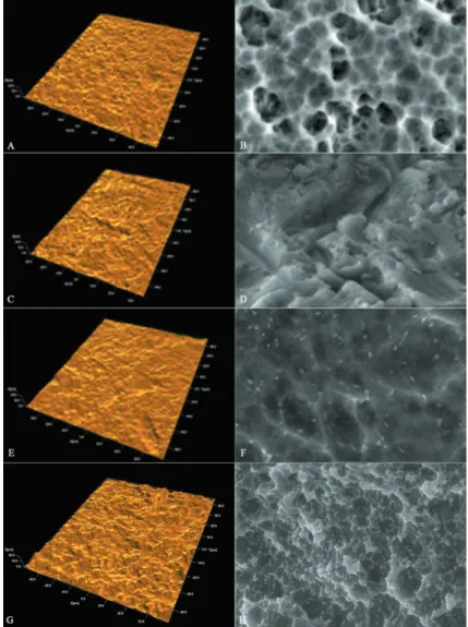
Figure 3- Interferometer images (left) and scanning electron microscopy (SEM) images 3,000X magniication (right). Note: A and B Neodent; C and D Titaniumix; E and F 3i Biomet; G and H Oneplant
Figure 2- a, b and c: Filter sequence in which the undulations and shapes have been removed
Note: a: Nanotite original; b: Nanotite 50X50 μm Gaussian ilter; c: Nanotite 50X50 μm Gaussian ilter low pass18
and 0.05 μm vertically.
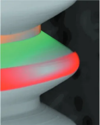
To evaluate an implant surface, three measurements are taken in different areas, i.e.,
the tops, valleys, and lanks of the threads (Figure
1) totaling nine measurements for each unit. In addition, a minimum of three samples were evaluated for each implant16.
The undulations from the machining process and the shape are considered separately to adequately describe the roughness obtained with the treatment.
A standard iltering process, with a 50X50 μm Gaussian ilter was used to make the separation
and evaluate the micrometric roughness (Figure 2). Surfascan software (Somicronic Instrument, Lyon, France) was used for this purpose, which also provides visual images and numerical descriptions.
The following parameters were used for the numerical description of the surface topography, which should preferably be in 3D:
Sa: represented the arithmetic mean of the roughness area from the mean plane.
Sds: represented the density, i.e., the number of peaks per unit of area.
Sdr: hybrid parameter that represented the ratio
between the developed surface area and a lat
reference area.
According to Albrektsson and Wennerberg2 (2004)implants can be divided into four different categories, depending on the surface roughness measured by the value of Sa: smooth (Sa<0.5
μm); minimally rough (Sa between 0.5-1.0 μm), moderately rough (Sa between 1.0-2.0 μm); rough (Sa>2.0 μm).
Images were also captured by scanning electron
microscopy on the top, lank and valley of the threads, with magniications at 65X, 350X, 1,000X,
3.000X and 5.000X. The purpose of these images was to allow a qualitative analysis of the changes obtained by the surface treatment, as well as to visualize the roughness characteristics and check the maintenance of the standard throughout the entire implant body.
Statistical evaluation
The implants were evaluated for signiicant
differences in surface topography at the micrometric level. The statistical analyses were performed with the software GraphPad Prism 5.0 (GraphPad Software, San Diego, CA, USA). The data was
analyzed by one-way ANOVA (Kruskall-Wallis test)
with the signiicance level set at p<0.05, and the
application of the Dunn’s multiple comparisons test.
RESULTS
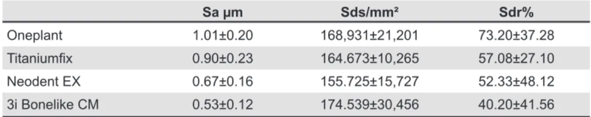
The evaluated implants showed a signiicant
variation in roughness values: Sa for Oneplant
was 1.01 μm; Titaniumfix reached 0.90 μm; implants from Neodent 0.67 μm, and Biomet 3i of Brazil 0.53 μm. Moreover, the SEM
images showed very different patterns for the surfaces examined (Table 1).
Figure 3 shows the interferometer and SeM images for the implants in this group:
DISCUSSION
When implants irst began to be manufactured
in developing countries, such as Brazil and Korea, the majority of the companies adopted models and surface treatments of well-known implants. Since then, no independent assessment has been made of the quality of the implant surfaces, by means of characterizing the roughness obtained, and their clinical performance.
Among the evaluated parameters, the most important are as follows: Sa, which represent the arithmetic mean, in 3D, for the height of the peaks and valleys of surface roughness; Sds, which represents the density of peaks per unit
area; and inally, Sdr, which is the intersection
of the irst two parameters and represents the
surface enlargement2,3,12,23. The analysis of these
factors, and prior knowledge of their inluence on
the repair process, makes it easier to signal the
behavior of a given surface. There is strong scientiic
evidence about the importance of Sa in the healing process2,10,25,29.
The type of titanium used needs to be added to these variables because its hardness directly affects the roughness obtained with the treatment. In other words, the same treatment can produce different roughness patterns in different grades of titanium10,23.
For this group of implants, with the same type of treatment - blasting and acid etching - different results were found. The Oneplant, from Korea showed a Sa value of 1.01 μm. All of the Brazilian
Sa µm Sds/mm² Sdr%
Oneplant 1.01±0.20 168,931±21,201 73.20±37.28
Titaniumix 0.90±0.23 164.673±10,265 57.08±27.10
Neodent EX 0.67±0.16 155.725±15,727 52.33±48.12
3i Bonelike CM 0.53±0.12 174.539±30,456 40.20±41.56
implants presented Sa values of less than 1.0
μm, with the Titaniumix reaching 0.90 μm, while those from Neodent presented a mean of 0.67 μm.
Moreover, the Bonelike implants from Biomet 3i of Brazil, which uses titanium alloy (Ti6Al4V), presented Sa values of only 0.53 μm. In this particular case,
the inluence of the titanium alloy hardness on the
low roughness should be highlighted, irrespective of which treatment would be appropriate for the materials to produce the desired roughness, and not the opposite.
When evaluating the Sdr to compare the surface enlargement, different values were also found: Oneplant had an Sdr value of 73.20%; while
Titaniumix showed 57.08%; Neodent a Sdr value of 52.33%; and Bonelike CM, a Sdr value of 40.20%, bearing in mind that an Sdr value of about 50% promotes strong bone formation26-28.
For comparison, the SLA implants from Straumann, with the same type of treatment, an
extensive scientiic documentation and good clinical
performance have shown a Sa value of 1.53 μm and a Sdr value of 74.52%22.
The long-term clinical results obtained with minimally or moderately rough implants have been shown to be very similar under normal conditions5. Moreover, when placed under adverse conditions, such as in bone grafts and type IV bone or for short implants, among others, comparative studies have
shown a signiicant superiority of the implants with
a moderately rough surface12,14,15.
Thus, using an implant without knowing its real roughness, the bone repair can be weaker than expected, which is highly undesirable, especially in critical cases, where more is expected of the surface properties of the implants.
In the literature, the surface topography of blasted and etched surfaces varies greatly because of the different measuring equipment and evaluation techniques, but mainly because the blasting and etching procedures may differ as well. In blasting, the types of particles used, along with the size and impact velocity, are directly responsible for the results. In acid etching, the type of acid, exposure time, and temperature are essential factors for surface characterization10. However, in the vast majority of studies, blasted and etched implants
are moderately rough, between 1.0-2.0 μm, which
is considered ideal and shows better results2,10,24,25. When analyzing the SeM images, it is quite clear how the same type of treatment may lead to surfaces with very different characteristics, and therefore, the need for clinical studies that prove
the eficiency of each surface obtained, must be
reinforced.
Further clinical studies are needed to show the factors that these differences may really represent. However, similar surface treatments do
not necessarily lead to the same results. According to Wennerberg and Albrektsson24,25 (2009, 2010) even the roughness of a machined surface, as well as blasted, acid etched and anodized surfaces, may vary considerably. They also point out that many studies and companies leave out the surface topographic characterization in the false belief that their treatment alone will determine the roughness of this surface24.
When the surface topography is altered, its chemical and/or physical characteristics can change at the same time, even accidentally24. Often, there are no short, medium, and long-term studies to evaluate the effects of these changes on the repair processes and longevity of implant osseointegration. This occurs mainly in implants, such as those from Brazilian companies that use surface treatments established by other companies, but do not conduct clinical studies to evaluate their clinical performance. It is not practicable to endorse the results of other implants only because they have the same type of surface treatment.
CONCLUSION
The results, with a significant variation in roughness values among the implants with the same type of surface treatment showed that the characterization of the surface of each implant is very important for a real evaluation of the obtained results.
Therefore, even if established treatments are used on the surface of these implants, it is very important that each company conducts its own laboratory and clinical experiments to validate them.
REFERENCES
1- Albrektsson T, Brånemark PI, Hansson HA, Lindström J.
Osseointegrated titanium implants. Requirements for ensuring a long-lasting, direct bone-to-implant anchorage in man. Acta Orthop Scand. 1981;52:155-70.
2- Albrektsson T, Wennerberg A. Oral implant surfaces: part 1 - review focusing on topographic and chemical properties of different surfaces and in vivo responses to them. Int J Prosthodont. 2004;17:536-43.
3- Albrektsson T, Wennerberg A. Oral implant surfaces: part 2 - review focusing on clinical knowledge of different surfaces. Int J Prosthodont. 2004;17:544-64.
4- Anselme K, Bigerelle M. Topography effects of pure titanium substrates on human osteoblast long-term adhesion. Acta Biomater. 2005;1:211-22.
5- Balshe AA, Assad DA, eckert Se, Koka S, Weaver AL. A retrospective study of the survival of smooth- and rough-surface dental implants. Int J Oral Maxillofacial Implants. 2009;24:1113-8. 6- Braceras I, De Maeztu MA, Alava JI, Gay-escoda C. In vivo
7- Brunette MD. Principles of cell behaviour on titanium surfaces and their application to implanted devices. In: Brunette DM, Tengvall P, Textor M, Thomsen P, editors. Titanium in Medicine. Berlin: Spinger Verlag; 2001. p. 485-512.
8- Chang PC, Lang NP, Giannobile WV. evaluation of functional dynamics during osseointegration and regeneration associated with oral implants. Clin Oral Implants Res. 2010;21:1-12. 9- Cooper LF. A role for surface topography in creating and maintaining bone at titanium endosseous implants. J Prosthet Dent. 2000;84:522-34.
10- elias CN, Meirelles L. Improving osseointegration of dental implants. expert Rev Med Devices. 2010;7:241-56.
11- elias CN, Oshida Y, Lima JH, Muller CA. Relationship between surface properties (roughness, wettability and morphology) of titanium and dental implant removal torque. J Mech Behav Biomed Mater. 2008;1:234-42.
12- Friberg B, Jemt T. Rehabilitation of edentulous mandibles by
means of ive TiUnite implants after one-stage surgery: a 1-year
retrospective study of 90 patients. Clin Implant Dent Relat Res. 2008;10:47-54.
13- Mendonça G, Mendonça DB, Aragão FJ, Cooper LF. Advancing dental implant surface technology - from micron- to nanotopography. Biomaterials. 2008;29:3822-35.
14- Pinholt eM. Brånemark and ITI dental implants in the human bone-grafted maxilla: a comparative evaluation. Clin Oral Implants Res. 2003;14(5):584-92.
15- Rocci A, Martignoni M, Gottlow J. Immediate loading of Brånemark System TiUnite and machined-surface implants in the posterior mandible: a randomized open-ended clinical trial. Clin Implant Dent Relat Res. 2003;5(Suppl 1):57-63.
16- Rosa MB, Albrektsson T, Francischone Ce, Schwartz Filho HO, Wennerberg A. Micrometric characterization of the implant
surfaces from the ive largest companies in Brazil, the second
largest worldwide implant market. Int J Oral Maxillofac Implants. In press 2012.
17- Shalabi MM, Gortemaker A, Van't Hof MA, Jansen JA, Creugers NH. Implant surface roughness and bone healing: a systematic review. J Dent Res. 2006;85:496-500.
18- Shalabi MM, Wolke JG, Jansen JA. The effects of implant
surface roughness and surgical technique on implant ixation in
an in vitro model. Clin Oral Implants Res. 2006;17:172-8.
19- Strassburger C, Kerschbaum T, Heydecke G. Inluence of
implant and conventional prostheses on satisfaction and quality of life: a literature review. Part 2: qualitative analysis and evaluation of the studies. Int J Prosthodont. 2006;19:339-48.
20- Sul YT, Johansson C, Albrektsson T. Which surface properties enhance bone response to implants? Comparison of oxidized magnesium, TiUnite, and Osseotite implant surfaces. Int J Prosthodont. 2006;19:319-28.
21- Sul YT, Johansson CB, Jeong Y, Wennerberg A, Albrektsson T. Resonance frequency and removal torque analysis of implants with turned and anodized surface oxides. Clin Oral Implants Res. 2002;13(3):252-9.
22- Svanborg LM, Andersson M, Wennerberg A. Surface characterization of commercial oral implants on the nanometer level. J Biomed Mater Res B Appl Biomater. 2010;92:462-9. 23- Wennerberg A, Albrektsson T. Suggested guidelines for the topographic evaluation of implant surfaces. Int J Oral Maxillofac Implants. 2000;15:331-44.
24- Wennerberg A, Albrektsson T. effects of titanium surface topography on bone integration: a systematic review. Clin Oral Implants Res. 2009;20(Suppl 4):172-84.
25- Wennerberg A, Albrektsson T. On implant surfaces: a review of current knowledge and opinions. Int J Oral Maxillofac Implants. 2010;25:63-74.
26- Wennerberg A, Albrektsson T, Andersson B. Bone tissue response to commercially pure titanium implants blasted with
ine and coarse particles of aluminum oxide. Int J Oral Maxillofac
Implants. 1996;11:38-45.
27- Wennerberg A, Albrektsson T, Andersson B, Krol JJ. A histomorphometric and removal torque study of screw-shaped titanium implants with three different surface topographies. Clin Oral Implants Res. 1995;6:24-30.
28- Wennerberg A, Albrektsson T, Johansson C, Andersson B. experimental study of turned and grit-blasted screw-shaped implants with special emphasis on effects of blasting material and surface topography. Biomaterials. 1996;17:15-22.