Article
J. Braz. Chem. Soc., Vol. 23, No. 1, 25-31, 2012. Printed in Brazil - ©2012 Sociedade Brasileira de Química
0103 - 5053 $6.00+0.00
A
*e-mail: [email protected]
Synthesis, Characterization and Photophysical Properties of
ESIPT Reactive Triazine Derivatives
Marcelo D. Kuplich, Fábio S. Grasel, Leandra F. Campo, Fabiano S. Rodembusch and Valter Stefani*
Laboratório de Novos Materiais Orgânicos, Instituto de Química, Universidade Federal do Rio Grande do Sul, Av. Bento Gonçalves 9500, CP 15003, 91501-970 Porto Alegre-RS, Brazil
Uma série de quatro compostos fotoativos derivados da triazina foi obtida a partir da substituição nucleofílica aromática no cloreto cianúrico. Os compostos foram caracterizados por espectroscopia de infravermelho (IR) e ressonância magnética nuclear (NMR de 1H e 13C), além de espectrometria
de massas de alta resolução (HRMS MALDI). A espectroscopia de absorção na região do UV-Vis e a emissão de fluorescência (no estado sólido e em solução) foram também utilizadas para estudar o comportamento fotofísico destes compostos. Os derivados obtidos são fluorescentes na região do azul-laranja por um mecanismo de transferência protônica intramolecular no estado excitado e apresentam um grande deslocamento de Stokes (6365-10290 cm-1). Os derivados sintetizados
neste trabalho reagiram com sucesso com fibras de celulose para dar novos compostos celulósicos fluorescentes.
Four new reactive fluorescent triazine derivatives were obtained from nucleophilic aromatic substitution of cyanuric chloride. The compounds were characterized by infrared spectroscopy (IR), nuclear magnetic resonance (13C and 1H NMR) and high resolution mass spectrometry
(HRMS MALDI). UV-Vis and steady-state fluorescence (in solution and in solid state) spectroscopies were also applied to characterize the photophysical behavior. The dyes are fluorescent by an intramolecular proton transfer mechanism (ESIPT) in the blue-orange region, with a large Stokes shift between 6365-10290 cm-1. The fluorescent cyanuric derivatives could
successfully react with cellulose fibers to give new fluorescent cellulosic materials. Keywords: triazine,benzazoles, cyanuric chloride, ESIPT, reactive dyes, cellulose
Introduction
The 2,4,6-trichloro-1,3,5-triazine (TCT), so-called cyanuric chloride, is an important triazine derivative and a well-known precursor in organic synthesis.1-11 This precursor have become a very attractive field of research by virtue of the applications that can be envisaged with the obtained products, such as polymers,12-15 non-linear optical materials16-18 and new photoactive materials.19-23 A particular interest has been reported on the use of cyanuric chloride in reactive dyes24 since they are able to bond hydroxy groups from the cellulose and amino or thiol from proteins or polyamides.25 In this way, several structures have been reported using TCT as molecular link between the chromophore or fluorescent dye and the organic matrix.26-29
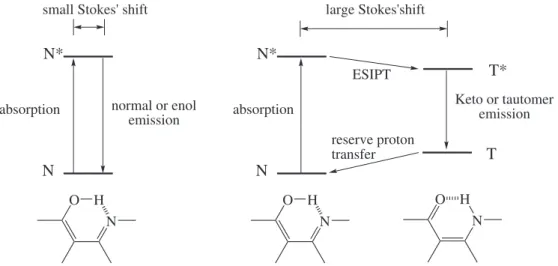
On the other hand, the benzazoles often show a large Stokes shift due to an excited state intramolecular proton
transfer (ESIPT) mechanism.30 This phenomenon has widespread implications as optical sensors,31-34 photoactive hybrid materials35,36 and new polymeric materials.37
Despite of the great attention of ESIPT-exhibiting dyes and the versatile applications of cyanuric chloride in organic synthesis as starting material to produce dyes, herbicides, polymers and compounds of pharmaceutical interest,38 this paper presents the synthesis and the photophysical characterization of new ESIPT reactive dyes based on the triazine moiety. Additionally, the obtained derivatives were tested as fluorophores for cellulosic materials.
Experimental
Materials and methods
Synthesis, Characterization and Photophysical Properties of ESIPT Reactive Triazine Derivatives J. Braz. Chem. Soc.
26
purified using standard procedures. Spectroscopic grade solvents (Merck) were used for fluorescence and UV-Vis measurements. Melting points (mp) were measured with a Gehaka PF 1000 apparatus and are uncorrected. Infrared spectra were recorded on a Shimadzu FTIR8300 in KBr pellets. 1H and 13C NMR spectra were performed on a VARIAN INOVA YH300 using tetramethylsilane (TMS) as the internal standard and DMSO-d6 (Aldrich)
as the solvent at room temperature. UV-Vis absorption spectra were performed on a Shimadzu UV-1601PC spectrophotometer. Steady state fluorescence spectra were measured with a Hitachi spectrofluorometer model F-4500. Spectrum correction was performed to enable measuring a true spectrum by eliminating instrumental response such as wavelength characteristics of the monochromator or detector using Rhodamine B as a standard (quantum counter). For the photophysical measurements in the solid state, the photoactive cellulose fibers were measured in bulk using a solid sample holder. In this device, the light beam was irradiated to the sample at an angle of ca. 30°, and the light beam from the sample was reflected at an angle of ca.
−60°. All experiments were performed at room temperature
in a concentration of 10-6 mol L-1. HRMS spectra were recorded with a Bruker Reflex III spectrometer.
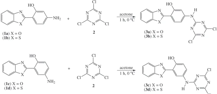
Synthesis of the triazine derivatives
The dyes 1a-d were prepared using a previously described methodology.39 The triazine benzazole derivatives
3a-d were prepared according to Figure 2. In a typical experiment, an equimolar amount of the amino derivative
1a-d and 2,4,6-trichloro-1,3,5-triazine were both dissolved in acetone. The dye solution was added dropwise into the triazine solution, cooled up to 0 ºC, followed by addition of a Na2CO3 solution (10%). The final solution was mixed
for 1 h. The crude product, which precipitates, was washed with water and cold acetone and dried at room temperature. The purification was made by recristallization using dioxane/water. The final yields were from 70 to 96%.
2-[4’-(N-4,6-Dichloro-1,3,5-triazin-2-yl)-2’-hydroxy phenyl] benzoxazole (3a)
Yield: 70%; mp > 350 ºC; IR (KBr) ν
max/cm-1 3296
ν(N-H), 3059 ν
arom(C-H), 1618 ν(C=N), 1537 and 1501
ν
arom(C=C), 1238 ν(Ar-O), 1188 ν(C-N), 748 ν(C-Cl); 1H NMR (300 MHz, DMSO-d
6) d/ppm 10.96 (s, 1H,
OH), 7.98 (d, 1H, H6’, Jo 8.7 Hz), 7.88-7.76 (m, 2H, H4 and H7), 7.66 (d, 1H, H3’, Jm 2.0 Hz), 7.50-7.38 (m, 2H,
H5 and H6), 7.21 (dd, 1H, H5’, Jm 2.0 Hz and Jo 8.7 Hz);
13C NMR (75.4 MHz, DMSO-d
6) d/ppm 162 (C2), 158 (C4’a), 154 (C4’b and C4’c), 153 (C2’), 149 (C8), 142 (C4’), 139 (C9), 128 (C5), 126 (C6 or C6’), 125 (C6’ or C6), 119 (C4), 112 (C3’ or C7), 111 (C7 or C3’), 108 (C5’), 106 (C1’); exact mass: 373.0133 g mol-1; the exact molecular mass for C16H9Cl2N5O2 m/z 373.012 was found by HRMS (MALDI).
2-[4’-(N-4,6-Dichloro-1,3,5-triazin-2-yl)-2’-hydroxy phenyl] benzothiazole (3b)
Yield: 80%; mp > 350 ºC; IR (KBr) ν
max/cm-1 3331
ν(N-H), 3069 ν
arom(C-H), 1612 ν(C=N), 1566 and 1481
ν
arom(C=C), 1238 ν(Ar-O), 1186 ν(C-N), 752 ν(C-Cl); 1H NMR (300 MHz, DMSO-d
6) d/ppm 11.19 (s, 1H, OH),
8.24 (d, 1H, H3’, Jm 2.7 Hz), 7.94-7.80 (m, 2H, H4 and H7), 7.72 (dd, 1H, H5’, Jm 2.7 Hz and Jo 9.0 Hz), 7.54-7.42 (m,
2H, H5 and H6), 7.19 (d, 1H, H6’, Jo 9.0 Hz);
13C NMR (75.4 MHz, DMSO-d6) d/ppm 170 (C2), 164 (C4’a), 162 (C4’b and C4’c), 155 (C2’), 149 (C9), 140 (C4’), 129 (C8), N*
T*
T
absorption Keto or tautomer emission
O N H O N H N
normal or enol emission N* absorption O N H N
small Stokes' shift large Stokes'shift
ESIPT
reserve proton transfer
128 (C4), 126 (C5 or C6), 125 (C5 or C6), 121 (C6’), 119 (C7 or C3’), 118 (C3’ or C7), 111 (C5’), 110 (C1’); exact mass: 388.9905 g mol-1; the exact molecular mass for C16H9Cl2N5OS m/z 388.990 was found by HRMS (MALDI).
2-[5’-(N-4,6-Dichloro-1,3,5-triazin-2-yl)-2’-hydroxy phenyl] benzoxazole (3c)
Yield: 83%; mp > 350 ºC; IR (KBr) ν
max/cm -1 3302
ν(N-H), 3065 ν
arom(C-H), 1616 ν(C=N), 1587 and 1501
ν
arom(C=C), 1238 ν(Ar-O), 1167 ν(C-N), 739 ν(C-Cl); 1H NMR (300 MHz, DMSO-d
6) d/ppm 10.98 (s, 1H, OH),
8.23 (d, 1H, H6’, Jm 2.7 Hz), 7.94-7.84 (m, 2H, H4 and H7), 7.61 (dd, 1H, H4’, Jm 2.7 Hz and Jo 9.0 Hz), 7.54-7.44 (m,
2H, H5 and H6), 7.17 (d, 1H, H3’, Jo 9.0 Hz);
13C NMR (75.4 MHz, DMSO-d6) d/ppm 170 (C2), 164 (C4’a), 162 (C4’b and C4’c), 155 (C2’), 149 (C8), 140 (C9), 129 (C4’), 126 (C5 or C6), 125 (C5 or C6), 121 (C4), 119 (C6’ or C3’), 118 (C3’ or C6’), 111 (C7), 110 (C1’); exact mass: 373.0133 g mol
-1; the exact molecular mass for C16H9Cl2N5O2m/z 373.012 was found by HRMS (MALDI).
2-[5’-(N-4,6-Dichloro-1,3,5-triazin-2-yl)-2’-hydroxy phenyl] benzothiazole (3d)
Yield: 96%; mp > 350 ºC; IR (KBr) ν
max/cm-1 3285
ν(N-H), 3063 ν
arom(C-H), 1556 and 1501 νarom(C=C), 1238
ν(Ar-O), 1198 ν(C-N), 758 ν(C-Cl); 1H NMR (300 MHz, DMSO-d6) d/ppm 11.09 (s, 1H, OH); 8.41 (d, 1H, H6’,
Jm 2.7 Hz). 8.15 (d, 1H, H4 or H7), 8.05 (d, 1H, H7 or H4), 7.60 (dd, 1H, H4’, Jm 2.7 Hz and Jo 9.0 Hz), 7.54 (t, 1H,
H5 or H6), 7.45 (t, 1H, H6 or H5), 7.13 (d, 1H, H3’, Jo 9.0 Hz);
13C NMR (75.4 MHz, DMSO-d
6) d/ppm 170 (C2), 169 (C5’a), 164 (C5’b or C5’c), 154 (C2’), 151 (C9), 135 (C5’), 129 (C8), 127 (C5 or C6), 127 (C5 or C6), 125 (C4), 122 (C7 or C4’), 122 (C4’ or C7), 122 (C6’), 118 (C1’), 117 (C3’); exact mass: 388.9905 g mol-1; the exact molecular mass for C16H9Cl2N5OS m/z 388.989 was found by HRMS (MALDI).
Dye incorporation into the cellulose fibers
In a typical experiment, the cellulose was stained by addition of the cotton (100 mg) into an aqueous solution of Na2SO4 (0.05 mol L-1) and 0.2 ml of sodium hydroxide (10% m/v) followed by the addition of the fluorescent dye (1 mg mL-1 in DMSO). The reaction mixture was allowed to react for 1 h at 45 ºC. The crude stained cotton was washed several times with water, followed by Soxhlet extraction using acetone for 3 h in order to remove the unreacted fluorescent dye. The percentages of the triazine dye incorporation were evaluated using the absorption data from UV-Vis spectroscopy. Since the extinction coefficients of the dyes are well-known, the percentages of the triazine dye incorporation, which were from 80 up to 95%, were determined by the reactive dye extinction coefficients at a specific wavelength from UV-Vis data of the washing solutions (Lambert-Beer Law).
Results and Discussion
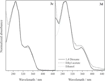
Photophysical characterization
Figures 3 and 4 show the normalized UV-Vis absorption spectra of the triazine derivatives (3a-d) in
1 h, 0 oC X N HO NH2 X N HO NH2 X N HO N N N N Cl Cl Cl N N N Cl Cl Cl acetone 1 h, 0 oC acetone H N N N Cl Cl + +
(1a) X = O (1b) X = S
(1c) X = O (1d) X = S
2
2
(3a) X = O (3b) X = S
(3c) X = O (3d) X = S X N HO N N H N N Cl Cl
Synthesis, Characterization and Photophysical Properties of ESIPT Reactive Triazine Derivatives J. Braz. Chem. Soc.
28
1,4-dioxane, ethyl acetate and ethanol. The relevant UV-Vis data are summarized in Table 1.
An absorption band maxima (λ
max) located around 358 and 368 nm (with molar extinction coefficient values (ε
max) in agreement with π-π* transitions) could be observed for the derivatives 3a and 3b, respectively. A small solvatochromic effect could be observed for these dyes (ca. 4 nm). The absorption maximum of 3b is red shifted in relation to 3a, which can be explained by the better electron delocalization. This is allowed by the sulfur atom in relation to the oxygen. The same photophysical behavior was observed for derivatives 3c and 3d, with an absorption maximum located ca. 338 and 352 nm, respectively.
The intense absorption bands observed at 280-310 nm for derivatives 3c-d indicate that the substitution in the phenolic ring is decisive for the photophysics of these dyes and can be associated to a difference of planarity of the dyes.40 A non-planar structure does not allow a
more effective electronic delocalization among the two π
systems (phenolic and benzoxazolic rings). In this way, the
para derivative showed to be less planar than their metha
analogue. The intense bands at 280-310 nm are related to the oxazole chromophore.41,42 The difference between the molecular planarity of these dyes is confirmed taking the molar extinction coefficient values into account (see Table 1). A less planar structure usually presents a lower probability for the π-π* transition.
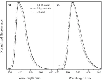
Figures 5 and 6 present the normalized fluorescence emission spectra of these dyes. The curves were obtained using the absorption maxima as the excitation wavelengths. The relevant data are also summarized in Table 1.
The derivatives 3a-d present one main band located at
ca. 471, 506, 514 and 546 nm, respectively. In solution, these dyes are fluorescent in the blue-green (3a-b) and green-yellow (3c-d) regions. The fluorescence emission bands are ascribed to the ESIPT band (T* emission) since
Table 1. Relevant UV-Vis data of the triazine derivatives 3a-d
Dye Solvent λabs / nm εmax × 104 / (mol-1 L cm-1) λ
em / nm DλST / (nm/cm-1)
3a 1,4-dioxane 358 4.4 474 116/6836
ethyl acetate 356 3.1 471 115/6859
ethanol 360 5.6 467 107/6365
3b 1,4-dioxane 370 5.2 510 140/7419
ethyl acetate 366 4.4 505 139/7520
ethanol 367 4.9 504 137/7407
3c 1,4-dioxane 339 1.4 515 176/10081
ethyl acetate 341 1.4 515 174/9908
ethanol 335 1.3 513 178/10358
3d 1,4-dioxane 356 1.2 547 191/9809
ethyl acetate 350 1.9 547 197/10290
ethanol 350 1.1 544 194/10189
a Stokes shift higher than 100 nm could be detected for all dyes.43
Figure 7 shows the normalized fluorescence emission spectra in the solid state of the dyes 3a-d, as well as the dyes covalently bounded into the cellulose matrix. The absorption maxima in solution were used as excitation wavelength in the solid state measurements. As already observed in solution, the derivatives 3a-d present one main band located from 459 to 537 nm, which comprises the blue-orange region.
Concerning the location of the fluorescence emission maxima in solution (Table 1), the dyes in the solid state present blue shifted bands (3a: 459 nm, 3b: 508 nm,
3c: 491 nm and 3d: 536 nm), indicating that the studied solvents can better stabilize the keto tautomer in the excited state. Additionally, comparing dyes 3a-c in the solid state and the cellulosic material after the staining process, it can be observed small changes in the fluorescence emission maxima due to the interaction with the cellulose matrix.
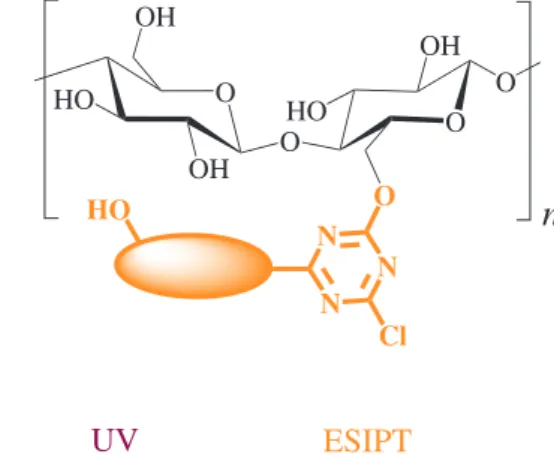
However, after the staining process using dye 3d, a dual fluorescence emission can be observed in the cellulose. The band located at higher wavelengths is ascribed to the ESIPT band and, the blue shifted one is ascribed to the normal relaxation (N* emission).44 This photophysical behavior confirms a conformational equilibrium in the ground state for this dye. Since the hydroxyl moieties in the cellulose matrix could stabilize through intermolecular hydrogen bond, additional conformers can be related to the normal emission.44 A pictorial scheme with the inter (left) and intramolecular (right) hydrogen bonds between the ESIPT dye and the cellulose is presented in Figure 8. Although the synthesized triazine dyes present two reactive chlorine atoms, the reactive dye is shown with only one replaced chlorine atom by the cellulose matrix. This is because the used temperature for the incorporation of the triazine derivatives into the cellulose fibers was below the needed temperature to replace the last chlorine atom.24
Conclusions
Four new fluorescent cotton reactive dyes were synthesized, purified and characterized by infrared spectroscopy, nuclear magnetic resonance (13C and 1H NMR), high resolution mass spectrometry (HRMS MALDI), UV-Vis and steady-state fluorescence spectroscopies (in solution and in the solid state). The triazine derivatives are fluorescent in the blue-orange region with a Stokes shift between 6365-10290 cm-1. The fluorescent cyanuric derivatives could Figure 6. Normalized fluorescence emission spectra of 3c-d.
Figure 5. Normalized fluorescence emission spectra of 3a-b.
Figure 7. Normalized solid-state fluorescence emission spectra of the derivatives 3a-d (bottom) and covalently bounded into cellulose fibers (top). This measurement treats the fibers in bulk using a solid sample holder with the light beam irradiated to the sample at an angle of
Synthesis, Characterization and Photophysical Properties of ESIPT Reactive Triazine Derivatives J. Braz. Chem. Soc.
30
successfully react with cellulose fibers to produce new ESIPT fluorescent cellulosic materials. A dual fluorescence emission could be observed in the stained cotton using the dye 3d, which indicates a conformational equilibrium in solution in the ground state. The emission at long wavelength (ESIPT band) is related to the ESIPT band and, the blue shifted ones are due to conformational forms with a normal relaxation.
Supplementary Information
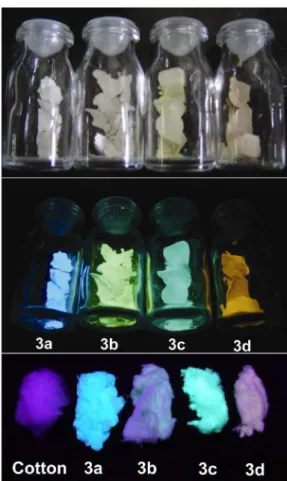
A color picture of the new ESIPT fluorescent cellulose fibers, as well as the dyes in solid state, are shown in the Supplementary Information (Figure S1), and other data free of charge at http://jbcs.sbq.org.br as pdf file.
Acknowledgements
We are grateful for financial support and scholarships from the Brazilian agencies CNPq (Conselho Nacional de Desenvolvimento Científico e Tecnológico) and Fundação de Amparo à Pesquisa do Estado do Rio Grande do Sul (FAPERGS).
References
1. Blotny, G.; Tetrahedron 2006, 62, 9507 2. Blotny, G.; TetrahedronLett. 2003, 44, 1499.
3. Zhang, P.; Yu, Y.-D.; Zhang, Z.-H.; Synth. Commun. 2008, 38, 4474.
4. Haval, K. P.; Synlett 2006, 13, 2156.
5. Giacomelli, G.; Porcheddu, A.; De Luca, L.; Curr. Org. Chem. 2004, 8, 1497.
6. Duan, H. D.; Wang, L. Z.; Qin, D. W.; Li, X. M.; Wang, S. X.; Zhang, Y.; Synth. Commun. 2011, 41, 380.
7. Mikhaylichenko, S. N.; Patel, S. M.; Dalili, S.; Chesnyuk, A. A.; Zaplishny, V. N.; Tetrahedron Lett. 2009, 50, 2505.
8. Matloobi, M.; Schramm, H. W.; J. Heterocycl. Chem. 2010, 47, 724.
9. Courme, C.; Gresh, N.; Lenoir, C.; Vidal, M.; Garbay, C.; Florent, J. C.; Bertounesque, E.; Heterocycles 2010, 81, 867. 10. Bork, J. T.; Lee, J. W.; Khersonsky, S. M.; Moon, H. S.;
Chang, Y. T.; Org. Lett. 2003, 5, 117.
11. Moral, M.; Ruiz, A.; Moreno, A.; Diaz-Ortiz, A.; Lopez-Solera, I.; de la Hoz, A.; Sanchez-Migallon, A.; Tetrahedron 2010, 66, 121.
12. Jan, J. Z.; Huang, B. H.; Lin, J.-J.; Polymer 2003, 44, 1003. 13. Ren, S. J.; Zeng, D. L.; Zhong, H. L.; Wang, Y.; Qian, S.; Fang, Q.;
J. Phys. Chem. B 2010, 114, 10374.
14. Liu, B.; Qian, D. J.; Chen, M.; Wakayama, T.; Nakamura, C.; Miyake, J.; Chem. Commun. 2006, 30, 3175.
15. Zhong, H. L.; Xu, E. J.; Zeng, D. L.; Du, J. P.; Sun, J.; Ren, S. J.; Jiang, B.; Fang, Q.; Org. Lett. 2008, 10, 709.
16. Van Cott, K. E.; Amos, T.; Gibson, H. W.; Davis, R. M.; Heflin, J. R.; Dyes Pigm. 2003, 58, 145.
17. Mahler, J.; Rafler, G.; Opt. Mater. 1999, 12, 363.
18. Zhong, H. L.; Lai, H.; Fang, Q. A.; J. Phys. Chem. C 2011, 115, 2423.
19. Omer, K. M.; Ku, S. Y.; Chen, Y. C.; Wong, K. T.; Bard, A. J.;
J. Am. Chem. Soc. 2010, 132, 10944.
20. Iijima, T.; Lee, C. H.; Fujiwara, Y.; Shimokawa, M.; Suzuki, H.; Yamane, K.; Yamamoto, T.; Opt. Mater. 2007, 29, 1782. 21. Cai, Y.-Q.; Wu, W.; Wang, H.; Miyake, J.; Qian, D.-J.; Surf. Sci.
2011, 605, 321.
22. Chen, H. F.; Yang, S. J.; Tsai, Z. H.; W. Y. Hung, Wang, T. C.; Wong, K. T.; J. Mater. Chem. 2009, 19, 8112.
23. Rothmann, M. M.; Haneder, S.; Da Como, E.; Lennartz, C.; Schildknecht, C.; Strohriegl, P.; Chem. Mater. 2010, 22, 2403. 24. De Hoog, P.; Gamez, P.; Driessen, W. L.; Reedijk, J.;
Tetrahedron Lett. 2002, 43, 6783. 25. Gorensek, M.; Dyes Pigm. 1999, 40, 225.
26. Taylor, J. A.; Pasha, K.; Phillips, D. A. S.; Dyes Pigm. 2001,
51, 145. Figure 8. Scheme of interaction between the ESIPT dyes and the cellulose.
O HO OH O OH O HO OH O O
n
N N N Cl HO O HO O O OH O HO OH O On
N N N Cl H O H UVradiation emissionESIPT
Normal
27. Um, S. I.; Lee, J. K.; Kang, Y.; Baek, D. J.; Dyes Pigm. 2006,
70, 84.
28. Son, Y. A.; Hong, J. P.; Lim, H. T.; Kim, T. K.; Dyes Pigm.
2005, 66, 231.
29. Czajkowski, W.; Paluszkiewicz, J.; Stolarski, R.; Kaźmierska, M.; Grzesiak, E.; Dyes Pigm. 2006, 71, 224.
30. Seo, J.; Kim, S.; Park, S. Y.; J. Am. Chem. Soc. 2004, 126, 11154. 31. Jung, H. S.; Kim, H. J.; Vicens, J.; Kim, J. S.; Tetrahedron Lett.
2009, 50, 983.
32. Yang, C.-C.; Tian, Y.; Chen, C.-Y.; Jen, A. K.-Y.; Chen, W.-C.;
Macromol. Rapid Commun. 2007, 28, 894.
33. Wu, Y. K.; Peng, X. J.; Fan, J. L.; Gao, S.; Tian, M. Z.; Zhao, J. Z.; Sun, S.; J. Org. Chem. 2007, 72, 62.
34. Klymchenko, A. S.; Demchenko, A. P.; J. Am. Chem. Soc. 2002,
124, 12372.
35. Rodembusch, F. S.; Campo, L. F.; Rigacci, A.; Stefani, V.;
J. Mater. Chem. 2005, 15, 1537.
36. Kober, U. A.; Campo, L. F.; Costa, T. M. H.; Stefani, V.; Gallas, M. R.; J. Photochem. Photobiol., A 2007, 186, 24.
37. Rodembusch, F. S.; Leusin, F. P.; Bordignon, L. B.; Gallas, M. R.; Stefani, V.; J. Photochem. Photobiol., A 2005, 153, 81. 38. Joule, J. A.; Mills, K.; Heterocyclic Chemistry, 4th ed.; Blackwell
Science: Cambridge, 2000.
39. Campo, L. F.; Corrêa, D. S.; Araújo, M. A.; Stefani, V.;
Macromol. Rapid Commun. 2000, 21, 832.
40. Douhal, A.; Amat-Guerri, F.; Lillo, M. P.; Acuña, A. U.;
J. Photochem. Photobiol., A 1994, 78, 127.
41. Nagaoka, S. I.; Kusunoki, J.; Fujibuchi, T.; Hatakenaka, S.; Mukai, K.; Nagashima, U.; J. Photochem. Photobiol., A 1999,
122, 151.
42. Guallar, V.; Moreno, M.; Lluch, J. M.; Amat-Guerri, F.; Douhal, A.;
J. Phys. Chem. 1996, 100, 19789.
43. Santra, S.; Dogra, S. K.; Chem. Phys. 1998, 226, 285. 44. Rodembusch, F. S.; Campo, L. F.; Leusin, F. P.; Stefani, V.;
J. Lumin. 2007, 126, 728.
Submitted: August 1, 2011
Supplementary Information
S
I
J. Braz. Chem. Soc., Vol. 23, No. 1, S1-S8, 2012. Printed in Brazil - ©2012 Sociedade Brasileira de Química 0103 - 5053 $6.00+0.00
*e-mail: [email protected]
Synthesis, Characterization and Photophysical Properties of
ESIPT Reactive Triazine Derivatives
Marcelo D. Kuplich, Fábio S. Grasel, Leandra F. Campo, Fabiano S. Rodembusch and Valter Stefani*
Laboratório de Novos Materiais Orgânicos, Instituto de Química, Universidade Federal do Rio Grande do Sul, Av. Bento Gonçalves 9500, CP 15003, 91501-970 Porto Alegre-RS, Brazil
Figure S1. Triazine derivatives under normal light (top) and UV light (middle) and the respective ESIPT fluorescent cellulosic fibers under UV light (bottom).
Figure S2. Triazine derivatives under normal light (top) and UV light (bottom).
Figure S3. FTIR spectrum of the dye 3a in KBr pellets. Picture of the dyes and stained cellulose under normal light
and UV radiation
Kuplich et al. S3 Vol. 23, No. 1, 2012
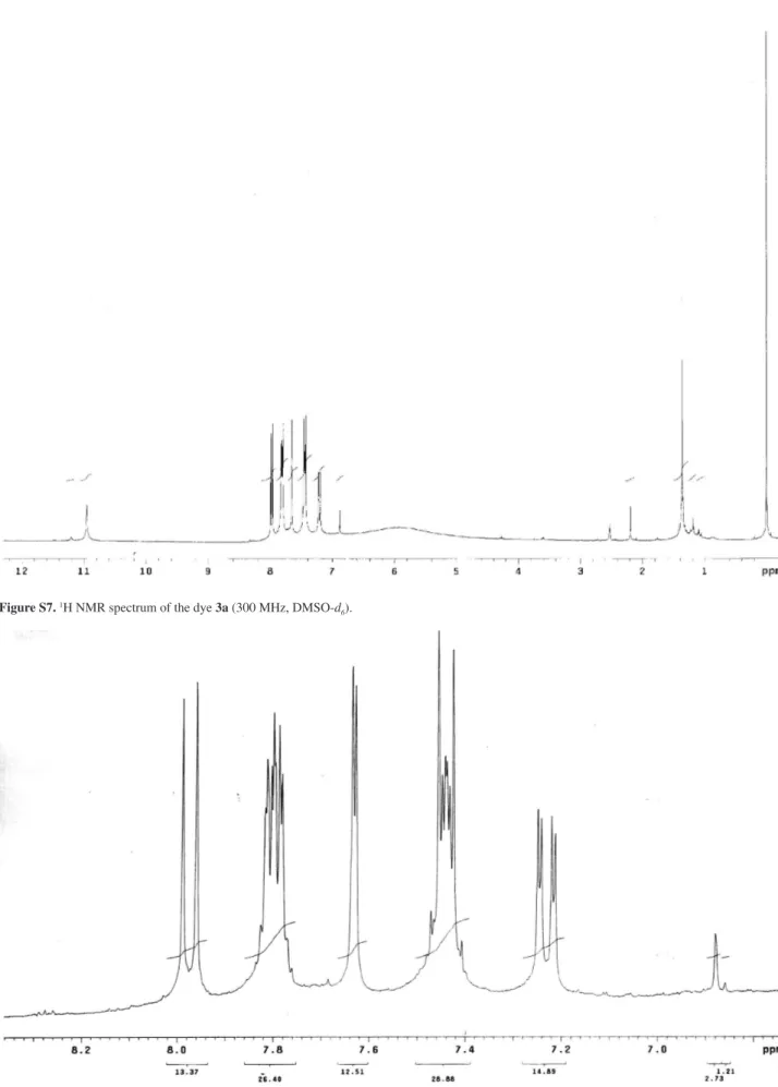
Figure S8. 1H NMR spectrum of the dye 3a (zoom of the aromatic region). Figure S7. 1H NMR spectrum of the dye 3a (300 MHz, DMSO-d
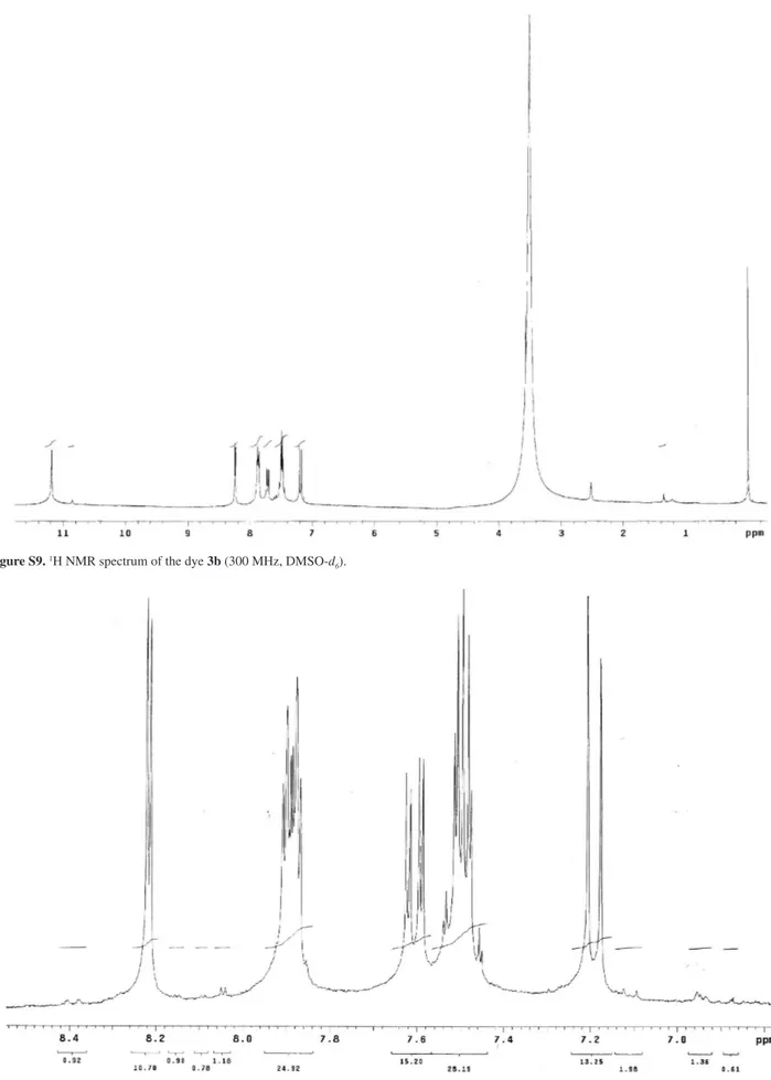
Figure S9. 1H NMR spectrum of the dye 3b (300 MHz, DMSO-d
6).
Kuplich et al. S5 Vol. 23, No. 1, 2012
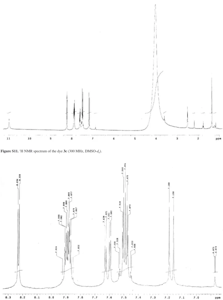
Figure S11. 1H NMR spectrum of the dye 3c (300 MHz, DMSO-d
6).
Figure S14. 1H NMR spectrum of the dye 3d (zoom of the aromatic region). Figure S13. 1H NMR spectrum of the dye 3d (300 MHz, DMSO-d
Kuplich et al. S7 Vol. 23, No. 1, 2012
Figure S15. 13C NMR (APT) spectrum of the dye 3a (75 MHz, DMSO-d
6).
Figure S16. 13C NMR (APT) spectrum of the dye 3b (75 MHz, DMSO-d
Figure S17. 13C NMR (APT) spectrum of the dye 3c (75 MHz, DMSO-d 6).
Figure S18. 13C NMR (APT) spectrum of the dye 3d (75 MHz, DMSO-d