O
RIGINALA
RTICLE Revista Brasileira de FisioterapiaEffects of different methods of antagonist
muscles pre-activation on knee extensors
neuromuscular responses
Efeitos da ordem de pré-ativação dos músculos antagonistas nas respostas
neuromusculares dos extensores do joelho
Rodrigo L. Carregaro1,3, Rafael R. Cunha3, Jefferson R. Cardoso2, Ronei S. Pinto4, Martim Bottaro3
Abstract
Background: Pre-activation of antagonistic muscles is used in different modalities of exercise and neuromuscular rehabilitation protocols, but its effectiveness is still controversial. Objective: To verify the impact of two different methods of pre-activation of knee antagonist muscles in the neuromuscular performance and electromyographic activity of knee extensors. Methods: Fifteen healthy men (23.9±4.2 years of age, 1.78±0.08 meters and 81.4±10.7 kg) performed, on different days, two protocols of isokinetic muscle contraction with 4 sets of 10 repetitions at 60°.s-1 and 1 minute between sets: (1) Reciprocal Contraction (RC): reciprocal concentric exercise of agonist/antagonist
muscles (knee flexion [KF] immediately followed by knee extension [KE]) and (2) Superset (SS): alternated concentric exercise of agonist/ antagonist muscles (KF set followed by a set of KE). A repeated measures ANOVA with least-significant difference post-hoc test was used to detect differences between protocols. Results: There were no significant differences between protocols (p>0.05) for peak torque (PT) and total work (Tw). On the SS protocol there was a significant decrease in Tw on the last two sets (p<0.05) while for RC the decrease occurred only in the last set. There were no significant differences of root mean square (RMS) between protocols, but the activation pattern was more uniform during the RC protocol. Conclusion: The results indicated that the peak torque was not influenced by the different pre-activation methods. However, the RC protocol appears to better maintain the total work training volume.
Keywords: electromyography;resistance training; dynamometer; muscle strength; rehabilitation; movement. Resumo
Contextualização: A pré-ativação de músculos antagonistas é utilizada em diferentes modalidades de exercício e em diferentes protocolos de reabilitação neuromuscular, porém suas respostas ainda são controversas. Objetivo: Verificar o impacto de duas diferentes estratégias de pré-ativação de músculos antagonistas no desempenho neuromuscular e na atividade eletromiográfica dos extensores do joelho. Métodos: Quinze homens sadios (23,9±4,2 anos; 1,78±0,08 m e 81,4±10,7 kg) realizaram, em dias distintos, dois protocolos de ações musculares isocinéticas com quatro séries de dez repetições a 60°.s-1 e intervalo de 1 minuto entre séries: 1) contração recíproca (CR): exercício
concêntrico recíproco de antagonistas/agonistas (uma repetição de flexão do joelho [FJ] imediatamente seguida por uma de extensão do joelho [EJ]) e 2) supersérie (SS): exercício concêntrico alternado dos antagonistas/agonistas (dez repetições de FJ seguidas por dez de EJ). Utilizou-se a ANOVA para medidas repetidas com teste post-hoc LSD (Least-significant diference) para verificar a diferença entre protocolos. Resultados: Não houve diferença significante (p>0,05) entre protocolos para o pico de torque (PT) e trabalho total (Tt). Em relação ao Tt, o protocolo SS apresentou quedas significantes nas duas últimas séries (p<0,05) enquanto, no CR, a queda ocorreu apenas na última série de exercício. Não houve diferenças no Root Mean Square (RMS) entre protocolos, mas o padrão de ativação foi mais uniforme durante o CR. Conclusão: Os resultados indicaram que a queda na força muscular não é influenciada pelas diferentes formas de pré-ativação da musculatura antagonista, no entanto parece que a utilização de CR permite uma melhor manutenção do volume de treinamento.
Palavras-chave: eletromiografia; treinamento de resistência; dinamômetro; força muscular; reabilitação; movimento.
Received: 02/10/2011 – Revised: 06/28/2011 – Accepted: 07/10/2011
1 Course of Physical Therapy, Universidade Federal de Mato Grosso do Sul (UFMS), Campo Grande, MS, Brazil 2 Physical Therapy Department, Universidade Estadual de Londrina (UEL), Londrina, PR, Brazil
3 Faculty of Physical Education, Universidade de Brasília (UnB), Brasília, DF, Brazil
4 School of Physical Education, Universidade Federal do Rio Grande do Sul (UFRGS), Porto Alegre, RS, Brazil
Correspondence to: Rodrigo Luiz Carregaro, CCBS/Curso de Fisioterapia, Unidade XII, Av. Costa e Silva, S/Nº, Campus Universitário UFMS, Cidade Universitária, CEP 79070-900, Campo Grande, MS, Brasil, e-mail: [email protected]
Introduction
Resistance exercise are considered an essential element in rehabilitation programs and physical conditioning1,2. Programs
that involve resistance exercises can also focus on the preven-tion of injuries3,4, such as in cases where joint instability
gener-ated by deiciency of the dynamic joint stabilizers predisposes to degeneration of synovial structures5.
Strength gains provided by resistance exercise programs are associated with important clinical beneit, and therefore, nu-merous methods of resistance training have been developed6,7.
Recently, the use of pre-activation of antagonist muscles be-fore activation of the agonists has received increasing atten-tion in rehabilitaatten-tion clinics and gyms. According to Júnior et al.8, a goal pursued by professionals that employ resistance
exercise for performance and rehabilitation is to link muscle actions so that the sequence of stimuli provides an efective muscle response. he characteristics of pre-activation of an-tagonistic muscles seem to positively inluence the generation of agonists’ strength. herefore, individuals performing these pre-activation exercises could potentially improve their motor performance and generate higher strength levels. However, evidence to support the use of pre-activation exercises are still scarce and controversial.
Among the types of agonists pre-activation, we can high-light the methods known as superset (SS)9-13 and reciprocal
contractions (RCs)14-19. he indings concerning the
efective-ness of these protocols are controversial and methodological variations makes it diicult to compare the beneits of both forms of exercises. Burke, Pelham and Holt9 found that the SS
protocol has positive efects on strength generation of the ago-nist; however this was not the case when the pre-activation of the antagonist was performed by means of simple sets and with maximal and prolonged contractions10.
Addition-ally, Bohannon, Gibson and Larkin15 showed no diference
in strength between the RC method and a traditional method (without pre-activation of the antagonist) in healthy subjects. On the other hand, other studies have shown beneits of RC16,17,19,
but the conclusions were based on exercises conducted with simple series, ie, only one set (deined as a group of repetitions developed continuously, without interruption) although resis-tance exercises are most commonly based on more than one set (multiple sets).
In one of the few studies that compared the two meth-ods, Bohannon14 showed a torque 10% higher during the RC
protocol compared to SS in participants with a history of stroke. In a recent study, Carregaro et al.20 reported no
sig-niicant diferences in torque production of the agonist, but found that RC leads to greater work capacity than the SS. It is important to note that most of the studies cited did not use
surface electromyography (EMG) to evaluate the diferences between the modalities, and to date, few studies have evalu-ated muscle activation10,13,18,19. Moreover, with the exception
of Robbins et al.13, all studies have focused on understanding
the efects of diferent execution speeds in strength genera-tion, making it diicult to compare performance between the modalities. hus, the objective of this study was to evaluate the impact of two diferent pre-activation methods of knee lexor muscles in the generation of torque, work and electro-myographic activation of the Vastus Medialis muscle (VM) during knee extension exercises (KE).
Methods
Sample
Fifteen healthy men (23.9±4.2 years of age; 1.78±0.08 me-ters and 81.4±10.7 kg) participated in the study. hey were in-structed not to perform any kind of strenuous physical activity for the lower limbs during the study period. Inclusion criteria were age between 18 and 35 years and be physically active (en-gaged in some kind of physical exercise, at least twice a week). Subjects were excluded if they had a history of trauma and any type of musculoskeletal surgery in the lower limbs and spine, cardiovascular disease and diagnosed arterial hypertension. All individuals who participated were informed about the research objectives and procedures and were invited to par-ticipate in the study duly approved by the Ethics Committee of the Faculty of Health of the Universidade de Brasília (FS/UnB), Brasília, DF, Brazil (protocol nº. 161/2008), signing an informed consent, in accordance with Resolution 196 of the CNS.
Evaluation procedures
Participants attended the Laboratory of Strength Training in three diferent occasions, with a minimum interval of 72 hours between each visit. On the irst day, participants familiarized themselves with the exercise protocols and performed two sets of four maximal repetitions at 60°.s-1 (in each protocol), with 1
minute interval between sets. Between the protocols, there was a period of 5 minutes of rest. A familiarization of the maximal voluntary isometric contraction (MVIC) was also performed, in which all patients performed two maximal contractions of 5 seconds, with an interval of 2 minutes between them.
On the second and third visits participants performed the exercise protocols, with four sets of ten repetitions each and speed of 60°.s-1, in the dominant limb (leg used to kick a
ball). Between sets, there was a rest interval of 1 minute21. he
protocols used were: 1) RC ( four series of reciprocal concen-tric exercise of antagonists and agonists, characterized by the movement of knee lexion (KF) immediately followed by knee extension (KE) in each repetition) and 2) SS ( four sets of exer-cise in alternate mode of antagonists and agonists, each set was characterized by ten concentric repetitions of KF with passive KE followed immediately by a set of ten concentric repetitions of KE with passive KF). In the 24 hours preceding the meetings, participants were instructed not to perform strenuous exercise and not to drink energy drinks. he order of the two protocols was randomized.
During the evaluations, participants were asked to position their arms against the chest, so that the arms did inluence the strength generation22. In addition, participants were instructed
to perform the movements with maximum strength through-out the full available range of motion. Before performing the procedures and exercises on the dynamometer, all subjects underwent a 10 minutes warm up session on a stationary bike with no load. Verbal encouragement and visual feedback using the computer screen were given in an attempt to achieve the maximum level of efort. All procedures were performed by the same investigator.
Isokinetic Dynamometer
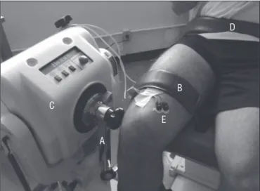
An isokinetic dynamometer Biodex System 3 (Biodex Medi-cal, Shirley, NY.) was used. Calibration was performed accord-ing to the speciications of the manufacturer’s manual, and the gravity correction was obtained through measuring the torque exerted by the resistance arm and the leg of subject (relaxed) in the position of terminal knee extension. Each subject was positioned on the chair allowing for a free and comfortable movement of knee lexion and extension (Figure 1). To prevent movements of knee hyperextension, a range of motion of 80° lexion/extension (excursion between 10° and 90°, considering 0° the full knee extension) was used. he hip position was stan-dardized at 100° of lexion (position of the chair) for all sub-jects. he lateral epicondyle of the femur was used as reference point for the knee rotation axis and used for aligning with the rotation axis of the device. he height of the chair, the backrest tilt, the height of the dynamometer and the adjustment of the resistance arm were recorded and replicated each day to guar-antee standardization between sessions.
Surface electromyography
he recording and processing of electromyographic signals were based on the recommendations of the International So-ciety of Electrophysiology and Kinesiology23 and of Soderberg
and Knutson24. he placement of the electrodes was based on
the guidelines of the project SENIAM (Surface Electromyogra-phy for the Non-Invasive Assessment of Muscles)25.
A portable surface electromyography of four channels (Miotool, Miotec Equipamentos Biomédicos Ltda, Brazil), with 14 bits resolution, noise level <2LSB and common mode rejection of 110 db was used. he channels were itted with a sampling of 2000 Hz, and a inal gain of 1000 times. he simple diferential active electrodes (input impedance of 1010 Ohm)
had polyethylene foam with hypoallergenic medical adhesive, solid stick gel, bipolar contact of Ag/AgCl and distance be-tween the poles of 20 mm. he muscle evaluated was the VM, and the reference electrode was attached to the bony promi-nence of the seventh cervical vertebra (C7). he VM muscle was chosen based on the study of Miller, Croce and Hutchins18,
that found that the VM had 1.5 times greater activation than the other muscles during a similar protocol to that used in the present study.
he electrode was positioned over the belly of the VM mus-cle and parallel to the musmus-cle ibers26 (Figure 1). Before placing
the electrodes, the area was shaved and light abrasion with 70% alcohol was performed. he placement of the electrodes was identiied on the irst day of testing and a high setting pen mark was made on the skin to ensure that the same position was used on the subsequent days.
On the second and third visits all subjects performed a MVIC with knee joint positioned at 60° (having as reference for 0° the full knee extension). he MVIC was characterized by two contractions of 5 seconds each, with interval of 2 minutes of rest between them. After 5 minutes of rest, subjects were in-structed to start the exercise protocols.
he electromyography analysis was conducted on the
Miograph 2.0 program (Miotec Equipamentos Biomédicos Ltda,
A) Lever arm; B) Thigh strap; C) Dynamometer; D) Waist strap; E) Electrode on VM muscle.
Figure 1. Experiment illustration on the isokinetic dynamometer.
A
B
C
D
E
Brazil). he electromyographic signal was iltered with a band-pass frequency between 20 Hz and 450 Hz (Butterworth ilter of 4th order) and was normalized using the MVIC. In the normalization procedure, the repetition of the MVIC with the higher Root Mean Square (RMS) value was used as reference, considering the central 3 seconds of the signal. To analyze the pattern of electromyographic activation of the VM muscle in the protocols, the mean RMS of the irst three and last three repetitions in each set of exercise was used.
Data analysis
he SPSS program (Statistical Package for Social Sciences), version 13.0, was used for all statistical analysis. he data are shown as mean±standard deviation; the normality of data was checked using the Shapiro-Wilk test. he dependent variables analyzed were peak torque (PT), total work (Tw) and RMS (dis-played as % of MVIC). he Analysis of Variance (ANOVA) 2 x 4 for repeated measures [protocols (RC and SS) x number of sets (1st, 2nd, 3rd, 4th)], with post-hoc least-signiicant diference (LSD) test was used for the PT and Tw variables. For the RMS, an ANOVA 2 x 4 x 6 for repeated measures [protocols (RC and SS) x number of sets (1st, 2nd, 3rd, 4th) x repetitions (1st, 2nd, 3rd, 8th, 9th, 10th)] was conducted. he Mauchly’s W sphericity test was conducted and when rejected, the analysis was always based on the Greenhouse-Geisser correction. he signiicance adopted was 5% (p<0.05).
Results
he values of PT and Tw generated during the execution of the RC and SS protocols are shown in Table 1. he comparison between the protocols showed no signiicant diference for PT and Tw (p>0.05).
In the intragroup analysis, there was a signiicant decrease in the strength generated in the last set of exercise for both protocols (p<0.05 - Table 1). However, despite the absence of statistical signiicance between the protocols, it is possible to notice that the RC protocol showed lower percentages of de-crease (2%, 5% and 10% in sets two, three and four) in relation to the SS protocol (3%, 7% and 14% in sets two, three and four), when compared with the irst set of exercise. In relation to Tw,
the SS protocol showed diferences in exercise volume char-acterized by signiicant decreases in the last two sets (p<0.05). For the RC protocol, the decrease occurred only in the last set (p<0.05). Despite the lack of diferences between protocols, the decrease in work in the last set reached 22% in SS, against a decrease of 14% in RC.
he indings relating to the RMS of the VM muscle are shown in Table 2. here was no statistically signiicant difer-ence between the protocols and the sets over the analyzed repetitions (p>0.05). It is possible to notice that the SS protocol showed signiicant variations (p<0.05) both in the initial and in the inal phase, for all series, indicating increase in the activa-tion of the VM muscle. he increases of activaactiva-tion were also signiicant in the RC (p<0.05), but concentrated in the irst two repetitions and maintaned until the end. here was a signii-cant interaction between protocols and repetitions (p<0.05), which may indicate speciic electromyographic patterns of the protocols, as illustrated in Figure 2.
Discussion
he present study hypothesized that diferent protocols of pre-activation of knee lexor muscles (antagonists) inluence the ability to generate strength and work of the extensors (ago-nists). he comparison between the modalities showed that both provide similar rates of generation of extensor strength. However, it appears that the RC method provides greater ca-pacity of work. he RC method showed, qualitatively, a curve with a more uniform distribution between sets of exercise, when compared to the SS. In addition, the RC showed a stron-ger increase of the RMS, especially in the initial phases, which may represent more eicient muscle recruitment.
In this study, we found no signiicant diferences of knee extensor torque between the protocols which difers from the results of Maynard and Ebben10, who found diferences in knee
extensor torque after a set of ive maximal repetitions of exer-cise in the SS modality at 60°.s-1. In this study, the torque
gener-ated in the SS condition was approximately 4% lower than the control (KE exercise without pre-activation of agonist). his lower strength production may be attributed to an increase in co-activation of the lexor muscles, previously fatigued. How-ever, comparisons with the study of Maynard and Ebben10 are
Knee extensor Peak Torque (N.m) Total Work (J)
Set 1 Set 2 Set 3 Set 4 Set 1 Set 2 Set 3 Set 4
SS 264.1±49.1 254.9±52.7 246.0±50.2 228.3±41.5* 2270.6±385.2 2104.3±347.4 1935.5±389.6* 1779.4±308.9*#
RC 261.4±37.0 257.1±37.2 248.3±37.3 234.1±32.3* 2262.7±327.6 2173.7±321.6 2052.9±295.5 1943.5±264.9*
Table 1. Values of knee extensor peak torque and total work (mean ± standard deviation).
Less than Set 1: *(p<0.05); Less than Set 2: # (p<0.05). RC=reciprocal contraction; SS=superset
limited by the fact that they used simple sets and compared the SS with another modality of exercise. Another aspect was the absence of a period of familiarization to the SS protocol. On the other hand, Baker and Newton11 reported increases in the
abil-ity to generate power after an intervention with a set of eight repetitions (seated rowing exercise like SS). In this case, Baker and Newton11 explained the gains in power as an increase in
the iring rate of the agonist muscles, caused by a neural stimu-lation inluenced by a previous contraction of the antagonists (called post-tetanic potentiation). Although the study11
evalu-ated the variable power and upper limb muscles (which may have a diferent speciicity than that of the lower limb muscles), it is possible that the same neural strategy occurred in our study and therefore, could explain the same performance and electromyography activation of the CR and SS modalities.
It is noteworthy that, in the present study, there was a de-crease of 14% on the last set of torque in the SS condiction, while, in the RC condition, the decrease was smaller (10%). Despite the lack of signiicance, this is an important practical observation, because a 4% diference between the protocols may represent beneits for an athlete or individual who is in process of rehabili-tation and needs strength to carry a speciic activity. In fact, Roy et al.17 suggested that the advantages stemming from reciprocal
actions are due to facilitatory stimulation of the Golgi tendon organs (GTOs) of the lexor muscle and muscles spindles of extensors muscle, assigned to prior lexion. heir indings show that the reciprocal modality tends to generate a greater torque of the knee extensors, which would explain lower decrease in the RC condition. Apparently, such response would be explained by a neuromuscular event caused by the action of the lexor muscle, which would activate the GTOs and their motoneurons network, while, at the same time, the muscle spindles of the ex-tensors (elongated) would facilitate and increase performance in the thereafter contraction. Kisner and Colby3 also suggested
that during a concentric activation of the agonist, the antagonist has a reciprocal inhibition that allows its relaxation and, conse-quently, may facilitate the action of the agonist. his response may represent beneits during the performance of exercises with
multiple sets, in the sense that the reciprocal inhibition may decrease the susceptibility to muscle fatigue over the repeti-tions and promote the maintenance of adequate levels of torque and work during the exercise session. However, this hypothesis must be conirmed by acute and chronic studies (longitudinal) through the use of indices based on the calculation of spectral moments, appropriate to monitoring the fatigue during dynamic muscle contractions27.
Despite the lack of statistical diference between protocols, the work generated by the RC showed higher values when compared to SS, especially in the last two sets. In turn, the intra protocols analysis showed that the RC performance was better, ie, had smaller decreases of work, unlike the SS. he decrease rates of RC and SS found in the last two sets (5% and 14%; 7% and 22%, respectively) showed that the work generated by two categories of resistance exercise in controlled conditions of speed, intensity and sets may impose important outcomes in training. Such indings points to the practical implications highlighted by Munn et al.28 and Kelly et al.29, who argued that
increases in work capacity can determine important strength gains during resistance exercise. Exercise programs should be dynamic to induce physiological responses and performance gains, and research should incorporate other variables and not just the efects of simple and multiple sets30, aspect much
dis-cussed in the literature28-32. Tran and Docherty33 showed that
the volume can also be expressed by the multiplication of rep-etitions and load used and also by the time under tension dur-ing muscle contraction. he longer the time under tension, the greater the deleterious efects on the excitation-contraction process of muscle ibers and, therefore, the greater the inten-sity of peripheral fatigue33. Although it was not measured in our
study, it is possible that in the RC protocol, the extensor group has not been exposed so intensely (as the SS) to factors related to peripheral muscle fatigue, which could explain the greater work decrease (interpreted as the ability to generate strength by a speciic distance) in the SS protocol.
In this study, there were no diferences in RMS values between protocols, partially corroborating Robbins et al.13, who found no
Rep1 Rep2 Rep3 Rep8 Rep9 Rep10
Set 1 SS 81.6±12.6 96.4±26.7* 100.3±20.4† 113.5±20.8‡ 107.3±18.2# 105.4±22.7 RC 89.9±19.5 102.2±25.6* 114.1±23.5† 108.8±23.2 112.9±24.7 106.0±18.3
Set 2 SS 79.4±15.8 94.4±21.5* 93.8±19.3† 112.4±25.1‡ 111.2±27.7# 103.8±21.6 RC 85.0±18.9 94.2±20.2* 106.3±19.4† 109.3±21.8 108.1±20.7 109.3±27.0
Set 3 SS 85.6±16.1 92.3±18.9* 96.0±14.9† 104.2±25.9‡ 102.7±19.5# 96.6±17.5 RC 90.7±24.6 101.8±21.1* 107.3±24.6† 107.4±23.2 109.5±23.3 102.9±24.8
Set 4 SS 91.5±19.5 93.7±17.2* 100.2±14.3† 110.3±23.3‡ 104.1±17.9# 104.2±14.3 RC 86.8±23.4 100.4±22.2* 106.9±21.5† 106.7±20.8 105.4±19.7 99.8±23.5
Table 2. Root mean square (RMS) values (presented as % MVIC) of 4 sets of each exercise protocol (RC: Reciprocal contraction; SS: Superset),
(data presented as mean ± standard deviation; Rep: Repetition).
Significant differences: *Rep1→Rep2: p=0.003; †Rep2→Rep3: p=0.006; ‡Rep3→Rep8: p=0.000; #Rep8→Rep9: p=0.03.
diferences in electromyography activation between the SS and a traditional method during an exercise with three series. Coburn et al.34 while applying a training of just three sessions with a
tra-ditional protocol of KE exercise ( four sets of ten repetitions at 30°.s-1 and 270°.s-1) also found no diference in RMS post-training,
which is an argument about the lack of diference between the
categories in our study. It is common consensus in the literature that the electromyographic signal amplitude is related to activa-tion of motor units, and the frequency domain relects the con-duction speed of the contraction potential24,35, characterizing the
EMG as an important tool to evaluate the neural adaptations after exercise. In the classic study by Moritani and deVries36, Figure 2. Temporal series with electromyography activation patterns (RMS in % MVIC) over the exercise repetitions (REP) for protocols RC (reciprocal contraction) and SS (superset).
70 80 90 100 110 120
REP1 REP2 REP3 REP8 REP9 REP10
RMS (%MVIC)
RC
Set 1 Set 2 Set 3 Set 4
70 80 90 100 110 120
REP1 REP2 REP3 REP8 REP9 REP10
RMS (%MVIC)
SS
Set 1 Set 2 Set 3 Set 4
increases in the amplitude of the electromyographic signal after two weeks of training were found, demonstrating that neural efects were responsible for increases in strength in the initial phase. he indings of Moritani and deVries36 and Coburn et al.34
suggests that perhaps the acute nature of our study was not suf-icient to determine diferences between the RC and SS modali-ties. However, the signiicant interaction between protocols and repetitions, coupled with the visual standard of curves, showed evidences that, in the initial third of the exercise, the RC category provided a more intense activation; that remained until the end. It is noteworthy that one of the limitations of this study was the non-evaluation of co-activation of antagonist muscles. Another limitation is the lack of a control group to better understand the neuromuscular responses of the knee extensors without a prior pre-load of the antagonists.
Among the clinical applications of the protocols, it is pos-sible to highlight the increased stability of the knee trough the strengthening of agonists and antagonists muscles37. he
antagonist muscles provide control of mechanical forces that cause joint instability that is generated during the action of the agonists, showing that the strengthening of the antagonist muscle groups can restore or increase muscular balance in a joint38. hus, the RC and SS modalities involve reducing the
risk of injuries while generating greater joint stability and, at the same time, promoting gains of muscle strength39. In this
sense, it is recommended that physical therapists and sports professionals include, in their strategies for treatment/train-ing, resistance exercises with alternate or reciprocal actions. Such strategies may inluence neuromuscular skills necessary for activities that require motor control of prime muscles and knee stabilizers3, help prevent joint imbalances40 and dynamic
stability in cases of anterior cruciate ligament deiciency37
herefore, it is suggested that longitudinal studies to better determine possible adaptations resulting from the RC and SS protocols are conducted. hen, it will be possible to elucidate which modalityis most efective for strength gains and muscle performance in diferent populations.
Acknowledgments
To the Fundação de Apoio à Pesquisa do Distrito Federal
(FAPDF), process nº. 2009/00212-2, and to the Conselho Nacio-nal de Desenvolvimento Cientíico e Tecnológico (CNPq) - (pro-cesses nº. 306114/2009-7 and 474740/2009-9 No) for inancial support.
References
1. Ratamess NA, Alvar BA, Evetoch TK, Housh TJ, Kibler WB, Kraemer WJ, et al. Progression models in resistance training for healthy adults. Med Sci Sports Exerc. 2009;41(3):687-708. 2. ACSM. American College of Sports Medicine (ACSM). Diretrizes do ACSM para os testes de
esforço e sua prescrição. 7ª ed. Rio de Janeiro: Guanabara Koogan; 2007.
3. Kisner C, Colby LA. Therapeutic exercise. Foundations and techniques. 5th ed. Philadelphia:
Davis Company; 2007.
4. Warburton DE, Nicol CW, Bredin SS. Prescribing exercise as preventive therapy. CMAJ. 2006;174(7):961-74.
5. Traete RF, Pinto KNZ, Mattiello-Rosa SM. Relação entre a lesão condral e o pico de torque após reconstrução do ligamento cruzado anterior do joelho: estudo de casos. Rev Bras Fisioter. 2007;11(3):239-43.
6. Fleck SJ, Kraemer WJ. Fundamentos do treinamento de força muscular. 3ª ed. Porto Alegre: Artmed; 2006.
7. Gentil P, Oliveira E, Fontana K, Molina G, Oliveira RJ, Bottaro M. Efeitos agudos de vários métodos de treinamento de força no lactato sanguíneo e características de cargas em homens treinados recreacionalmente. Rev Bras Med Esporte. 2006;12(6):303-7.
8. Júnior VAR, Bottaro M, Pereira MCC, Andrade MM, Júnior PRWP, Carmo JC. Análise eletromiográfica da pré-ativação muscular induzida por exercício monoarticular. Rev Bras Fisioter. 2010;14(2):158-65.
9. Burke DG, Pelham TW, Holt LE. The influence of varied resistance and speed of concentric antagonistic contractions on subsequent concentric agonistic efforts. J Strength Cond Res. 1999;13(3):193-7.
10. Maynard J, Ebben WP. The effects of antagonist prefatigue on agonist torque and electromyography. J Strength Cond Res. 2003;17(3):469-74.
11. Baker D, Newton RU. Acute effect on power output of alternating an agonist and antagonist
muscle exercise during complex training. J Strength Cond Res. 2005;19(1):202-5.
12. Kelleher AR, Hackney KJ, Fairchild TJ, Keslacy S, Ploutz-Snyder LL. The metabolic costs of reciprocal supersets vs. traditional resistance exercise in young recreationally active adults. J Strength Cond Res. 2010;24(4):1043-51.
13. Robbins DW, Young WB, Behm DG, Payne WR. The effect of a complex agonist and antagonist resistance training protocol on volume load, power output, electromyographic responses, and efficiency. J Strength Cond Res. 2010;24(7):1782-9.
14. Bohannon RW. Knee extension torque during repeated knee extension-flexion reversals and separated knee extension-flexion dyads. PhysTher. 1985;65(7):1052-4.
15. Bohannon RW, Gibson DF, Larkin P. Effect of resisted knee flexion on knee extension torque. PhysTher. 1986;66(8):1239-41.
16. Grabiner MD, Hawthorne DL. Conditions of isokinetic knee flexion that enhance isokinetic knee extension. Med Sci Sports Exerc. 1990;22(2):235-44.
17. Roy MA, Sylvestre M, Katch FI, Katch VL, Lagassé PP. Proprioceptive facilitation of muscle tension during unilateral and bilateral knee extension. Int J Sports Med. 1990;11(4):289-92. 18. Miller JP, Croce RV, Hutchins R. Reciprocal coactivation patterns of the medial and lateral
quadriceps and hamstrings during slow, medium and high speed isokinetic movements. J ElectromyogrKinesiol. 2000;10(4):233-9.
19. Jeon HS, Trimble MH, Brunt D, Robinson ME. Facilitation of quadriceps activation following a concentrically controlled knee flexion movement: the influence of transition rate. J Orthop Sports PhysTher. 2001;31(3):122-32.
20. Carregaro RL, Gentil P, Brown LE, Pinto RS, Bottaro M. Effects of antagonist pre-load on knee extensor isokinetic muscle performance. J Sports Sci. 2011;29(3):271-8.
21. Parcell AC, Sawyer RD, Tricoli VA, Chinevere TD. Minimum rest period for strength recovery during a common isokinetic testing protocol. Med Sci Sports Exerc. 2002;34(6):1018-22.
22. Stumbo TA, Merriam S, Nies K, Smith A, Spurgeon D, Weir JP. The effect of hand-grip stabilization on isokinetic torque at the knee. J Strength Cond Res. 2001;15(3):372-7.
23. Merletti R. Standards for reporting EMG data. International Society of Electrophysiology and Kinesiology; 1999.
24. Soderberg GL, Knutson LM. A guide for use and interpretation of kinesiologicelectromyographic data. PhysTher. 2000;80(5):485-98.
25. Hermens HJ, Freriks B, Disselhorst-Klug C, Rau G. Development of recommendations for SEMG sensors and sensor placement procedures. J ElectromyogrKinesiol. 2000;10(5):361-74. 26. Basmajian JV, Deluca CJ. Muscles Alive. Their functions revealed by electromyography. 5th ed.
Baltimore: Williams & Wilkins; 1985.
27. Dimitrov GV, Arabadzhiev TI, Mileva KN, Bowtell JL, Crichton N, Dimitrova NA.Muscle fatigue during dynamic contractions assessed by new spectral indices. Med Sci Sports Exerc. 2006;38(11):1971-9.
28. Munn J, Herbert RD, Hancock MJ, Gandevia SC. Resistance training for strength: effect of number of sets and contraction speed. Med Sci Sports Exerc. 2005;37(9):1622-6.
29. Kelly SB, Brown LE, Coburn JW, Zinder SM, Gardner LM, Nguyen D. The effect of single versus multiple sets on strength. J Strength Cond Res. 2007;21(4):1003-6.
30. Galvão DA, Taaffe DR. Single- vs. multiple-set resistance training: recent developments in the controversy. J Strength Cond Res. 2004;18(3):660-7.
31. Rhea MR, Alvar BA, Burkett LN, Ball SD. A meta-analysis to determine the dose response for strength development. Med Sci Sports Exerc. 2003;35(3):456-64.
32. Humburg H, Baars H, Schröder J, Reer R, Braumann KM. 1-set vs. 3-set resistance training: a
crossover study. J Strength Cond Res. 2007;21(2):578-82.
33. Tran QT, Docherty D. Dynamic training volume: a construct of both time under tension and volume load. J Sports Sci Med. 2006;5(4):707-13.
34. Coburn JW, Housh TJ, Malek MH, Weir JP, Cramer JT, Beck TW, et al. Neuromuscular responses to three days of velocity-specific isokinetic training. J Strength Cond Res. 2006;20(4):892-8.
35. Hassani A, Patikas D, Bassa E, Hatzikotoulas K, Kellis E, Kotzamanidis C. Agonist and antagonist muscle activation during maximal and submaximal isokinetic fatigue tests of the knee extensors. J ElectromyogrKinesiol. 2006;16(6):661-8.
36. Moritani T, deVries HA. Neural factors versus hypertrophy in the time course of muscle strength gain. Am J Phys Med. 1979;58(3):115-30.
37. Solomonow M, Baratta R, Zhou BH, Shoji H, Bose W, Beck C, et al. The synergistic action of the anterior cruciate ligament and thigh muscles in maintaining joint stability. Am J Sports Med. 1987;15(3):207-13.
38. Baratta R, Solomonow M, Zhou H, Letson D, Chuinard R, D’Ambrosia R. Muscular coactivation. The role of the antagonist musculature in maintaining knee stability. Am J Sports Med. 1988;16(2):113-22.
39. Andersen LL, Magnusson SP, Nielsen M, Haleem J, Poulsen K, Aagaard P. Neuromuscular activation in conventional therapeutic exercises and heavy resistance exercises: implications for rehabilitation. PhysTher. 2006;86(5):683-97.
40. Poletto PR, Santos HH, Salvini TF, Coury HJCG, Hansson GA. Peak torque and knee kinematics during gait after eccentric isokinetic training of quadriceps in healthy subjects. Rev Bras Fisioter. 2008;12(4):331-7.