O
RIGINALA
RTICLE Revista Brasileira de FisioterapiaAbdominal muscle electrical activity during
labor expulsive stage: a cross-sectional study
Atividade elétrica muscular abdominal durante os esforços expulsivos
do parto: um estudo transversal
Belisa D. R. Oliveira1, Armèle Dornelas de Andrade2, Andréa Lemos2, Vitor C. Brito3, Manuela L. Pedrosa1, Thayse N. S. Silva1
Abstract
Background: During the second stage of labor, the progression of the fetal expulsion depends on many factors related to maternal and fetal parameters, including the voluntary abdominal pushing. Objectives: This study aimed to correlate the maternal and fetal parameters that may influence the voluntary maternal pushes during the second stage of labor by using surface electromyography. Methods: The electromyographic activity of the rectus abdominis and external oblique muscles were measured during the second stage of labor in 24 Brazilian pregnant women. The diastasis of the rectus abdominis, the body mass index and the uterine fundal height were analyzed as maternal parameters and the fetal weight, cephalic circumference, APGAR scores and arterial pH and CO2 were analyzed as fetal parameters. The oxytocin usage and the expulsive phase duration were considered. Results: A negative correlation between the rectus abdominis diastasis and the rectus abdomini muscle electromyographic parameters was found (r=-0.407 p=0.04). No statistically significant correlations were found among the rectus abdominis and external oblique muscles electromyography and the other maternal or fetal parameters, as well as among expulsive phase duration and the oxytocin usage. Conclusions: This study suggests that the rectus abdominis diastasis may be an influential parameter in generating voluntary pushes during the second stage of labor, however it cannot be considered the only necessary parameter for a successful labor.
Keywords: physicaltherapy; natural childbirth; electromyography; second labor stage; rectus abdominis; external oblique.
Resumo
Contextualização: Durante o segundo estágio do parto, a progressão da expulsão fetal depende de vários fatores ligados a parâmetros maternos e fetais, dentre eles, o esforço abdominal voluntário. Objetivos: Correlacionar os parâmetros maternos e fetais que podem influenciar os esforços voluntários durante a fase do segundo estágio do parto por meio da eletromiografia de superfície. Métodos: As atividades eletromiográficas dos músculos retoabdominal e oblíquo externo foram medidas durante o segundo estágio do parto em 24 gestantes. A diástase do músculo retoabdominal, o índice de massa corpórea e a altura de fundo de útero foram analisados como parâmetros maternos, e o peso fetal, o perímetro cefálico, os índices de Apgar e o pH e pCO2 arterial foram analisados como parâmetros fetais. O uso de ocitocina e o tempo do período expulsivo foram considerados. Resultados: Encontrou-se uma correlação negativa entre a diástase umbilical e os parâmetros eletromiográficos do músculo retoabdominal (p=0,04; r=-0,407). Não se encontrou correlação significativa entre a eletromiografia dos músculos retoabdominal e oblíquo externo e os demais parâmetros maternos e fetais, bem como entre o tempo do período expulsivo e o uso da ocitocina. Conclusões: O presente estudo sugere que a diástase umbilical pode ser um parâmetro influente na geração de esforços voluntários durante o período expulsivo do parto, porém não deve ser considerada de forma isolada para o sucesso do andamento do trabalho de parto.
Palavras-chave: fisioterapia; parto normal; eletromiografia; segundo estágio; retoabdominal; oblíquo externo.
Received: 01/28/2011 – Revised: 06/16/2011 – Accepted: 07/16/2011
1 Physical Therapy Department, Faculdade Integrada do Recife (FIR), Recife, PE, Brazil 2 Physical Therapy Department, Universidade Federal de Pernambuco (UFPE), Recife, PE, Brazil
3 Department of Animal Morphology and Physiology, Universidade Federal Rural de Pernambuco (UFRPE), Recife, PE, Brazil
Correspondence to: Belisa Duarte Ribeiro de Oliveira, Rua Marquês do Paraná, 160 - 1202, Espinheiro, CEP 52021-050, Recife, PE, Brazil, e-mail: [email protected]
Introduction
he progression of the fetus through the birth canal de-pends on the uterine contractions as well as on the voluntary abdominal contraction. However, just one study evaluated the abdominal musculature action in the normal labor¹.
Pregnant women are encouraged to use the abdominal pushing along with uterine contractions, in order to reduce the duration of the expulsive stage of labor. This stage, gen-erally defined as active pushing or directed pushing occurs by the abdominal muscles contraction together with the diaphragm during a strong air exhalation with the glottis closed². Similar to the defecation, the abdominal muscula-ture function and its integrality during the labor process are not completely understood³.
Some authors4 described the maternal anatomy (the
ma-ternal pelvis bone formation) as a factor that inluences the process of normal labor. In the obstetric practice, however, this parameter does not predict the success of a normal delivery5.
On the other hand, other authors suggest that the rupture of the amniotic sac increases the pressure on the uterus that may contribute to the reduction of the second stage duration6-8.
Another factor that constitutes the maternal anatomy refers to the lexibility of the pelvic loor musculature. hese muscles contractions help the rotation and the lexion of the fetus head in the passage through the birth canal9.
Some authors2,4,9 state that the fetus has an active role on
labor and some of its characteristics like weight, cephalic cir-cumference and the presentation inside mother’s pelvis may alter the physiological course of the expulsive phase, such as the duration of the second stage. However, the maternal and fetal variables that may afect abdominal voluntary contrac-tion during labor are not clear in the literature.
his study aims through the electromyography surface to verify which maternal and fetal parameters can inluence the voluntary abdominal pushing during the second stage and how these contractions can inluence some parameters of labor.
Methods
Sample
Participants were recruited sequentially and by conve-nience and included a total of 24 pregnant women in the second stage of labor. his study was previously approved by the Ethics of Research with Human Subjects Committee of the Instituto de Medicina Integral Professor Fernando Figueira, Recife, PE, Brazil (number CAAE: 0162.0.099.000-07). Pregnant women that wanted to participate were informed about the study objectives and procedures and then they signed the consent form. he maternal, fetal and birth characteristics are listed on Table 1. Pregnant women between the 37th and 40th gestational
week, conirmed by the date of the last menstruation or by an ultrasonography during the 1st trimester; with longitudinal
fetal presentation during labor, aged from 18 to 35 years and Body Mass Index (BMI) adequate to their gestational age (i.e. from 18.5 to 29 Kg/m2) were included. We excluded high-risk
pregnant women as well as women with a BMI of 29 Kg/m2
or more. We did not considered fetal data from children with fetal distress, such as the ones sufering from umbilical cord compressions or meconium, for example10,11.
Maternal data
Rectus Abdominis Muscle Diastasis (RAMD)
he RAMD data was obtained when the patient was on the irst stage of labor, by using a digital paquimeter (JOMARCA®
) with 0.02 mm accuracy. he measurement was performed passively on three levels, starting from the umbilical scar and measured 4.5 cm above supra-umbilical diastasis (SUD), 4.5 cm below infra-umbilical diastasis (IUD) and on the umbilical scar level umbilical diastasis (UD), as suggested by Hsia and Jones12.
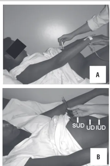
During the measurements, the pregnant was positioned on supine position, with the hips lexed at 90°, the knees lexed at 120° and the feet rested on the plinth. Women were asked to lex the torso with the arms stretched out in a way that the spines of the scapula were lifted from the bed, until the mo-ment that the examiner could ind, by palpation the RAMD and mark it with a demographic pencil. After the pregnant woman returned to the resting position, the digital paquimeter was then placed perpendicularly to the torso in order to perform the measurement (Figure 1).
Electromyography measurement protocol
In the beginning of the second stage, when the pregnant had 10 cm cervical dilatation, the electromyographic signal for the abdominal muscle activity was obtained. The surface
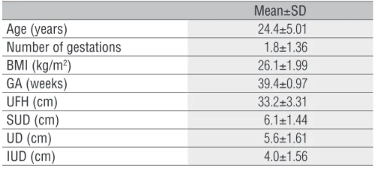
Mean±SD
Age (years) 24.4±5.01
Number of gestations 1.8±1.36
BMI (kg/m2) 26.1±1.99
GA (weeks) 39.4±0.97
UFH (cm) 33.2±3.31
SUD (cm) 6.1±1.44
UD (cm) 5.6±1.61
IUD (cm) 4.0±1.56
Table 1. Maternal Characteristics related to age, number of gestations, body mass index (BMI), gestational age (GA), uterus fundus height (UFH), supra-umbilical diastasis (SUD), umbilical diastasis (UD) and infra-umbilical diastasis (IUD).
electrodes were positioned on a rectus abdominis muscle bundle, 5 cm above and 3 cm to the sides of the umbilical scar, following Vera-Garcia, Grenier and McGill13
reccomen-dations. In those cases that the diastasis was higher than 3 cm to the sides of the umbilical scar, the medial edge of the muscle was taken as reference position to place the elec-trode. To obtain the external oblique muscle activity data the electrode was placed on the 8th rib, in the direction of
the muscular fibers14 and a reference electrode at the wrist
of each patient was used to eliminate external interference (30 mm x 45 mm x 1 mm) (Figure 2). In order to normalize the electromyography signal, the patient was asked to relax for 10 seconds and the electromyography data related to this period were stored in a computer software (DATAQ® for windows).
Later on, the electromyography signals were captured from the moment that the patient started the spontaneous expulsive efforts in second phase of labor. Those signals were captured for 15 minutes, being the strongest abdomi-nal push sigabdomi-nal captured during 5 seconds in each contrac-tion used for the data analysis. The electrodes were removed from the patient’s abdomen and discarded immediately after 15 minutes of signal capturing. Labor process data, like the expulsive period duration and the usage of oxytocin were considered.
In order to extract the electromyographic measurements an analog-to-digital converter module was used, using 4000 Hz frequency, 1000 times total internal gain, common mode re-jection ratio of >120Db, a 20 Hz high-pass ilter and a 500 Hz low-pass ilter. Two cardiology-type surface electrodes made of silver (Ag) and silver chloride (AgCl) were used – each of them with 4.5 cm in length and the distance between their compo-nents was 2 cm. he software Aqdados® was used to analyze
the signals and the Root Mean Square (RMS) of each contrac-tion was used as a parameter to measure the activity level of electromyography signal.
Measurements from the Uterus fundus height (UFH), Ges-tational Age (GA) and Body Mass Index (BMI) were taken from medical records.
Neonatal data
Arterial gasometry
To collect the arterial blood from the umbilical vein, a 10 cm segment of the umbilical cord was clamped and isolated for further analysis. A previously heparinized 1ml seringe was used to collect a blood sample of 1 ml from the umbilical vein, being this sample immediately analyzed in a
digital gasometer. Figure 2. Electrodes position.
A) Rectus abdomini muscle - 5 cm above and 3 cm to the sides of the umbilical scar; B) External oblique muscle - On the 8th rib, in the direction of the muscular fibers; C) Reference electrode.
Figure 1. Rectus abdominis muscle diastasis measurement.
A
A) The pregnant flexing the torso with the arms stretched out in a way that the spines of the scapula went out of the bed; B) Starting from the umbilical scar and measuring 4.5 cm above (SUD), 4.5 cm below (IUD) and on the umbilical scar level (UD), it was used a digital paquimeter.
B
APGAR index, Cephalic circumference and newborn weight
he data referring to the APGAR from the irst and the ifth minutes, cephalic circumference and birth weight were acquired from the hospital medical records.
Labor data
Labor parameters like expulsive period duration and oxyto-cin usage by the pregnant women were collected.
Statistical analysis
All the tests were applied with a signiicance level of 95% (p<0.05). To test the association of the dependent and indepen-dent variables we used the Spearman’s correlation coeicient. In order to compare the duration of second stage of labor Mann-Whitney test was used. he SPSS 13.0 for Windows and the Excel 2003 software were used for all analysis.
Results
he mean age of the sample in this study was 24.4 (SD 5.01) years and the mean gestational age was 39.4 (SD 0.97) weeks (Table 1). he mean second stage of labor duration of the stud-ied population was 41.7 (SD 32.26) minutes. Duration of the second stage of labor was similar in multiparae and nuliparae (43.69; SD=27.58 min for the nuliparae and 37.75 SD=42 min for the multiparae) (p=0.284).
No statistically significant correlation was found be-tween the electromyographic parameters of the rectus
abdominis (RA) and external oblique (EO) muscles and the most of maternal parameters – BMI (RA: p=0.063; r=0.094/ EO: p=0.757; r=-0.067); uterus fundus height (RA: p=0.787; r=-0.058/ EO: p=0.353; r=0.198), supra umbilical diastasis (RA: p=0.124; r=-0.323/ EO: p=0.074; r=0.371), umbilical dia-stasis (EO: p=0.612 ; r=0.109) and infra-umbilical diadia-stasis (RA: p=0.082; r=-0.362/ EO: p=0.227; r=0.256), except for the negative correlation found between the electromyographic activity of the rectus abdominis muscle and the umbilical diastasis (p=0.04; r=-0.407) - Figure 3.
he other fetal parameters were also not correlated with the electromyographic data of both muscles - fetal weight (RA: p=0.059; r=-0.390/ EO: p=0.978; r=0.006); one-minute APGAR score (RA: p=0.808; r=-0.052/ EO: p=0.923; r=0.021); ive-minute APGAR score (RA: p=0.165; r=0.263/ EO: p=0.549; r=-0.129); arterial pH (RA: p=0.869; r=-0.035/ EO: p=0.487; r=-0.149) and arterial pCO2 (RA: p=0.675; r=-0.09/ EO: p=0.366; r=-0.193). No
correlation was found between the duration of second stage and the electromyographic activity of both muscles (RA: p=0.989; r=-0.003/ EO: p=0.740; r=-0.072).
A significant correlation was also found between the supra-umbilical diastasis and the fetal cephalic circumfer-ence (p=0.036; r=0.431), although this parameter was not associated with electromyographic parameters of the rec-tus abdominis and external oblique muscles (RA: p=0.07, r=-0.37; EO: p=0.34; r=0.20). The use of oxytocine was not associated with electromyography of the studied muscula-ture (RA: p=0.815; EO: p=0.640).
Discussion
The uniqueness of the importance of this study is that the umbilical diastasis may interfere in the maternal vol-untary pushing, and, to date, its potential impact has not been well studied. This is the first study that has considered the relationship between the abdominal muscle electro-myographic activity and the abdominal diastasis. The ab-dominal muscle is an important factor to the increase of the intra-abdominal pressure during the second stage of labor. In clinical practice, pregnant women are encouraged to use these muscles to help fetal expulsion4.
Physiological functions in which the abdominal pressure increases (such as defecation or delivery), the action of these muscles is related to relexes that occur due to the stretching of speciic muscle receptors. Located in the pelvic loor, these re-ceptors send aferent impulses through the pelvic nerve to the medulla when triggered, stimulating motor neurons that are responsible for the abdominal muscles contraction3. Although
being triggered by a relex, the maternal voluntary pushing
Figure 3. Correlation between the rectus abdomini electromyographic activity (RMS) and umbilical diastasis.
7.00
6.00
5.00
4.00
3.00
2.00
1.00
RMS rectus abdomini
Umbilical diastasis
2.00 3.00 4.00 5.00 6.00 7.00 8.00 9.00
Spearman’s correlation: r=-0.407; p=0.048.
eforts during the second stage of labor are an important factor to predict the success of labor15.
A pilot study about surface electromyography of the ab-dominal muscles demonstrated that the transmission of the intrauterine pressure through the birth canal, and the recruit-ing the abdominal musculature with the voluntary pushes dur-ing the second stage of labor are essential to the fetal mobility through the uterus1. It was observed an increase of 62% in the
intrauterine pressure in pregnant women that had used vol-untary eforts during the spontaneous uterine contractions when compared to those who did not performed those pushes. However, other factors that are beyond maternal desire and medical care may inluence the efectiveness of pushing and be strongly related to the optimization of expulsive eforts2.
Some factors may contribute to the pushing eiciency, like fetus weight, the absence of augmentations, BMI and the myo-metrium thickness. Studies have shown that the myomyo-metrium thickness was the factor that most contributed for the pushing eiciency. his thickness speciically around 6 mm eases the transfer of the abdominal wall pressure strength to the uterine wall, helping the fetal expulsion2.
Although BMI has been suggested as an inluencing factor in the labor process, little is known about the real inluence on the eiciency of the pushing. BMI may be linked to this relationship, since the fat excess inside the abdominal wall makes the transference of force from the abdomen to the uterus diicult, which may be associated with high oxytocin index in obese mothers16,17. In our study, it was not possible
to detect a correlation between BMI and pushing eiciency, because the sample had only pregnant women with normal BMI as an inclusion criterion.
Besides those previously given factors, there are authors18,19
who believe that imbalance in the abdominal wall musculature (external and internal oblique, transverse abdominis and rectus abdominis) inluence its functions, hampering the increase of the intra-abdominal pressure, as well as impairing the genera-tion of efective pushing on the second stage of labor.
During the third trimester of gestation the fetus size along with a larger abdominal wall distension of the pregnant woman promote a biomechanical disadvantage to the abdomi-nal muscles, giving it relatively lower contraction power when compared to non-pregnant woman. his may interfere in other abdominal wall functions, impairing its strength generation capacity20. In this phase of the gestation the rectus abdominis
muscle line of action sufers a modiication in its insertion angle, thus altering this line of action.
he uterus fundus height (UFH) may be used as a param-eter to evaluate the abdominal wall distention, since it is a measurement that starts on the pubic symphysis ending up on the uterine fundus21. During pregnancy there is an increase of
about 115% in the rectus abdominis musculature length at 38 weeks18. Based on this length tension relationship, it is known
that an overload on the muscle iber is able to interfere in the capacity of producing normal tension. According to our ind-ings, however, there was no correlation between UFH and the electromyographic activity for the rectus abdominis muscle. Even being overloaded by a large distention the muscle is highly adaptable and sarcomeres are acquired according to the muscle length. Studies in animals show that sarcomeres are added to the muscle ibers when they are stretched for more than three weeks, thus increasing its strength. here are reports of an increase in the muscular length in humans when the muscles are progressively stretched22. One can
therefore infer that the absence of correlations between the electromyography data and the UFH is due to prolonged and progressive stretching that the abdominal musculature un-dergoes during pregnancy under hormonal as well as due to mechanic inluences.
he biomechanical changes in the abdominal muscles that occur during gestation, including the rectus abdominis muscle diastasis, probably cannot afect the muscle activity by them-selves. Since the muscular adaptations are not isolated, it is possible that the entire muscle group torque generation capac-ity is already compromised. In our study a negative correlation between the UD and the electromyography activity of the rectus abdominis muscle was observed, which may indicate a compro-mise in the strength generation capacity of this muscle during excessive efort. he irst investigation about the RAMD began in 198818 when a study investigated whether the RAMD inluence
the expulsive phase of labor. he anterior abdominal wall trauma increases the diiculty to increase the intra-abdominal pressure, which is necessary to the fetus expulsion, decreasing the expul-sive eforts during the second stage of labor. With a large dia-stasis during expulsive period, the increase of intra abdominal pressure would cause the ejection of the uterus ahead, through the space between the rectus abdominis muscle bundles, in-stead of expelling the fetus through the birth canal, what clearly is a biomechanical disadvantage as it alters the longitudinal axis between the fetus and the pelvis, impairing the voluntary eforts’ optimization19.
An important question would be how to identify the cut-of values considered pathological for a rectus abdominis muscle diastasis. he literature12,22 is not conclusive about
the possible physiopathologic repercussions of this diasta-sis. In fact, there is no actual scientiic evidence to point out the exact numerical value of a pathological diastasis and its biomechanical repercussions. he only parameters described in literature are the criteria established in a study23, which
deined a RAMD of 3 cm as pathological, but without any further biomechanical basis.
here was no correlation between the infra-umbilical dia-stasis and the electromyographic indings in this study. As for the morphologic and anatomic characteristics of the abdomi-nal region, the rectus abdominis tendinous band, known as linea alba, is stronger below the umbilical scar. In this region the aponeurosis from the four abdominal wall muscles cross right in front of the rectus abdominis muscle. Both sides of this muscle resemble a “V” when close to their insertion in the pubis, along with the other muscles, strengthening this area and decreasing its distention24.
Studies about the electromiographyc activity of abdomi-nal muscles in pregnant, suggest that during pregnancy the maternal organism have compensatory mechanisms that impaired physiologic functions, even with important biome-chanical alterations. his fact may be described in the results of our study, in which, even with lower electromyographic activity in pregnant women that had larger diastasis, we did not found important alterations when we correlated the elec-tromyography and the other parameters related to the birth labor and to the newborn22.
The duration of the second stage of labor, the APGAR scores, the arterial pH and the pCO2 of neonates blood were not correlated to a minor effectiveness of the push, from where it was suggested having any loss in the physiologi-cal of the labor mechanism, lower pushing efficiency, thus having no damage in the physiological mechanism of labor. Some studies25-28 that correlated fetal indices with the push
efficiency found correlations between the voluntary push decrease and lower arterial PH and O2 saturation in new-borns. Those studies, however, were performed in mothers that were anesthetized during delivery, therefore being part of the exclusion criteria of this study, in which no motor block technique was used.
his study suggests that the umbilical diastasis may act as an inluent parameter in the generation of voluntary pushes during the expulsive period of labor. Although this parameter should not be considered by itself in the study as a predictive factor of the voluntary abdominal efort in the expulsive pe-riod of labor, we suggest a link between this factor and other parameters described in literature (that were not evaluated in our study), like intra-uterine pressure and myometrium thickness. his association will make possible to develop, in future studies and with a largest sample, more detailed infor-mation about the expulsive period dynamics in order to help the obstetricians, nurses and physical therapists to optimize pregnant woman’s expulsive eforts during labor.
Acknowledgments
he Brazilian agency Conselho Nacional de Desenvolvimento Cientíico e Tecnológico (CNPq) and the Obstetrics Group of
Instituto de Medicina Integral Professor Fernando Figueira (IMIP) for their assistant and support.
References
1. Demaria F, Porcher R, Sheik-Ismael S, Amarenco G, Benifla JL. [Recording expulsive forces during childbirth using intercostal muscle electromyogram: a pilot study]. Gynecol Obstet Fertil. 2005;33(5):299-303.
2. Buhimschi CS, Buhimschi IA, Malinow AM, Kopelman JN, Weiner EP. The effect of fundal pressure manoeuvre on intrauterine pressure in the second stage of labour. BJOG. 2002; 109(5):520-6.
3. Shafik A, El Sibai O, Shafik IA, Shafik AA. Electromyographic activity of the anterolateral abdominal wall muscles during rectal filling and evacuation. J Surg Res. 2007; 143(2):364-7.
4. Liao JB, Buhimschi CS, Norwitz ER. Normal labor: mechanism and duration. Obstet Gynecol Clin North Am. 2005;32(2):145-64, vii.
5. Buhimschi C, Boyle M, Garfield RE. Electrical activity of the human uterus during pregnancy as recorded from the abdominal surface. Obstet Gynecol. 1997;90(1):102-11.
6. Chen DC, Ku CH, Huang YC, Chen CH, Wu GJ. Urinary nitric oxide metabolite changes in spontaneous and induced onset active labor. Acta Obstet Gynecol Scand. 2004; 83(7):641-6.
7. Okawa T, Vedermikov YP, Saade GR, Garfield RE. Effect of nitric oxide on contractions of uterine and cervical tissues from pregnant rats. Gynecol Endocrinol. 2004;18(4):186-93.
8. Väissänen-Tommiska M, Nuutila M, Ylikorkala O. Cervical nitric oxide release in women postterm. Obstet Gynecol. 2004;103(4):657-62.
9. Ponkey SE, Cohen AP, Heffner LJ, Lieberman E. Persistent fetal occiput posterior position: obstetric outcomes. Obstet Gynecol. 2003;101(5 Pt 1):915-20.
10. Atalah ES, Castilho CL, Castro Santoro R, Aldea AP. Propuesta de un Nuevo estándar de evaluación nutricional en embarazadas. Rev Méd Chile. 1997;125(12):1429-36.
11. Ministério da Saúde. Pré-natal e puerpério.: Atenção qualificada e humanizada. Brasília: Ministério da Saúde; 2005.
12. Hsia M, Jones S. Natural resolution of rectus abdominis diastasis. Two single case studies. Aust J Physiother. 2000;46(4):301-7.
13. Vera-Garcia FJ, Grenier SG, McGill SM. Abdominal muscle response during curl-ups on both stable and labile surfaces. Phys Ther. 2000;80(6):564-9.
14. Ng JK, Kippers V, Richardson CA. Muscle fibre orientation of abdominal muscles and suggested surface EMG electrode positions. Electromyogr Clin Neurophysiol. 1998;38(1):51-8.
15. Cheng YW, Hopkins LM, Caughey AB. How long is too long: Does a prolonged second stage of labor in nulliparous women affect maternal and neonatal outcomes? Am J Obstet Gynecol. 2004;191(3):933-8.
16. Jensen H, Agger AO, Rasmussen KL. The influence of pregnancy body mass index on labor complications. Acta Obstet Gynecol Scand. 1999;78(9):799-802.
17. Buhimschi CS, Buhimschi IA, Malinow AM, Weiner CP. Intrauterine pressure during the second stage of labor in obese women. Obstet Gynecol. 2004;103(2):225-30.
18. Boissonnault JS, Blaschak MJ. Incidence of diastasis recti abdominis during the childbearing year. Phys Ther. 1988;68(7):1082-6.
19. Thornton SL, Thornton SJ. Management of gross divarication of the recti abdominis in pregnancy and labour. Physioterapy. 1993;79(7):457-8.
20. Fast A, Weiss L, Ducommun E, Medina E, Butler JG. Low back pain in pregnancy. Abdominal muscles, sit-up performance, and back pain. Spine (Phila Pa 1976). 1990;15(1):28-50.
21. Freire DMC, Paiva CSM, Coelho EAC, Cecatti JG. Curva da altura uterina por idade gestacional em gestantes de baixo risco. Rev Bras Ginecol Obstet. 2006;28(1):3-9.
22. Gilleard WL, Brown JM. Structure and function of the abdominal muscles in primigravid subjects during pregnancy and the immediate postbirth period. Phys Ther. 1996;76(7):750-62.
23. Noble E. Essential Exercises for the Childbearing year. Boston: Houghton Mifflin Co; 1982.
24. Thompson AM. Maternal behavior during spontaneous and directed pushing in the second stage of labour. J Adv Nurs. 1995;22(6):1027-34.
25. Spencer JA, Koutsoukis M, Lee A. Fetal heart rate and neonatal condition related to epidural analgesia in women reaching the second stage of labor. Eur J Obstet Gynecol Reprod Biol. 1991;41(3):173-8.
26. Aldrich CJ, D’Antona D, Spencer JA, Wyatt JS, Peebles DM, Delpy DT, et al. The effect of maternal pushing on fetal cerebral oxygenation and blood volume during the second stage of labour. Br J Obstet Gynaecol. 1995;102(6):448-53.
27. Myles TD, Santolaya J. Maternal and neonatal outcomes in patients with a prolonged second stage of labor. Obstet Gynecol. 2003;102(1):52-8.
28. Tracy S, Sullivan E, Wang YA, Black D, Tracy M. Birth outcomes associated with interventions in labour amongst low risk women: a population-based study. Women Birth. 2007;20(2):41-8.