From the Liver Unit, University of São Paulo School of Medicine, São Paulo – Brazil.
SIZE OF GASTROESOPHAGEAL VARICES: ITS
BEHAVIOR AFTER THE SURGICAL TREATMENT OF
PORTAL HYPERTENSION
Edna Strauss, Paulo Sakai, Luiz Carlos da Costa Gayotto, Rita Antonelli Cardoso, Sonia Forsterand Silvano Raia
RHCFAO/2991 STRAUSS E et al. - Size of gastroesophageal varices: Its behavior after the surgical treatment of portal hypertension. Rev. Hosp. Clín. Fac. Med.
S. Paulo 54 (6):193-198, 1999.
SUMMARY: The size of gastroesophageal varices is one of the most important factors leading to hemorrhage related to portal hypertension. An endoscopic evaluation of the size of gastroesophageal varices before and after different operations for portal hypertension was performed in 73 patients with schistosomiasis, as part of a randomized trial: proximal splenorenal shunt (PSS n=24), distal splenorenal shunt (DSS n=24), and esophagogastric devascularization with splenectomy (EGDS n=25). The endoscopic evaluation was performed before and up to 10 years after the operations. Variceal size was graded according to Palmer’s classification: grade 1 – up to 3 mm, grade 2 – from 3 to 6 mm, grade 3 – greater than 6 mm, and were analyzed in four anatomical locations: inferior, middle or superior third of the esophagus, and proximal stomach. The total number of points in the pre-operative grading minus the number of points in the post-operative grading gave a differential grading, allowing statistical comparison among the surgical groups. Good results, in terms of disappearance or decrease of variceal size, were observed more frequently after PSS than after DSS or EGDS — 95.8%, 83.3%, and 72%, respectively. When differential grading was analyzed, a statistically significant difference was observed between PSS and EGDS, but not between proximal and distal splenorenal shunts. In conclusion, shunt surgeries were more efficient than devascularization in diminishing variceal size.
DESCRIPTORS: Hepatosplenic Schistosomiasis. Portal hypertension. Esophageal varices. Surgical treatment. Variceal size.
Nowadays, there are many options besides surgery for the elective treat-ment of portal hypertension. Endo-scopic methods as sclerotherapy1, and
band ligation2, are widely used either
isolated or in different combinations3
whereas N-butyl 2 cyanoacrylate4 can
also be used for obliteration of the va-rices. Medical therapies comprise dif-ferent types of drugs such as beta blockers5 or many others6, and a
vas-cular approach is transjugular porto-systemic shunting (TIPPS)7. The
ulti-mate goal in any type of treatment for portal hypertension is the elimination of the gastroesophageal varices, or at least reduction of variceal size, since the presence of gastroesophageal va-rices is one of the major factors impli-cated in re-bleeding8.
Surgical treatment aiming at devia-tion of portal blood to the systemic
cir-culation would be a preferential choice if severe side effects, such as hepatic encephalopathy, could be avoided. In an attempt to maintain good results without undesirable side effects, tech-nical modifications have been intro-duced. The splenorenal shunt was sup-posed to be as effective as portocaval shunt, with lower incidence of porto-systemic encephalopathy; whereas se-lective decompression of the gastroe-sophageal venous plexus through a dis-tal splenorenal shunt could also be an effective option. Theoretically, devas-cularization of the gastroesophageal area tends to maintain a high portal
pressure and therefore a good perfu-sion to the liver, with the inconvenience of re-appearance of gastroesophageal varices, with a higher risk of re-bleed-ing9.
In a previous paper, we published the clinical results of a randomized trial comparing the long-term efficacy of these three types of surgery10.
PATIENTS AND METHOD
Seventy-three patients with hepatosplenic schistosomiasis and por-tal hypertension, randomly assigned to three types of surgery to prevent re-bleeding, were evaluated endoscopi-cally for gastroesophageal status. This protocol was approved by the Ethical Committee of the hospital, and a writ-ten informed consent was obtained from each patient.
Two shunt procedures namely proximal splenorenal shunt (PSS) and distal splenorenal shunt (DSS) were compared to esophagogastric devascu-larization with splenectomy (EGDS). The inclusion criteria for entering the study were: a) diagnosis of Mansoni Schistosomiasis based on epidemio-logical, clinical, and parasitological data and confirmed by histopathologi-cal analysis of the wedged liver biopsy taken at the time of operation; b) age from 18 to 55 years; c) minimum in-terval of 15 days between last hemor-rhage and operation; d) absent or eas-ily controlled ascites; e) absence of chronic alcoholism, liver failure, cir-rhosis, peptic ulcer, diabetes, renal fail-ure, and portal thrombosis at angiog-raphy; f) minimum follow-up of 12 months, g) absence of endoscopic, pharmacologic, or any other kind of treatment for the portal hypertension during the whole follow-up period.
Varices, present in all patients be-fore the operation, were classified ac-cording to: a) their anatomical location – inferior, middle or superior part of esophagus, and proximal stomach and b) their size — grade 0 = no varices, grade 1 = varices diameter up to 3 mm, grade 2 = varices diameter from 3 to 6 mm, and grade 3 = varices diameter greater than 6 mm11. According to the
protocol, endoscopic evaluation was performed before the surgical proce-dure and every one or two years until 5 or 10 years of follow-up. All patients had a first evaluation one year after
sur-gery, and only 7 of them (9.6%) had only two endoscopic examinations. The latest evaluation, usually 5 or 10 years after surgery, was considered for statistical analysis. The mean period of time between surgery and the final evaluation was 5.83 3.05; 6.00 +/-3.05, and 6.12 +/-2.77 years respec-tively for PSS, EGDS and DSS.
Under comparative assessment, the gastroesophageal varices could : I) dis-appear, II) decrease III) remain un-changed, or IV) increase during fol-low-up. The post-operative endoscopic evaluation was also compared to pre-operative data by summing up the grades given to the size of varices — 0 to 3 — in the four different anatomic sites. The total number of points in the pre-operative grading (P) minus the to-tal number of points in the final grad-ing (E) was used for statistical com-parisons among the three types of
op-erations. The statistical method applied was variance analysis with multiple amplitude comparisons using the test of Ryan-Einot-Gabriel-Welsch12, and
using SAS (Statistical Analysis Sys-tem) software.
RESULTS
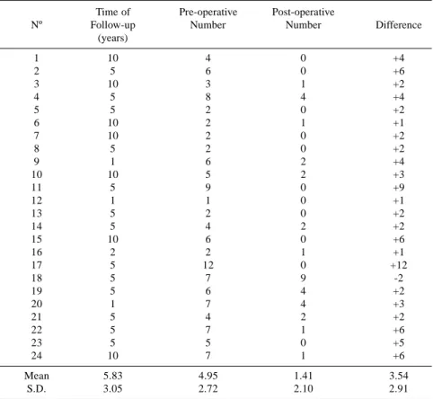
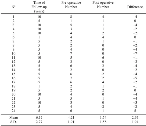
In Tables 1, 2, and 3 the time of the latest endoscopic evaluation, total num-ber of points before (P) and after sur-gery (E), as well as the difference be-tween them, is depicted respectively to PSS, EGDS, and DSS.
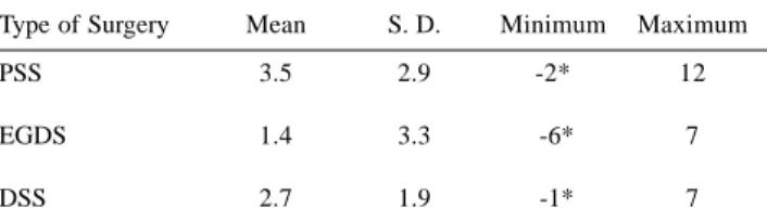
When the differential gradings in the three groups of patients were com-pared, a statistically significant differ-ence was obtained. The positive values for PSS were the highest (3.5 +/-2.9) and significantly different from EGDS (1.4 +/-3.3 ). Nevertheless, when DSS
Table 1 – Results of the endoscopic evaluations in the group of patients who received a proximal splenorenal shunt: time of latest endoscopy, total number of points in the pre-operative grading, post-operative grading and its difference.
Time of Pre-operative Post-operative
Nº Follow-up Number Number Difference
(years)
1 10 4 0 +4
2 5 6 0 +6
3 10 3 1 +2
4 5 8 4 +4
5 5 2 0 +2
6 10 2 1 +1
7 10 2 0 +2
8 5 2 0 +2
9 1 6 2 +4
10 10 5 2 +3
11 5 9 0 +9
12 1 1 0 +1
13 5 2 0 +2
14 5 4 2 +2
15 10 6 0 +6
16 2 2 1 +1
17 5 12 0 +12
18 5 7 9 -2
19 5 6 4 +2
20 1 7 4 +3
21 5 4 2 +2
22 5 7 1 +6
23 5 5 0 +5
24 10 7 1 +6
Mean 5.83 4.95 1.41 3.54
is compared either to PSS or EGDS, a significant difference could not be found (Table 4).
Good results, in terms of disappear-ance or decreasing of the variceal size, were observed more frequently after PSS than DSS or EGDS (Table 5). In-creasing of variceal size, on the other hand, was observed in 20% of the cases after EGDS and in only 4.2% (one case ) after the two shunt proce-dures.
Figure 1 illustrates the pre-opera-tive and post-operapre-opera-tive variceal sizes in the four anatomic sites. Gastric varices, present in 33.3%, 20.8%, and 12.0% before respectively, PSS, DSS, and EGDS, were present in 0%, 16.6% and 28.0% in the follow-up period.
DISCUSSION
The identification of risk factors for gastroesophageal bleeding is of utmost importance, not only for prophylaxis of the first hemorrhage episode, but also for patients with risk of re-bleeding. There is a consensus that bleeding seems to be high in patients with me-dium to large varices, although other endoscopic, hemodynamic, and clini-cal factors have also been associated with risk of gastroesophageal bleed-ing13,14.
More recently, metabolic variables, such as poor nutritional status, low se-rum albumin, and decreased clotting factors were independently associated with a higher risk of bleeding in cirrho-sis15. Although interesting, these
clini-cal parameters may be related more to the hepatic insufficiency of severe cir-rhosis than to portal hypertension itself. On the other hand, the endoscopic risk factors, such as size of varices or red whale markings would be applicable to portal hypertension due to other etiolo-gies besides cirrhosis.
The hepatosplenic form of Mansoni’s schistosomiasis is an excel-Table 2 - Results of the endoscopic evaluations in the group of patients who
underwent esophagogastric devascularization with splenectomy: time of latest endoscopy, total number of points in the pre-operative grading, post-operative grading and its difference.
Time of Pre-operative Post-operative
Nº Follow-up Number Number Difference
(years)
1 10 3 2 +1
2 5 9 8 +1
3 10 8 7 +1
4 10 4 4 0
5 10 9 2 +7
6 5 1 5 -4
7 5 5 8 -3
8 10 2 4 -2
9 10 5 2 +3
10 10 3 2 +1
11 1 6 1 +5
12 5 5 8 -3
13 3 4 3 +1
14 10 6 2 +4
15 2 6 1 +5
16 5 6 0 +6
17 5 2 0 +2
18 3 4 4 0
19 5 6 12 -6
20 5 4 3 +1
21 5 5 3 +2
22 5 6 5 +1
23 5 5 0 +5
24 5 6 1 +5
25 1 4 3 +1
Mean 6.00 4.96 3.60 1.36
S.D. 3.05 1.98 3.01 3.32
Table 3 - Results of the endoscopic evaluations in the group of patients who received a distal splenorenal shunt: time of latest endoscopy, total number of points in the pre-operative grading, post-operative grading and its difference.
Time of Pre-operative Post-operative
Nº Follow-up Number Number Difference
(years)
1 10 8 4 +4
2 5 1 1 0
3 10 5 1 +4
4 10 3 1 +2
5 10 4 2 +2
6 1 4 4 0
7 5 2 1 +1
8 5 2 0 +2
9 5 4 0 +4
10 5 7 0 +7
11 10 5 6 +1
12 5 3 0 +3
13 5 6 2 +4
14 5 2 0 +2
15 5 6 2 +4
16 5 7 2 +5
17 5 6 4 +2
18 1 2 1 +1
19 5 2 2 0
20 10 4 0 +4
21 5 5 1 +4
22 10 3 0 +3
23 5 4 2 +2
24 5 6 1 5
Mean 6.12 4.21 1.54 2.67
Table 4 - Differential grading of gastroesophageal varices between pre-operative and post-operative period.
Type of Surgery Mean S. D. Minimum Maximum
PSS 3.5 2.9 -2* 12
EGDS 1.4 3.3 -6* 7
DSS 2.7 1.9 -1* 7
F = 3.86; p = 0.026; S.D. = standard deviatio; * = Negative numbers correspond to an increase of variceal size when compared to pre-operative status.
MULTIPLE COMPARISON OF AMPLITUDE
Operations Conclusion
PSS X EGDS *
PSS X DSS N.S.
EGDS X DSS N.S.
* = statistically significant at the level of 5%. N.S. = not significant.
pre operative
evolutive
Esophagus Lower third
0 2 4 6 8 10 12 14
PSS DSS EGDS
0 2 4 6 8 10 12 14
PSS DSS EGDS
0 2 4 6 8 10 12 14
PSS DSS EGDS
Esophagus Middle third
0 2 4 6 8 10 12 14
PSS DSS EGDS
0 2 4 6 8 10 12 14
PSS DSS EGDS
0 2 4 6 8 10 12 14
PSS DSS EGDS
Esophagus Upper third
0 2 4 6 8 10 12 14
PSS DSS EGDS
0 2 4 6 8 10 12 14
PSS DSS EGDS
0 2 4 6 8 10 12 14
PSS DSS EGDS
Gastric
0 2 4 6 8 10 12 14
PSS DSS EGDS
0 2 4 6 8 10 12 14
PSS DSS EGDS
0 2 4 6 8 10 12 14
PSS DSS EGDS
1 2 3
Grade of Varices
Figure 1 - Illustration of variceal size in the four anatomic sites.
Table 5 - Comparison of the variations of the size of gastroesophageal varices before and after different operations
Type of PSS EGDS DSS TOTAL
Surgery Varices
Disappear 11 (45.8%) 3 (12%) 7 (29.2%) 21
Decrease 12 (50.0%) 15 (60%) 13 (54.1%) 40
Unchanged 0 2 (8%) 3 (12.5%) 5
Increase 1 (4.2%) 5 (20%) 1 (4.2%) 7
Total 24 25 24 73
PSS = proximal splenorenal shunt.
lent model for the evaluation of thera-peutic procedures in portal hyperten-sion. Upper gastrointestinal hemor-rhage, occurring as a consequence of gastroesophageal varices, is the most outstanding clinical feature, whereas alterations of liver function are usually slight, if present at all.9,16 Thus it is
pos-sible to evaluate the consequences of portal hypertension separately from those of hepatic insufficiency. This evaluation is difficult to accomplish in patients with cirrhosis.
A common and easily reproducible method for comparing variceal size be-fore and after a therapeutic procedure is to evaluate whether varices have dis-appeared, decreased, remained un-changed, or increased. The different percentages obtained in each group, as shown in Table 5, point to a better per-formance of PSS and worse results af-ter EGDS, although these percentages do not allow statistical comparisons.
A major problem in comparing va-riceal size to evaluate effectiveness of a therapeutic procedure is the subjec-tivity of the procedure. Quantification
of variceal size alterations would be an ideal approach, if possible. Since a di-rect measure is not usually performed, a numerical value (from 0 to 3) was obtained using a known classification, in which the terms grade 1, grade 2 and grade 3 correspond to well known and accepted criteria of variceal size in mil-limeters11.
To our knowledge this is the first time, a statistical analysis has been per-formed to compare variceal size. It was interesting to verify that after the three surgical treatments, a good average re-sult was achieved in terms of diminish-ing variceal size. Since we have sub-tracted from the pre-operative values those obtained during the follow-up, as shown in Tables 1, 2, and 3 and in Fig. 1, a positive number is indicative of smaller varices. On the other hand, in every type of surgery we also obtained negative numbers, corresponding to patients who developed larger varices during the follow-up.
Presence of gastric varices is a common finding in portal hyperten-sion17. In that study, gastric varices
were found in 12 to 33.3% of the cases in the three groups of patients in the pre-operative period. When patients underwent shunt surgeries, a decrease in size after distal splenorenal shunt and disappearance after a proximal splenorenal shunt were observed.
As shown in Fig. 1 the percentage of gastric varices has increased after esophagogastric devascularization, similarly to what may happen after sclerotherapy, when high levels of por-tal hypertension persist. Hemodynamic studies have recently shown that por-tal pressure can either decrease or in-crease after sclerotherapy, depending on presence or absence of spontaneous collateral circulation in each patient18.
Similarly to what we have observed in the long-term clinical comparison among the three types of surgery, it was not possible to isolate the effects of DSS from EGDS. On the other hand EGDS, the surgery with the best results in terms of survival and lack of side ef-fects, was the less effective in terms of improving the gastroesophageal varices status.
RESUMO RHCFAP/2991
STRAUSS E e col. - Variações no cali-bre das varizes esôfago-gástricas após tratamentos cirúrgicos de hipertensão portal. Rev. Hosp. Clín. Fac. Med. S. Paulo 54 (6): 193-198, 1999.
Um dos mais importantes fatores que levam à hemorragia digestiva por hipertensão portal é o calibre das vari-zes esôfago-gástricas. Visamos, no pre-sente trabalho, avaliar endoscopi-camente as variações de calibre antes e após diferentes cirurgias de hiperten-são portal, realizadas em 73 pacientes
com esquistossomose hépato-esplê-nica, no contexto de um estudo contro-lado e aleatorizado, sendo 24 deles submetidos a Anastomose Espleno-Re-nal (AER), 24 a Descompressão Por-tal Seletiva (DPS) e 25 a Desconexão Azigo-Portal com Esplenectomia (DAPE). As avaliações endoscópicas foram realizadas antes e até 10 anos após as cirurgias. O calibre das varizes foi classificado, segundo Palmer como de grau 1- até 3mm, grau 2 de 3 a 6 mm e grau 3 quando maiores do que 6mm de diâmetro, analizadas em qua-tro localizações anatômicas a saber:
diferenciais de gradação demonstrou diferença estatisticamente significante favorecendo a AER em relação à DAPE, não havendo diferenças entre AER e DPS. Em conclusão, as
cirur-gias de anastomose (“shunt”) foram mais eficientes do que a desvascula-rização, em termos de diminuir o cali-bre de varizes esôfago-gástricas.
DESCRITORES: Esquistossomose hépato-esplênica. Hipertensão por-tal. Varizes esofágicas. Tratamento cirúrgico. Calibre de varizes.
REFERENCES
1. INFANTE RC, ESNAOLA S & VELLENEUVE JP - Role of endoscopic variceal sclerotherapy in the long-term management of variceal bleeding: a meta-analysis. Gastroenterology 1989; 96:1087-1092. 2. STIEGMANN GV, CAMBRE T & SUN J - A new endoscopy elastic
band device. Gastrointest. Endos. 1986; 32:230-233.
3. STRAUSS E - Band ligation, sclerotherapy, both or ...brains? Am J
Gast. 1997; 92(6):920-923.
4. SOEHENDRA N, GRIMM H & MAM VC. - N-butyl-2–cyannoacrylate: a supplement to endoscopic sclerotherapy. Endoscopy 1987;
19:221-224.
5. LEBREC D, POYNARD T, CAPRON JP, et al. - Nadolol for prophylaxis of gastrointestinal bleeding in patients with cirrhosis. A randomized trial. J. Hepatol 1988; 7:118-125.
6. D’AMICO G, PAGLIARO L & BOSCH J. The treatment of portal hypertension: a meta-analytic review. Hepatology 1995; 22:332-354.
7. SHIFFMAN ML, JEFFERS L, HOOFNAGLE JH et al. - The role of transjugular intrahepatic portosystemic shunt for the treatment of portal hypertension and its complications. Hepatology 1995;
22:1591-1597.
8. LEBREC D, DE FLEURY P, RUEFF B et al. - Portal hypertension, size of esophageal varices and risk of gastrointestinal bleeding in alcoholic cirrhosis. Gastroenterology 1980; 79:1139-1144. 9. RAIA S, MIES S & MACEDO AL. - Portal hypertension in
schistosomiasis. Clin Gastroenterol 1985; 14:57-82.
10. RAIA S, SILVA LC, GAYOTTO LC et al. Portal hypertension in schistosomiasis: a long-term follow-up of a randomized trial comparing three types of surgery. Hepatology 1994; 20:398-403.
11. PALMER ED & BRICK IB - Correlation between the severity of esophageal varices in cirrhosis and their propensity toward hemorrhage. Gastroenterology 1956; 30:85-90.
12. RANSEY PH - Power differences between pairwise multiple comparisons. J Amer Statist Ass 1978; 73:479-487.
13. BEPPU K, INOKUCHI K, KOYANAGI N, et al. Prediction of variceal hemorrhage by esophageal endoscopy. Gastrointest Endosc 1981;
27:213-218.
14. THE NORTH ITALIAN ENDOSCOPIC CLUB FOR THE STUDY AND TREATMENT OF ESOPHAGEAL VARICES - Prediction of the first variceal hemorrhage in patients with cirrhosis of the liver and esophageal varices. A prospective multicenter study. N
Engl J Med 1988; 319:983-989.
15. MOLLER S, BENDTSEN F, CHRISTENSEN E et al. - Prognostic variables in patients with cirrhosis and oesophageal varices without prior bleeding. J Hepatol 1994; 21:940-946.
16. EL-ROOBY A - Management of hepatic schistosomiasis.
Semin Liver Dis 1985; 5:263-276.
17. SARIN SK, LOHOTI D, SAXENA S P, et al. - Prevalence, classifi-cation and natural history of gastric varices: a long-term follow-up study in 568 portal hypertension patients. Hepatology 1992;
16:1343-1349.
18. TOYONAGA A, IWAO T, SUMINO M, et al. - Portal pressure after prophylactic sclerotherapy in patients with high-risk varices.
J Hepatology 1994; 21:515-520.