Kinesiographic study of complete denture movement related to
mucosa displacement in edentulous patients
Estudo cinesiográfico da movimentação da prótese total
resultante da deformação da fibromucosa em desdentados totais
Marco Antonio Compagnoni* Raphael Freitas de Souza** Cláudio Rodrigues Leles***
ABSTRACT:The mucosa that covers the residual ridges of edentulous patients may present some distortion or dis-placement when occlusal loading is applied in complete dentures. This distortion and movement of the denture can re-sult in acceleration of residual ridge resorption and loss of retention and stability. The aim of this study was to analyze the pattern of upper complete denture movement related to underlying mucosa displacement. A sample of 10 complete denture wearers was randomly selected, which had acceptable upper and lower dentures and normal volume and re-silience of residual ridges. The kinesiographic instrument K6-I Diagnostic System®was used to measure denture movements, according to the method proposed by Maedaet al.7, 1984. Denture movements were measured under the following experimental conditions: (A) 3 maximum voluntary clenching cycles and (B) unilateral chewing for 20 sec-onds. The results showed that under physiological load, oral mucosa distortion has two distinct phases: a fast initial displacement as load is applied and a slower and incomplete recovery when load is removed. Intermittent loading such as chewing progressively reduces the magnitude of the denture displacement and the recovery of the mucosa is gradu-ally more incomplete.
DESCRIPTORS:Mouth mucosa; Denture, complete.
RESUMO:Devido às características de compressibilidade da fibromucosa, a incidência de cargas sobre a prótese total resulta na movimentação da prótese em direção ao rebordo, a qual pode resultar no aumento da reabsorção do rebordo residual, na desadaptação interna e na perda de retenção da prótese. O objetivo do presente estudo é avaliar o padrão de movimentação da prótese total superior em função da deformação da fibromucosa. Foram selecionados 10 pacien-tes usuários de própacien-teses totais bimaxilares, com retenção satisfatória e relações oclusais corretas e rebordos alveola-res com volume e grau de alveola-resiliência normais. Para a análise da movimentação da prótese total superior foi utilizado o sistema eletrônico K6-I Diagnostic System®, sendo o dispositivo eletromagnético fixado na região vestibular dos incisi-vos centrais da prótese superior, de acordo com uma adaptação do método sugerido por Maedaet al.7(1984). Foram obtidos registros gráficos em duas condições experimentais: (A) 3 ciclos de apertamento máximo voluntário e (B) mas-tigação unilateral simulada por um período de 20 segundos. Os resultados mostraram um padrão uniforme de movi-mentação durante o apertamento máximo e a mastigação simulada. Sob a ação de cargas, a fibromucosa apresenta uma deformação rápida e uma recuperação mais lenta e incompleta, de forma que a ação repetida da aplicação de car-ga reduz gradualmente a quantidade de movimentação e retorno da prótese; na masticar-gação simulada não há a comple-ta recuperação da fibromucosa durante os ciclos mastigatórios.
DESCRITORES:Mucosa bucal; Prótese total.
*Chairman Professor; **Graduate Student – Department of Dental Materials and Prosthodontics, Araraquara Dental School, São Paulo State University.
***Professor, Department of Prevention and Oral Rehabilitation, School of Dentistry, Federal University of Goiás.
INTRODUCTION
When occlusal pressure is applied to a complete denture, there is a displacement or distortion of the mucous membrane and a consequent move-ment of the denture. This displacemove-ment is related to changes in blood circulation and tissue ele-ments of connective tissues5,8
, and depends on the histological and morphological characteristics of
the mucous membrane that is in close contact with the prosthesis.
Kydd et al.5observed that sustained loading can
compress the tissues up to 45% of its original thickness. This deformation is viscoelastic in na-ture, which means that its mechanical response depends on the rate of loading, its magnitude, du-ration and previous loading history10
The distortion of the masticatory mucosa and the related movement of the denture can result in acceleration of residual ridge resorption and loss of retention and stability of the denture. Chong2
ob-served a close relation between internal adaptation and movement of the maxillary denture during masticatory function. Well-fitted dentures exhib-ited a lesser amount of movement than did poorly fitted ones. The range of well-fitted dentures move-ment varied from 0 to 1.4 mm on the chewing side and from 0.1 to 1.6 on the other side.
The thickness and displaceability of the muco-sal support for dentures should be considered when recording impressions. Mobile tissue pres-ents problems of support and stability which are dealt with either by surgical reduction in the thick-ness of these tissues or by using special impres-sion techniques which distribute the load in a par-ticular manner.
Clinical assessment of the supporting tissue in complete denture patients is important for preop-erative diagnosis and treatment planning. Such clinical assessment tends to differ considerably between dentists and most assessment is subjective9
. Therefore, it is important to make clin-ical mucosal displacement reliable and accurate. The aim of the present study was to observe the be-havior of complete denture displacement and tissual deformation under occlusal loading in dif-ferent clinical situations. The viability of a kinesiographic instrument to measure this dis-placement was also analysed.
MATERIAL AND METHOD
Ten complete denture wearers were selected ac-cording to the following criteria: (1) recently in-stalled upper and lower conventional complete dentures, (2) good retention and satisfactory inter-nal adaptation of the dentures, (3) normal volume and resilience of residual ridges, and (4) informed consent of the patients. Their ages ranged from 52 to 77 years, six were female and four male. Fur-thermore, the initial project was approved by the Ethical Committee, Araraquara Dental School. All participants were informed on the nature of the in-vestigation and agreed to take part in it.
A kinesiographic instrument (K6-I Diagnostic System, Myotronics Research Inc., Seattle, WA, USA) connected to a personal computer system (Microsoft Windows 95, Version 4.1, Microsoft Corporation, Redmond, WA, USA), was used for patient evaluation and graphic recordings.
Patients were erectly seated, with the Frankfurt plane parallel with the horizontal plane. The sen-sory array was positioned according to the manu-facturer’s instructions (Figure 1) and the magnet was attached to the labial midline surface of the maxillary complete denture, as proposed by Maeda
et al.7
.
Linear vertical movements of the maxillary den-ture were registered under the following experi-mental conditions:
1. Maximum voluntary clenching (MVC) - 3 cycles of three-seconds MVC and a rest period of five-seconds between each cycle.
2. Simulated mastication (SM) - unilateral chew-ing (preferred side) of a test food for a 20 sec-onds period of time. The material used as a food substitute was a 10 x 10 x 5 mm polysulfide specimen (Regular Permlastic,
, Kerr Corpora-tion, Orange, CA, USA). The material had a soft rubber consistency and was maintained throughout the chewing cycle.
The sets of measurements were transferred to an image editor software (PhotoImpact SE, Ver-sion 3.02 (1997), Unlead Systems Inc., Taipei, Tai-wan) for further assessment. The measurement scale was converted from pixels to millimeters by selecting reference points on x and y axis and
paring them to the millimeter scale of the kine-siographic tracings. Assuming that tracings pre-sented the same magnification, a conversion scale was established by using the following formula:
a = 0.0435. b
Where:
a- distance between two selected points (in mil-limeters).
b- number of pixels.
Descriptive statistics and correlation analysis were performed at a 95% level of confidence.
RESULTS AND DISCUSSION
Graph 1 illustrates a common graphic pattern of all subjects in both experimental conditions. The displacement/time ratio selected was 1 mm/2 s, expressed in the vertical and horizontal axes, respectively. According to this graphic pat-tern, the following assumptions could be made:
Experiment 1
Immediately after denture clenching an instan-taneous and fast upper displacement of the den-ture occurred. As long as the clenching force was sustained for 3 seconds, the altered position of the denture was maintained constant. Subsequently, when the clenching force was interrupted, there was a gradual recovery of denture initial position. During this recovery, two distinct phases were clearly recognized: an initial fast recovery and a late delayed and gradual recovery that did not complete until the next clenching cycle. This typi-cal behavior is illustrated in Graph 2.
These findings were similar to the observations in dogs reported by Wills, Manderson10
(1977). Mu-cosa behavior under abrupt loading showed an im-mediate elastic displacement. If this loading is maintained there is a tendency to a subsequent
gradual displacement. When load is removed, an instantaneous recovery takes place (elastic recov-ery), followed by a slow constant recovery (viscoelastic recovery) that can last up to three hours until its initial condition, as long as blood and extracellular fluids are expelled in a slow and steady way. In another animal study, Picton, Wills8
(1978) also observed these viscoelastic properties of the mucosa under tissue-supported prosthetic baseplates.
In the present study, the measurements of the graphic registrations varied widely and are de-scribed in Tables 1 and 2. The respective means are shown in Graphs 3 and 4.
These values are comparable to those of previ-ous studies8,10
. The overall amount of denture dis-placement (0.82 mm) was greater than its related recovery (0.75 mm). Moreover, as stated by Picton, Wills8
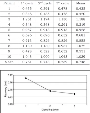
(1978), if the cycles of loading are repeated in a less than 1.5 minutes time interval, mucosa re-covery tends to be progressively more incomplete. This occurrence is clearly verified when comparing the decreasing quantity of displacement and re-covery between the three clenching cycles.
The level of displacement and recovery varied widely between patients. The possible explanation is related to anatomical and physiological charac-teristics of the mucosa that influence denture sup-port, prosthetic features like denture extension, internal adaptation, occlusal relations, and forces produced by occlusion1,3
. Ismail4
(1971) reported a vertical displacement of dentures that ranged from 0.5 mm to 2.5 mm (mean = 0.94 mm). Maedaet al.7
(1984), using a kinesiographic instrument in two patients observed a vertical displacement from 0.5 mm to 1.6 mm resultant from maximal clench-ing sustained for 2 seconds.
GRAPH 2 - Theoretical model of vertical denture
move-ment during maximum voluntary clenching.
GRAPH 1 -Example of graphic registration of both
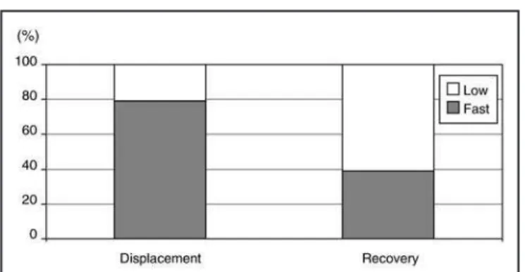
The rapid phase of the displacement represents the greatest part of the total displacement and the slower part of denture recovery corresponds to the greatest part of overall recovery (Graph 5). This pe-culiarity is due to marked differences in velocity of diffusion and return of blood and extracellular flu-ids. During maximal clenching this effect is more apparent because the magnitude of loading is di-rectly proportional to the extent of compression of cellular bodies around tissue10.
The variation in the displacement and recovery times with the loading cycles can be explained on the basis of fluid flow of the tissues within the pal-atal mucosa, which is determined by the pressure pattern under the denture baseplate.
The recovery of the mucosa is also prolonged and of variable duration6
. This would indicate that there is not a simple recovery mechanism when load is removed. The most likely explanation is that fluid (blood and extra-cellular fluid) is ex-pelled from loaded areas and the recovery is
con-trolled by the rate at which these fluids return to the loaded area10
.
Experiment 2
Reproducible graphs of vertical movement of upper denture during simulated chewing was ob-tained in 6 of the 10 selected patients. A common pattern was observed in all patients and this model is schematically illustrated in Graph 1. Since there is not enough time between two chewing cycles, a residual displacement is maintained throughout the masticatory activity. Consequently, there is an incomplete recovery of compression of the mucosa. The values of vertical displacement and residual displacement are described in Table 3. A positive correlation was observed between number of chewing cycles and amount of residual displace-ment. As the number of chewing cycles increase, a significant increasing in mucosa residual displace-ment is observed (r = 0.68; p < 0.01), as shown in Graph 6.
TABLE 2 - Recovery movement of the upper complete
denture after maximum voluntary clenching (in millime-ters).
Patient 1st
cycle 2nd
cycle 3rd
cycle Mean
1 0.435 0.391 0.478 0.435
2 0.348 0.435 0.478 0.420
3 1.261 1.174 1.130 1.188
4 0.348 0.348 0.261 0.319
5 0.957 0.913 0.913 0.928
6 0.696 0.696 0.652 0.681
7 0.913 0.826 0.826 0.855
8 1.130 1.130 0.957 1.072
9 0.478 0.522 0.652 0.551
10 1.043 1.000 1.043 1.029
Mean 0.761 0.743 0.739 0.748
TABLE 1 -Vertical denture movement during maximum
voluntary clenching (in millimeters).
Patient 1stcycle 2ndcycle 3rdcycle Mean
1 0.522 0.435 0.435 0.464
2 0.435 0.435 0.435 0.435
3 1.478 1.304 1.174 1.319
4 0.435 0.348 0.304 0.362
5 0.957 0.870 0.870 0.899
6 0.870 0.596 0.696 0.720
7 1.130 0.957 0.870 0.986
8 1.435 1.261 1.261 1.319
9 0.609 0.609 0.565 0.594
10 1.174 1.087 1.043 1.101
Mean 0.904 0.790 0.765 0.820
GRAPH 4 -Mean vertical denture recovery according to
the clenching cycle.
GRAPH 3 - Mean vertical denture displacement
Picton, Wills8
(1978) also reported that chewing activity causes a progressive intrusion of the den-ture base, although this effect tends to become stable within 20 or 30 chewing cycles. This intru-sion can last up to 10 minutes until its complete recovery8
.
The kinesiographic method was employed in previous studies to evaluate viscoelastic properties of the mucosa2,7
, and was considered to be useful for documentation of denture displacement under different conditions of occlusal loading.
As denture displacement related to supporting tissues distortion could disturb denture balance, clinical variables related to the displaceability of the mucosa should be properly assessed by den-tists, even though objective assessment at chairside is extremely difficult9.
These results can also be applied to im-plant-supported overdentures or removable par-tial dentures that have mucosa-borne support. The considerably higher fluid component of mu-cosa can lead to increased horizontal loading un-der implants or teeth and clinical management may include impression procedures that consider tissue resiliency and displaceability.
CONCLUSION
Under the present experimental conditions there was a consistent movement of the upper den-ture as a result of mucosa displacement during maximum voluntary clenching and simulated chewing, and the following conclusions could be drawn:
•Under occlusal loading, the mucosa revealed a fast and immediate displacement and a gradual and incomplete recovery.
•Repeated loading cycles gradually reduced the amount of displacement and recovery of the mucosa.
•During simulated chewing cycles there was not a complete recovery of the mucosa between each chewing stroke.
ACKNOWLEDGEMENTS
This study was supported by Fundação de Amparo à Pesquisa do Estado de São Paulo (FAPESP) - Grant #99/11727-0.
GRAPH 5 -Comparison between faster and slower
pha-ses during displacement and recovery of denture.
TABLE 3 -Values of vertical denture movement and
re-sidual displacement during simulated chewing.
Patient
Number of chewing
cycles
Maximal vertical displacement
of denture
Residual displacement (mm)
Mean SD
1 27 0.9 0.504 0.10
2 27 0.7 0.192 0.05
3 28 1.5 0.637 0.13
5 39 1.4 0.234 0.35
8 25 1.5 0.553 0.24
10 23 1.7 0.081 0.23
Mean 28 1.28 0.367 0.18
GRAPH 6-Meanresidual displacement of denture as a
REFERENCES
1. Bearn E. Effect of different occlusal profiles on the mastica-tory forces transmitted by complete dentures. Br Dent J 1973;134:7-10.
2. Chong LC. Movement of maxillary complete dentures – a kinesiographic study. J Dent 1983;11:257-63.
3. Cutright D, Brudvik JS, Gay WD, Selting WJ. Tissue pres-sure under complete maxillary dentures. J Prosthet Dent 1976;35:160-70.
4. Ismail YH. Denture movement under occlusal forces [abs-tract 445]. J Dent Res 1971;50:164.
5. Kydd W, Stroud W, Moffett BC Jr, Tamarin A. The effect of mechanical stress on oral mucoperiosteum of dogs. Arch Oral Biol 1969;14:921-31.
6. Lytle R. Soft tissue displacement beneath removable parti-al and complete dentures. J Prosthet Dent 1962;12:34-43. 7. Maeda Y, Okada M, Makishi A, Nokubi T, Okuno Y, Aoki T.
Using mandibular kinesiograph for measuring complete denture movements – a preliminary report. J Osaka Univ Dent Sch 1984;24:123-9.
8. Picton D, Wills D. Viscoelastic properties of the periodontal ligament and mucous membrane. J Prosthet Dent 1978;40:263-72.
9. Sato Y, Tsuga K, Yoshida M, Kubo T. Factors influencing the clinical composite assessment of denture-supporting tissues. Int J Prosthodont 2002;15:49-54.
10. Wills DJ, Manderson RD. Biomechanical aspects of the support of partial dentures. J Dent 1977;5:310-8.