ABSTRACT
Paget’s disease of bone is a focal disorder of bone remodeling accom-panied initially by an increase in bone resorption, followed by a disorga-nized and excessive formation of bone, leading to pain, fractures and deformities. It exhibits a marked geographical variation in its prevalence. In Brazil it predominantly affects persons of European descent. The major-ity of the reported cases of the disease in Brazil are from Recife, owing to its peculiar mixed European colonization over approximately four cen-turies. The etiology is complex and involves both genetic and environ-mental factors. The disease is often asymptomatic and diagnosis is usu-ally based on biochemical markers of bone turnover, radionuclide bone scan and radiological examination. Bisphosphonates, in particular zole-dronic acid, are regarded as the treatment of choice for Paget’s disease of bone. (Arq Bras Endocrinol Metab 2006;50/4:814-822)
Keywords:Paget’s disease of bone; Prevalence; Bone resorption; Bispho-sphonate; Calcitonin
RESUMO
Doença de Paget Óssea.
Doença de Paget óssea é uma desordem focal da remodelação óssea, inicialmente acompanhada de um aumento da reabsorção óssea, seguida de desorganizada e excessiva formação óssea, levando a dor, deformidades e fraturas. Exibe uma variável distribuição geográfica em sua prevalência. No Brasil acomete predominantemente pacientes de descendência européia. Recife, devido à sua peculiar colonização mista européia por cerca de 4 séculos, tem a maioria dos casos relata-dos no Brasil. A etiologia é complexa e envolve fatores ambientais e genéticos. A doença é freqüentemente assintomática e o diagnóstico é feito usualmente através dos marcadores bioquímicos do turnover ósseo associado a cintilografia óssea e dos sinais típicos do exame radi-ológico. Os bisfosfonatos representam o tratamento de escolha na doença de Paget óssea, particularmente o ácido zolidrônico. (Arq Bras Endocrinol Metab 2006;50/4:814-822)
Descritores: Doença de Paget óssea; Prevalência; Reabsorção óssea; Bisfosfonato; Calcitonina
P
AGET’S DISEASE OF BONE (PDB) was first described in 1876 by Sir James Paget (1). It is a focal, progressive disorder of bone remodeling (2). Initially, there is an excessive osteoclastic bone resorption, followed by a secondary increase in osteoblastic activity resulting in a mosaic pattern of lamellar bone. Normal bone is replaced by a disorganized, hypertrophic and softened osseous structure that is prone to deformity and fracture. Other pathologic features include an excess of fibrous connective tissue inLuiz Griz
Gustavo Caldas
Cristina Bandeira
Viviane Assunção
Francisco Bandeira
Department of Medicine, Division of Endocrinology, Agamenon Magalhães Hospital, University of Pernambuco, Recife, PE, Brazil.
the marrow spaces and a marked increase in blood ves-sels. The main sites affected are the vertebrae, long bones, pelvis and skull (3).
EPIDEMIOLOGY
In countries where the disease is prevalent, up to 3% of the population over the age of 40 are affected. It is common in England, the United States, Australia and New Zealand and rare in Scandinavia, Asia and Africa. Recent studies from England, New Zealand and the USA have shown that there has been a decrease in the prevalence and severity of Paget’s disease of bone (PDB) (4,5).
The frequency of PDB in most of South Amer-ica is low (6-8). However, a number of cases have been reported in Argentina and Brazil. Two large series of patients have been published in Buenos Aires (9).
In Brazil the disease predominantly affects patients of European descent (10). Recife, due to its peculiar mixed European colonization over about 4 centuries, has the majority of reported cases of PDB in Brazil for a reason dating back to Dutch rule in the Recife area during the seventeenth century. Amster-dam was a place of great religious tolerance at a time of anti-semitism, which is why Jews from many Euro-pean countries moved to that part of the continent and then to the new colony in Brazil (11). Moreover, most Portuguese who moved to the Recife area during the Dutch period were in fact European Jews who had converted to Christianity in an attempt to escape the Inquisition, and found a new and tolerant environ-ment as a result of the religious policy adopted by the Dutch in Pernambuco, where the first synagogue in the Americas was built. This is very different from the rest of Brazil, where migration at that time was of truly Portuguese origin. In fact only small series of cases of PDB have been reported from other parts of Brazil (12,13). A recent publication involving 103 cases of PDB at a reference center in the city of Recife (14) reveals that most cases occurred in patients of Euro-pean descent. Five of the patients were black, four were of mixed Portuguese and Indian blood, six were second generation Jews of Ucranian origin, while thir-teen were of Italian, three of English, two of French and fifty-eight of Portuguese and Dutch descent, and seven of uncertain origin. Although the etiology of PDB is unknown, the disease may be caused by genet-ic factors and/or slow virus infection. A family history is present in about 15% of the patients and first-degree relatives of the patients have a sevenfold increase in risk
of developing the disease (15,16). Several susceptibili-ty loci have been linked to the disease, including SQSTM1 (encoding sequestosome 1 or p62) on chro-mosome 5q35 and TNFRSF11A (encoding RANK) on chromosome 18q21-22. The SQSTM1, also known as p-62 or sequestosome 1, is located on chro-mosome 5q35 and is a signaling protein that appears to be involved in the pathogenic mechanisms as it increases osteoclast activity. Chance mutations (P392L) of this gene have been detected in over 30% of familial PDB (17).
The role of SQSTM1/ p62 has not yet been fully clarified. There is evidence that the mutations may reduce the ability to sequester cytoplasmatic proteins, lead to changes in the nuclear factor kB (NF-kB) and result in increased osteoclastogenesis. The action of osteoprotogerin on the RANK recep-tor has also been described (18,19). Biological (hybridization in situ and immunohistochemistry) studies have suggested the possibility of infection of the osteoclasts by a virus, particularly the para-mixovirus, as the causation of PDB, but this virus has yet to be isolated (20-22) and some viral com-ponents have been detected as nuclear inclusions in the osteoclasts of affected patients. These corre-spond to measles virus nucleocapsid protein and res-piratory syncial virus, and more recently the full-length sequence for the measles virus nucleocapisid gene in bone marrow was obtained from patients with PDB.
CLINICAL AND RADIOLOGICAL PRESENTATION
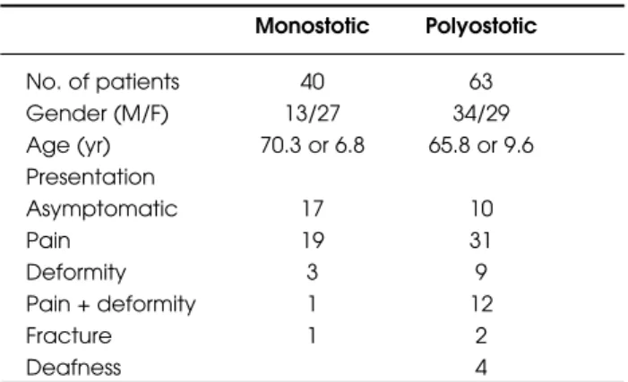
In the 103 patients of our cohort (14), the female/male ratio was 1.2:1, and ages ranged from 48 to 86 years. Forty patients presented with monostotic (27 women and 13 men, aged 70.3 ± 6.8 years), and sixty-three with polyostotic disease (29 women and 34 men, aged 65.8 ± 9.6 years). Most patients presented with symptoms (almost 50% with joint or bone pain), and 36% were asymptomatic (table 1). This suggests that our Institutional prevalence, as a reference center, may be underestimating the true prevalence in the general population, even with the use of biochemical screening.
Dutch ancestry, and seven had an uncertain ancestry. Blue eyes were present in 19 patients (18.5%) and a family history in 8 (7.8%).
Paget’s disease of bone is usually asympto-matic. In most cases it is detected accidentally through radiological findings or by the serum increase in alkaline phosphatase while other clinical conditions are being investigated (23). Pain and deformity are the most common presentations. The pain arises from the pagetic lesion itself or, more frequently, from indirect complications such as degenerative arthritis, nervous compression or osteosarcoma. Other major causes of pain are increased vascularization, distor-tions of the periosteum resulting from the disorgani-zation of bone remodeling and focal mechanical trau-mas (24). Hypertrophy of bone in the subchondral region may damage the cartilage, leading to osteoarthritis. Distinguishing pain of pagetic origin from that resulting from osteoarthritis is difficult. A response to the specific treatment for PDB may clari-fy this question.
The deformity affects mainly the long bones, skull and clavicles.
Pathological and/or traumatic fractures may arise as a result of pagetic lesions and the fissures may be complete or incomplete. Fractures of the femur occur more frequently than those of the tibia.
Diagnosis is mostly determined by radiogra-phy, the widening of bone being the most visible radi-ological feature. Other radiradi-ological findings include thickening of the cortex, osteolytic areas and osteosclerosis (25).
Lytic lesions arise in the initial phase and may acquire a focal aspect (osteoporosis circunscriptis) or a candle flame appearance. Next, areas of sclerosis appear, leading to the mixed aspect of lytic and scle-rotic areas. The radiological findings are usually
char-acteristic, but occasionally a differential diagnosis needs to be made with lytic or sclerotic metastases. The radionuclide bone scan is the most sensitive test in identifying PDB lesions, but is not specific and should be followed by plain X-rays. It is recommended that a bone scan be performed in all patients as part of the initial investigation. This procedure is intended to determine the distribution of the disease, identifying the sites affected and determining their potential for developing complications.
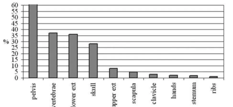
From our cohort the skeletal involvement was as follows: vertebrae: 38 patients (37%), long bones of the lower extremities: 37 patients (36%) and of the upper extremities: 8 patients (7.8%), pelvis: 64 patients (62%), skull 29 patients (28%), scapula: 5 patients (4.8%), clavicle: 3 patients (2.9%), metacarpals: 2 patients (2%) sternum: 2 patients (1.9%), and ribs: 1 patient (figure 1). Figures 2 and 3 show the percent-age of patients according to sites of skeletal involve-ment with respect to monostotic or polyostotic pre-sentation. More patients in the polyostotic group had involvement of pelvis, skull and vertebrae (56 versus 16, 20 versus 9, and 45 versus 4 respectively). Also infrequently affected sites like hands, scapula, clavicle and ribs were also seen more often in the polyostotic than in the monostotic presentation.
LABORATORY FEATURES AND MONITORING
In the biochemical evaluation of PDB the markers of bone remodeling are extremely useful. Clinically, the one most commonly used is serum alkaline phos-phatase. This marker, however, may be normal in around 10% of patients, as well as being subject to metabolic interference in patients with liver disease. In these cases, bone specific alkaline phosphatase or uri-nary markers of bone resorption may be helpful (26).
Markers such as C-Telopeptide (CTX) and N-telopeptide (NTX) have been employed. Telopeptides are type I collagen degradation products and may be measured by immunoassays, reflecting the degree of bone resorption. They are used as markers of the activ-ity of the disease and in the assessment of the response to therapy. In the terminal C telopeptides the α -aspar-tic acid present is converted to the βform of aspartic acid as the bone ages. This may be measured in human serum using monoclonal antibodies that recognize all the fragments of type I collagen that contain the β-8A octapeptide in duplicate (sβ-CTX) (27).
It has been postulated that the pagetic bone matrix decreases the degree of β-isomerization of
C-Table 1. Age, gender and clinical presentation of 103
patients with Paget’s Disease of Bone.
Monostotic Polyostotic
No. of patients 40 63
Gender (M/F) 13/27 34/29
Age (yr) 70.3 or 6.8 65.8 or 9.6 Presentation
Asymptomatic 17 10
Pain 19 31
Deformity 3 9
Pain + deformity 1 12
Fracture 1 2
terminal telopeptide, and some studies have demon-strated that the urinary excretion of non-isomerized forms (α-CTX) and urinary NTX, which may not be isomerized, are better predictors of disease activity than serum alkaline phosphate and urinary β-CTX (27,28). Also, anti-β-CTX antibodies tend to react more intensively with normal bone while the pagetic woven bone tends to stain predominately with anti α -CTX antibodies (29).
However, by using newer techniques for mea-surements of β-CTX in serum by electrochemolumi-nescence, it is possible to detect disease activity in those patients with normal serum AP. Pyridinoline and deoxypyridinoline, also employed as markers of bone remodeling, constitute the cross-links of the helicoidal structure of type 1 collagen whose concentration in the urine is proportional to the activity of the osteoclasts. Deoxipiridinoline is more sensitive than piridinoline.
In our study conducted at the Pernambuco Osteoporosis Center, serum alkaline phosphatase (AP) remains the most practical tool to evaluate and
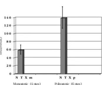
moni-tor disease activity in pagetic patients and is elevated in the majority of them. In the monostotic/polyostotic patients, serum AP was 2.2 ± 1.9 / 5.9 ± 2.8 (mean ± SD) times the upper limit of normal respectively. Eight patients had normal serum AP activity (seven had high urinary N-terminal-telopeptide [NTX] levels, and one had high serum β-isomerized C-terminal-telopeptide levels [β-CTX]). Ten patients had their urinary NTX measured, and 15 patients had their serum β-CTX measured. All of them showed values above 95o
cen-tile for normal individuals (figure 4 and 5).
COMPLICATIONS
The most frequent complications include the follow-ing: pathological fractures, bone deformities, degener-ative arthritis, loss of hearing, basilar invagination, nerve root or cord compression, and rarely hypercal-cemia during immobilization, increased cardiac output in cases with severe bone involvement and
osteosarco-Figure 1.Skeletal involvement in 103 patients with Paget’s Disease of Bone.
Figure 2.Skeletal involvement in 40 patients with monostotic
ma, the latter being a rare complication that occurs in only 0.7–1% of the cases (30). Deafness may be of the conduction type, owing to the involvement of the ossicles of the middle ear, of central origin, from com-pression of the auditory nerve, or a mixed form.
Calcium and phosphate in the blood are normal in the majority of patients with PDB. Nevertheless, hypercalcemia may occur as a result of prolonged immobilization. Primary hyperparathyroidism has been reported in patients with PDB, but it is unclear whether there is any relation between these two dis-eases, and compensatory secondary hyperparathy-roidism may also occur.
TREATMENT
The purpose of treatment is to restore normal bone metabolism, relieve bone pain and prevent future com-plications, particularly deformities of bone, secondary osteoarthritis, fracture and compression of nerve struc-tures, prepare for orthopedic surgery to reduce bleed-ing and control hypercalcemia due to immobilization. Calcitonin was the first osteoclast inhibitor to be used and nowadays represents the second treatment option in PDB. It suppresses bone turnover and alle-viates bone pain, but is more expensive, less effective, and causes more side effects than the bisphosphonates. It is given in an initial daily subcutaneous dose of 100 U for 3–6 months, after which the dose should be reduced (31). The normalization of alkaline phos-phatase is unusual and only occurs in patients with a
small increase in bone turnover. The suppression of disease activity does not persist for very long, even with the continuation of the drug. Antibodies develop in around 30–60% of the patients (32,33). Side effects occur in approximately 10% of the patients and include nauseas, a metallic taste and flushing.
The bisphosphonates represent the treatment of choice in PDB. They are pyrosphosphate analogues, whose oxygen bridge is replaced by a carbonate bound to several lateral chains. Their characteristic bindings of phosphorate-carbon-phosphorate (P-C-P) make them resistant to the hydrolysis of the phosphatases, allowing them to bind to the calcified bone matrix. They bind to the bone surfaces, preferably in areas of high bone turnover. When taken orally, they are poor-ly absorbed, ranging from 0.5 to 3%, especialpoor-ly in the presence of food or even of small amounts of calcium salts. For this reason they should be administered on an empty stomach. They may produce side effects in the upper gastrointestinal tract, such as heartburn, dyspepsia and esophageal ulcers, and caution should be exercised in administering them to patients with duodenitis and gastritis. They are contraindicated in those suffering from esophageal disease. Other side effects that may occur are acute febrile reaction and, more rarely, uveitis and rash. Rare occurrences of osteonecrosis of the mandible and maxilla have been reported in patients receiving new generation of bis-phosphonates (34,35). Most osteonecrosis cases have occurred in cancer patients. No cases were reported with zolidronic acid in the treatment of Paget’s dis-ease. The first-generation bisphosphonates, such as
Figure 4.Serum β-CTX in 15 patients with Paget’s Disease of
Bone. Reference range for premenopausal women: 80–450 pg/ml.
nmol/mmolCr
pg/ml
Figure 5.Urinary NTX in 10 patients with Paget’s disease of
etidronate, are weak antiresorptives, while the second and third generation ones, which contain nitrogen in their molecules (amino bisphosphonates) are much more powerful. They inhibit farnesil pyrophosphatase synthase, an enzyme essential for the prenilation of small G proteins along the cholesterol synthesis path-way which, when inhibited, triggers failure of the resorptive function, leading to cell death (36).
The first bisphosphonate to be used in PDB was etidronate. New, more potent biphosphonates have proved to be more effective, leading to more pro-longed periods of remission. Etidronate was used for the first time in the treatment of PDB in 1971 (37). The recommended dose is 5 mg/kg/day (average dose 400 mg/day) for 6 months (37,38). In general, patients whose disease is very active show a moderate clinical and biochemical improvement and a rapid relapse after the medication is suspended, tending to become more resistant after a repeated course of ther-apy. Histological studies of bone have shown osteo-malacia in both pagetic and nonpagetic bone follow-ing treatment with 10–20 mg/day per day, but not with a dose of 5 mg/day (39,40).
Pamidronate is 10 to 100 times more potent than etidronate and produces a reduction of bone remodeling in 60–70% of patients (41,42). It is used parenterally in a single IV infusion of 60 mg in cases in which there is little disease activity (alkaline phos-phatase 2–3 times above the normal maximum value). Larger doses (90–180 mg), which may be given for three or four days, may be preferable in patients with more pronounced disease activity (43). The maximum dose given in a single day is 90 mg, diluted in a saline or glucose solution for 4–6 hours.
Oral alendronate is more effective than etidronate in the treatment of PDB. It may be used in a dose of 20–40 mg/day for 6 months (44). In a dose of 40 mg/day for 6 months it leads to a 77% decrease in alkaline phosphatase, compared with the 44% decrease produced by etidronate (45). The normaliza-tion of alkaline phosphatase is also more frequent in patients treated with alendronate than in those treated with etidronate (63.4% vs. 17%).
Tiludronate is recommended in a dose of 400 mg/day for 3 months, normalizing alkaline phos-phatase in 35% of patients (46). It is more effective than etidronate and does not cause demineralization of bone (47).
Clodronate is more potent than etidronate and does not lead to defects of mineralization. It should be given intravenously in a daily dose of 300 mg for 5 days (48), but is usually less effective than pamidronate.
Risedronate in a daily dose of 30 mg for 2 months leads to the normalization of alkaline phos-phatase in 73% of patients, compared with a 15% decrease in those on a daily dose of 400 mg of etidronate for 6 months. Sixteen months after the ces-sation of medication, 53% of the patients on rise-dronate remain in remission, compared with 14% of those on etidronate (49). In one study patients with resistance to calcitonin and pamidronate, associated with severe bone involvement, risedronate caused a significant reduction in the levels of serum alkaline phosphatase (50).
Ibandronate is another bisphosphonate that has also been used safely and efficaciously in PDB in an intravenous dose of 2 mg (51).
Zoledronic acid, also known as zoledronate, is 10,000 times more potent than etidronate and 100 times more potent than pamidronate (52). It is used intravenously for 15–20 minutes. Patients with resis-tance to other bisphosphonates usually respond to zole-dronic acid (53), which is highly effective in reducing the biochemical markers of bone remodeling (54).
Zoledronic acid may lead to more rapid and prolonged remission in the treatment of PDB than risedronate (55). When evaluated for 6 months after a single 5 mg infusion for 15 minutes, resulting in the normalization of alkaline phosphatase or in a fall of at least 75%, it leads to a 96% decrease in alkaline phos-phatase, compared with a 74.3% decrease with a daily 30 mg dose of risedronate for 3 months. More patients in the zoledronic acid group achieved normal alkakine phosphotase than those in the risedronate group (88.6% vs. 57.9%).
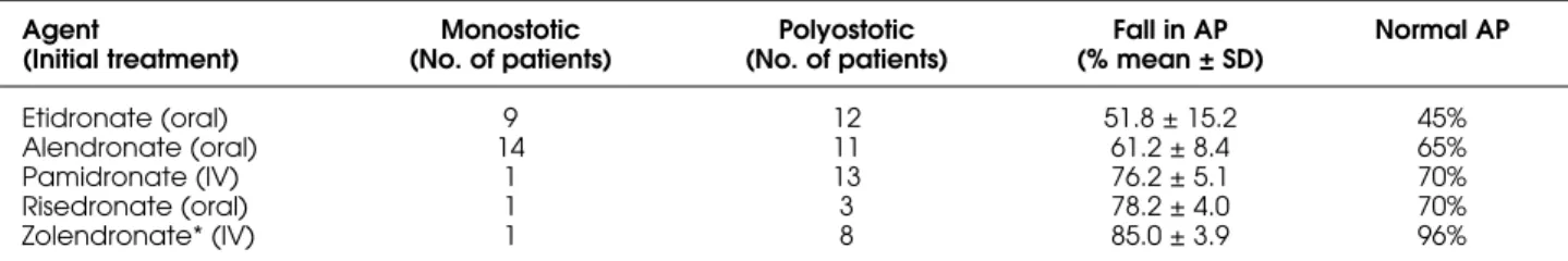
All patients treated with bisphosphonates should be given a calcium and vitamin D supplement. Table 2 shows the initial therapeutic responses with calcitonin and the most commonly used bisphos-phonates in a group of patients from our cohort. The fall in serum AP, after a median follow-up of 6 months, was as follows: 51.8 ± 15% with oral etidronate, 61.2 ± 8.4% with oral alendronate, 76.2 ± 5.1% with intra-venous pamidronate, 78.2 ± 4.0 with oral risedronate, and 85.0 ± 3.9% with intravenous zoledronic acid.
A surgical procedure is indicated in certain situ-ations, such as a hip replacement, severe osteoarthritis, osteotomy for the correction of a deformed tibia, occipital craniotomy for decompression of the posteri-or fossa in patients with platybasia and fposteri-or the decom-pression of nerves.
zoledro-nic acid in order to decrease hypervascularity, thereby reducing bleeding during surgery.
Regarding the control of pain, when the pain is directly attributable to PDB, it is usually relieved by antiresorptive treatment (bisphosphonate or calci-tonin). When pain is the result of bone deformity or secondary osteoarthritis, acetominofen, nonsteroid anti-inflammatories or cox-2 inhibitors may be of use.
FOLLOW-UP
The patient is considered to be in remission when the normal levels of biochemical markers, such as alkaline phosphatase, have been reached and in partial remission when there is a fall of over 75% three to six months after the start of treatment. Alkaline phosphatase should be measured every four to six months after the course of therapy and a new treatment should be given if the alka-line phosphatase is above normal or above the previous lowest value. Bone resorption markers such as C-telopeptide exhibit a high sensitivity, especially in indi-viduals with normal alkaline phosphatase (56).
REFERENCES
1. Paget J. On a form of chronic inflammation of bone (osteitis deformans). Medico-cirurgical Transactions of
London 1887;60:37-63.
2. Kanis JA. Pathophysiology and treatment of Paget’s
dis-ease of bone. London: Carolina Academic Press/ Martin
Dunitz, 1991.
3. Bandeira F, Caldas G, Griz L, Macedo G, Marinho C, Bandeira C. Paget’s disease of bone – Characteristics of 49 patients from a single institution in Recife, Brazil. J
Bone Miner Res 1999;14(suppl. 1):S539.
4. Cooper C, Schafheutle K, Denninson E, Kellingray S, Guyer P, Barker D. The epidemiology of Paget’s disease in Britain: Is its prevalence decreasing? J Bone Miner Res
1999;14:192-7.
5. Tiegs RD, Lohse CM, Wollan PC, Melton LJ. Long-term trends in the incidence of Paget’s disease of bone. Bone
2000;27:222-5.
6. Palazzi N, Cuadra C, Rondon E. Enfermedad de Paget óseo: a propósito de una casuística venezolana. Med
Intern (Caracas) 1993;9:161-9.
7. Sanchez A, Zuniga LR, Iglesias A. Enfermedad ósea de Paget. Acta Med Colomb 1996;21:255-61.
8. Gonzalez G, Brusco F, Arteaga L, Rodriguez J, Jaco-belli S, Massardo L, et al. Enfermedad de Paget en Chile: una serie de 15 pacientes. Rev Med Chile 2003;131:491-7.
9. Acotto CG, Mautalen CA. European origin of patients with Paget’s disease of bone in the Buenos Aires area.
Euro J Epid 2001;17:409-11.
10. Bandeira F, Alencar S, Caldas G, Griz L, Macedo G, Mar-inho C, et al. Paget’s disease of bone revisited – a study on 84 patients. Arq Bras Endocrinol Metab 2002;46(suppl. 1):S364.
11. Kauffman TN. The Jewish presence in
Pernambuco-Brazil. Lost footsteps, history recovered. 1st ed, Recife:
Editora Bagaço, 2005. p. 271.
12. Sathler M. Aspectos dinâmicos da doença de Paget.
Rev Bras Ortop 1995;30:330-6.
13. Ribeiro ML, Carvalho EA, Fernandes JL. Avaliação por imagem da doença de Paget do osso. Radiol Bras
1999;32:69-74.
14. Bandeira F, Griz L, Caldas G, Macedo G, Marinho C, Moutelik M, et al. A single center experience of 103 cases. Paget’s disease of bone in Brazil. Proceedings of
the International Symposium on Paget’s Disease of Bone/Fibrous Dysplasia: Advances and Challenges 2006. The Paget’s Foundation, National Institute of Health, pp 53.
15. Siris ES. Epidemiologic aspects of Paget’s disease: Fami-ly history and relationship to other medical conditions.
Semir Arthritis Rheum 1994;23:222.
16. Siris ES, Ottoman R, Flaster E, Kelsey JL. Familial aggrega-tion of Paget’s disease of bone. J Bone Miner Res
1991;6:495.
17. Roodman GD, Windle J. Paget disease of bone. J Clin
Invest 2005;115:200-8.
18. Friedrichs WE, Sakamuri VR, Bruder JM, Cundy T, Cornish J, Singer F, et al. Sequence analysis of measles virus nucleocapsid transcripts in patients with Paget’s dis-ease. J Bone Miner Res 2002;17:145-51.
Table 2.Initial (6 months) therapeutic responses in 77 patients with Paget’s Disease of Bone. Three patients were treated with
SC, with 37% fall in AP. One patient was treated with IV Clodronate, with 60% fall in AP.
Agent Monostotic Polyostotic Fall in AP Normal AP
(Initial treatment) (No. of patients) (No. of patients) (% mean ± SD)
Etidronate (oral) 9 12 51.8 ± 15.2 45%
Alendronate (oral) 14 11 61.2 ± 8.4 65%
Pamidronate (IV) 1 13 76.2 ± 5.1 70%
Risedronate (oral) 1 3 78.2 ± 4.0 70%
Zolendronate* (IV) 1 8 85.0 ± 3.9 96%
19. Janssens K, Van Hul W. Molecular genetics of too much bone. Hum Mol Genet 2002;11:2385-93.
20. Helfrich MH, Hobson RP, Grabowski PS, Zurbriggen A, Cosby SL, Dickson GR, et al. A negative search for a paramyxoviral etiology of Paget’s disease of bone: mol-ecular, immunological, and ultrastructural studies in UK patients. J Bone Miner Res 2000;15:2315-29.
21. Walsh JP. Paget’s disease of bone. Med J Aust
2004;181:262-5.
22. Rima BK, Gassen U, Helfrich MH, Ralston SH. The pros and cons of measles virus in Paget’s disease. J Bone Miner
Res 2002;17:2290-2.
23. Langston AL, Ralston SH. Management of Paget’s dis-ease of bone. Rheumatology 2004;43:955-9.
24. Hosking D, Meunier PJ, Ringe JD, Reginster J, Gennari C. Fortnightly review: Paget’s disease of bone: diagnosis and management. Br Med J 1996;312:491-4.
25. Delmas PD, Meunier PJ. The management of Paget’s disease of bone. N Engl J Med 1977;336:558-66.
26. Alvarez L, Peris P, Pons F, Guanabens N, Herranz R, Monegal A, et al. Relationship between biochemical markers of bone turnover and bone scintigraphic indices in assessment of Paget’s disease activity. Arthritis
Rheum 1997;40:461-8.
27. Alexandersen P, Peris P, Guanabens N, Byrjalsen I, Alvarez L, Solberg H, et al. Non-isomerized c-telopeptide fragments are highly sensitive markers for monitoring dis-ease activity and treatment efficacy in Paget’s disdis-ease of bone. J Bone Miner Res 2005;20:588-95.
28. Garnero P, Fledelius C, Gineyts E, Serre CM, Vignot E, Delma PD. Decreased b-isomerization of C-telopeptides of a1 chain of type I collagen in Paget’s disease of bone. J Bone Miner Res 1997;12:1407-15.
29. Delmas PD. Biochemical markers of bone turnover in Paget’s disease of bone. J Bone Miner Res
1999;14(suppl. 2):66-9.
30. Hadjipavlou A, Lander P, Srolovitz H, Enker IP. Malignant transformation in Paget’s disease of bone. Cancer
1992;70:2808.
31. Singer FR. Clinical efficacy of salmon calcitonin in Paget’s disease of bone. Calcif Tissue Int 1991;49(suppl. 12):57.
32. Singer FR, Aldred JP, Neer RM, Krane SM, Potts JT jr, Bloch KJ. An evaluation of antibodies and clinical resistance to salmon calcitonin. J Clin Invest 1972;51:2331-8.
33. Dietrich FM, Fisher JA, Bijvoet OLM. Formation of anti-bodies to synthetic human calcitonin during treatment of Paget’s Disease. Acta Endocricol 1979;92:468-76.
34. Ferrugia MC, Summerlin DJ, Kroviak E, Huntley T, Free-man S, Borrowdale R, et al. Osteonecrosis of mandible/maxilla and use of new bisphosphonates.
Laryngoscope 2006;115-20.
35. Hoff AO, Toth B, Altundag K, Guarneri V, Nooka A, Desrouleaux, et al. Osteonecrosis of the jaw in patients receiving intravenous bisphosphonates therapy. J Bone
miner Res 2005;20(suppl. 1):S53
36. Rassel RRG, Benford HL, Fleisch HA, et al. The pharma-cology of bisphosphonates and new insights into their mechanism of action. J Bone Miner Res 1999;14(suppl. 2).
37. Smith R, Russell RGG, Bishop M. Bisphosphonates and Paget’s disease of bone. Lancet 1971;1:945-47.
38. Alman RD, Johnston CC, Khairi MRA, Wellman H, Scrafini NA, Sankey RR. Influence of disodium etidronate on clin-ical and laboratory manifestations of Paget’s disease of bone. N Engl J Med 1973;289:1379-84.
39. Meunier PJ, Revault A. Treatment of Paget’s disease with etidronate disodium. In: Singuer FR, Wallach S (eds).
Paget’s disease of bone: Clinical assessment, present
and future therapy. New York: Elsvier, 1991. pp. 86-99.
40. Alexandre CM, Collins B. Disodium etidronate in Paget’s disease: 11-year study of 93 patients. Arthritis Rheum
1983;26(suppl.):S9.
41. Frijlink WB, Bijvoet OLM, Heynen G. Treatment of Paget’s disease with (3-amino-1-hydroxypropylidene)-1, 1-bis-phosphonate (A.P.D.). Lancet 1979;1:799-803.
42. Cantrill JA, Anderson DC. Treatment of Paget’s disease of bone. Clin Endocrinol 1990;32:507-18.
43. Thiebaud D, Jaeger P, Gobelet C. A single infusion of the bisphosphonate (AMP) as treatment of Paget’s disease of bone. Am J Med 1988;85:207-12.
44. Reid IR, Siris E. Alendronate in the treatment of Paget’s disease of bone. Int Clin Pract 1999;101:62-6.
45. Siris ES, Weinstein RS, Altman R, Conte JM, Favus M, Lom-bardi A, et al. Comparative study of alendronate and etidronate for the treatment of Paget’s disease of bone.
J Clin Endocrinol Metab 1996;81:961-7.
46. McClung MR, Tou CK, Goldstein NH, Picot C. Tiludronate therapy for Paget’s disease of bone. Bone
1995;17(suppl.):493S-6.
47. Roux C, Gennari C, Farrerons J, Devogelaer JP, Mulder H, Kruse HP, et al. Comparative prospective, double-blind, multicenter study of the efficacy of tiludronate and etidronate in the treatment of Paget’s disease of bone. Arthritis Rheum 1995;38:851-8.
48. Yates AJ, Percival RC, Gray RE, Atkins RM, Urwin GH, Hamdy NA, et al. Intravenous clodronate in the treat-ment of Paget’s disease of bone. Lancet 1985;1:1474-7.
49. Miller PD, Brown JP, Siris ES, Hoseyni MS, Axelrod DW, Bekker PJ. A randomized, double-blind comparison of risedronate and etidronate in the treatment of Paget’s disease of bone. Paget Risedronate / Etidronate Study Group. Am J Med 1999;106:513-20.
50. Singuer FR, Clemens TL, Eusebio RA, Bekker PJ. Rise-dronate, a highly effective oral agent in the treatment of patients with severe Paget’s disease. J Clin Endocrinol
Metab1998;83(6):1906-10.
51. Grauer A, Heichel S, Knaus J, Dosch E, Ziegler R. Iban-dronate treatment in Paget’s disease of bone. Bone
1999;24(suppl.5):87S-9.
52. Fleisch H. Bisphosphonates in bone disease from the
53. Chung G, Keen RN. Zoledronate treatment in active Paget disease. Ann Rheum Dis 2003;62:275-6.
54. Bandeira F, Griz L, Caldas G, Saraiva W, Carvalho W, Rosado VA. Serum C-telopeptide and alkaline phos-phatase changes following a single intravenous infusion of zoledronic acid in patients with Paget’s disease. J
Bone Miner Res 2003;18:(suppl):S389.
55. Reid IR, Miller P, Lyles K, Fraser W, Brown J, Saidi Y, et al. Comparison of a single infusion of zoledronic acid with risedronate for Paget’s disease. N Engl J Med 2005; 353:898-908.
56. Bandeira F, Griz L, Caldas G, Bandeira C. Paget’s dis-ease of bone with normal serum alkaline phosphatase
activity: effects of bisphosphonates on clinical symp-toms and bone markers. J Bone Miner Res 2001;16 (suppl):S301.
Address for correspondence:
Francisco Bandeira Department of Medicine University of Pernambuco Division of Endocrinology Agamenon Magalhães Hospital Dilab laboratories
Rua da Hora 378