758
https://doi.org/10.1590/0004-282X20170143
IMAGES IN NEUROLOGY
Dissecting superior cerebellar artery
aneurysm: spontaneous resolution in a
long-term follow-up
Aneurisma dissecante de artéria cerebelar superior: resolução espontânea após
seguimento de controle
Marcos Vinicius Tadao Fujino
1, Rogério Iquizli
1, Thiago Abud
1, Carlos Eduardo Baccin
1, Gisele Sampaio Silva
1,2,
Orlando G. Barsottini
1,2, José Luiz Pedroso
1,2A 48-year-old woman presented with sudden posterior
neck pain 12 hours before admission. Past medical history
was unremarkable. Neurological examination was normal.
A brain CT scan and cerebrospinal luid were normal. A MRI
angiography (MRA) and digital angiography conirmed a dis
-secting superior cerebellar artery (SCA) aneurysm (Figure 1).
We decided for noninvasive therapy. Six months later, the
MRA showed complete resolution (Figure 2).
1Hospital Israelita Albert Einstein, São Paulo SP, Brasil;
2Universidade Federal de São Paulo, Departamento de Neurologia, São Paulo SP, Brasil.
Correspondence: José Luiz Pedroso; Departamento de Neurologia da UNIFESP; Av. Santa Catarina; 04378-200 São Paulo SP, Brasil; E-mail: zeluizpedroso@yahoo.com.br
Conflict of interest: There is no conflict of interest to declare.
Received 15 January 2017; Accepted 18 July 2017.
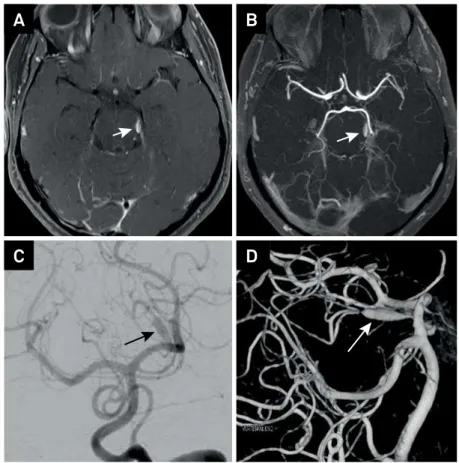
Figure 1.
Axial post-contrast brain MRI (vessel wall imaging) shows abnormal asymmetric vessel wall enhancement in left
superior cerebellar artery (A); axial 3D-TOF MRI angiography shows segmental ectasia in the left superior cerebellar artery (B).
Digital subtraction angiogram of the left vertebral artery and 3D reconstructions confirmeda dissecting superior cerebellar artery
aneurysm (lateral pontomesencephalic segment) (arrows) (C and D).
A
B
759
Fujino MVT et al. Spontaneous resolution of dissecting aneurysmFusiform aneurysms of the SCA related to dissection
are rare
1,2. Treatment strategies are usually aggressive and
include: aneurysm clipping, arterial bypasses and artery
oclusion
1,2,3. Our report suggests that noninvasive therapy
should be considered as an option for unruptured fusiform
aneurysms of the SCA related to dissection.
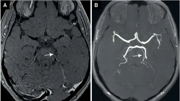
Figure 2.
Six-month follow-up. Axial post-contrast brain MRI (vessel wall imaging) shows no enhancement in left superior
cerebellar artery (A); axial 3D-TOF MRI angiography shows no artery aneurysm (B).
A
B
References
1. Lamis FC, De Paiva Neto MA, Cavalheiro S. Fusiform superior cerebell arartery aneurysm treated with STA-SCA by passand trapping. Surg Neurol Int. 2014;5(Suppl 4):S139-42. https://doi.org/10.4103/2152-7806.134806
2. Kang IH, Malla HP, Lee SH, Park CK, Choi SK. Revascularization as treatment of a ruptured fusiform aneurysm at the cortical segment of the superior
cerebellar artery: a case report and literature review. J Neurol Surg A Cent Eur Neurosurg. 2017;78(3):302-5. https://doi.org/10.1055/s-0036-1582436