DOI: 10.14260/jemds/2015/971
CASE REPORT
J of Evolution of Med and Dent Sci/ eISSN- 2278-4802, pISSN- 2278-4748/ Vol. 4/ Issue 38/ May 11, 2015 Page 6696
ENDOSCOPIC MARSUPIALISATION OF THORNWALDT’S CYST
Manish Ajit Patankar1, Yogesh Dabholkar2,Haritosh Velankar3, Yuvaraj Patil4
HOW TO CITE THIS ARTICLE:
Manish Ajit Patankar, Yogesh Dabholkar, Haritosh Velankar, Yuvaraj Patil. Endoscopic Marsupialisation of
Thornwaldt’s CYST . Journal of Evolution of Medical and Dental Sciences 2015; Vol. 4, Issue 38, May 11; Page: 6696-6699, DOI: 10.14260/jemds/2015/971
ABSTRACT: Thornwaldt’s cyst is almost always asymptomatic and usually diagnosed incidentally on nasal endoscopy examination. Radiological examination particularly MR imaging is useful to diagnose the cyst & differentiated from meningocele or menigoencephalocele. Various therapeutic approaches for removal of cyst have been described. We present a case of Thornwaldt’s cyst which was managed successfully by the endoscopic approach.
KEYWORDS: Thornwaldt’s cyst; nasopharyngeal Bursa; endoscopic marsupialization; nasoph-aryngeal mass.
INTRODUCTION: Thornwaldt’s cyst also known as pharyngeal bursa, is a rare midline cyst found in the nasopharynx.1 It is found in the midline of the posterior wall of nasopharynx. It has an incidence
of 3% in the adult population.2 Most cases are diagnosed during second & third decades of life, with
higher prevalence in males.2
Thornwaldt’s bursa also known as nasopharyngeal bursa is a recess in the midline of the nasopharynx which is produced by persistent notochord remnants. If the opening of the bursa is occluded, a benign midline nasopharyngeal mucosal cyst called Thornwaldt’s Cyst develops.
CASE REPORT: A 41 years old man presented with progressive nasal obstruction since last 4 years, which would aggravate on lying down. He also complained of a continuous dull occipital headache, a foreign body sensation in the nasopharynx and a sensation of ear fullness. On examination there was a post nasal drip with a foul smell.
A diagnostic nasal endoscopy revealed a smooth, yellowish, submucosal, cystic swelling in the midline in the nasopharynx.
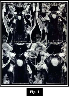
MRI neck (Plain & Contrast) revealed a 2.0x1.5x1.3 cm sized, abnormal well defined cyst in the midline of the posterior wall of the nasopharynx, appearing hyper intense on T2 weighted & STIR sequences and iso to hyper intense on T weighted sequences, suggestive of Thornwaldt’s cyst.
Histopathological examination showed a cyst lined by pseudo-stratified ciliated columnar epithelium, composed of dense fibro-collagenous tissue with a few areas of chronic inflammatory
cells & hemorrhage, consistent with Thornwaldt’s cyst. There was no evidence of atypia or
malignancy.
DISCUSSION: Thornwaldt’s cyst is a rare disease, frequently missed as it is asymptomatic. The diagnosis requires a high index of suspicion based on clinical presentation and is aided by investigations, particularly MRI and histopathological examination.
The German physician Gustav Ludwig Thornwaldt classified the symptoms into proximal and associated symptoms.
DOI: 10.14260/jemds/2015/971
CASE REPORT
J of Evolution of Med and Dent Sci/ eISSN- 2278-4802, pISSN- 2278-4748/ Vol. 4/ Issue 38/ May 11, 2015 Page 6697 chronic gastritis, reflex cough. Most common symptoms are persistent nasal discharge, dull
continuous occipital headache and halitosis.3
Our case also presented with a progressive nasal obstruction and purulent post nasal drip as proximal symptoms and dull occipital headache and blocking of the ear as associated symptoms.
MRI of nasopharynx is a sensitive method for detecting and evaluating cystic lesions of the nasopharynx.4Thornwaldt’s cyst has characteristic high intensity on T -weighted, and intermediate
to high signal intensity on T1 weighted MRI imaging.5
MRI reported an abnormal well defined lesion in the midline of the posterior wall of the nasopharynx which appears as a hyper-intense on T2W and STIR sequences and iso to hyper intense on T1W sequences.
By a transnasal endoscopy approach Thornwaldt’s cyst was drained & marsupialized. A
follow up endoscopy after two months did not reveal any recurrence.
Histopathological examination revealed a cyst lined by pseudo-stratified ciliated columnar epithelium, and composed of a dense fibro- collagenous tissue with lymphocytic infiltrates consistent with Thornwaldt’s cyst.
Thornwaldt’s cyst can be differentiated from adenoid retention cyst as Thornwaldt’s cyst wall has lymphocytic infiltrates & lack of lymphoid follicles whereas adenoid retention cyst has abundant lymphoid tissue, many inflammatory cells & germinal centers.6
Thornwaldt’s cyst is differentiated from Rathke’s pouch (diverticulum of nasopharyngeal
mucosa, a remnant of buccopharyngeal membrane invagination to form anterior lobe of pituitary) by the presence of cylindrical ciliated columnar epithelium as compared to internal stratified squamous epithelium found in Rathke’s pouch.
Asymptomatic cyst requires no treatment. The treatment of choice for Thornwaldt’s cyst is complete exenteration or marsupialization. Endoscopic approach provides excellent visualization of nasopharynx and avoids damage to the Eustachian tube openings.7 Thus, it is the preferred approach
for marsupialization of most cysts. Larger cysts require trans-palatal approach for removal.
CONCLUSION: Thornwaldt’s cyst being asymptomatic in many cases goes undetected. A thorough clinical history, physical examination and investigations like MRI and histopathology will confirm the diagnosis.
A trans-nasal endoscopic approach for marsupialization is a safe and effective surgery for Thornwaldt’s cyst.
REFERENCES:
1. AE EL- Shazly, S Barriat, P. P. Lefebvre. Nasopharyngeal bursitis: from Embryology to clinical presentation. American Journal of Clinical Hypnosis 2011; 53 (4): 331-4.
2. Marco Antonio Thomas Caliman, Erika Mucciolo Cabernite et al; Thornwaldts cyst -treatment
with diode laser ; Braz. j. Otorhinolaryngol Sao Paulo Sept/Oct 2013;79 (5).
3. Miller RH, Sneed WF; Thorwaldt’s bursa ; Clin Otolaryngol Allied sci : : -5.
4. Bouche. RM, Hendrix R A, Guttenplan MD; the diagnosis of Thorwaldt’s cyst , Trans Pa Acad Opthalmol otolaryngol 1990; 42: 1026-30.
DOI: 10.14260/jemds/2015/971
CASE REPORT
J of Evolution of Med and Dent Sci/ eISSN- 2278-4802, pISSN- 2278-4748/ Vol. 4/ Issue 38/ May 11, 2015 Page 6698 6. Miyahara H, Matsunaga T; Thornwaldt’s disease . Acta Otolaryngol suppl : : -9. 7. Koksal yucca, yasin kursad varsak; Thornwaldts cyst ; Eur J gen med 2012; 9 (suppl): 26-29.
Figure 1: Coronal view of MRI neck (plain+ contrast) shows 2.0x1.5x1.3 cm size well defined lesion in midline of posterior wall of nasopharynx appearing as hyper-intenseon T1w & T2w sequences.
Figure 2: Axial view of MRI neck Shows Hyperintense lesion in midline of posterior nasopharynx.
Fig. 1
DOI: 10.14260/jemds/2015/971
CASE REPORT
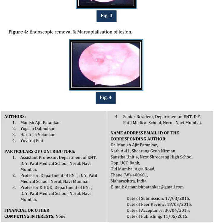
J of Evolution of Med and Dent Sci/ eISSN- 2278-4802, pISSN- 2278-4748/ Vol. 4/ Issue 38/ May 11, 2015 Page 6699 Figure 3: Endoscopic image of midline lesion of posterior Nasopharynx.
Figure 4: Endoscopic removal & Marsupialisation of lesion.
AUTHORS:
1. Manish Ajit Patankar
2. Yogesh Dabholkar
3. Haritosh Velankar
4. Yuvaraj Patil
PARTICULARS OF CONTRIBUTORS:
1. Assistant Professor, Department of ENT,
D. Y. Patil Medical School, Nerul, Navi Mumbai.
2. Professor, Department of ENT, D. Y. Patil
Medical School, Nerul, Navi Mumbai. 3. Professor & HOD, Department of ENT,
D. Y. Patil Medical School, Nerul, Navi Mumbai.
FINANCIAL OR OTHER
COMPETING INTERESTS: None
4. Senior Resident, Department of ENT, D.Y.
Patil Medical School, Nerul, Navi Mumbai.
NAME ADDRESS EMAIL ID OF THE CORRESPONDING AUTHOR:
Dr. Manish Ajit Patankar,
Nath A-41, Sheerang Gruh Nirman
Sanstha Unit 4, Next Shreerang High School, Opp. UCO Bank,
Old Mumbai Agra Road, Thane (W)-400601, Maharashtra, India.
E-mail: drmanishpatankar@gmail.com
Date of Submission: 17/03/2015. Date of Peer Review: 18/03/2015. Date of Acceptance: 30/04/2015. Date of Publishing: 11/05/2015.
Fig. 3